Diplopia Charting: Double Vision Test (www.healthkura.com)
•
6 gostaram•687 visualizações

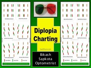
Diplopia Charting (Visit https://healthkura.com/diplopia-charting-double-vision-test/) All about Diplopia Charting » - Diplopia Charting Principle - Diplopia Chart Procedure - Diplopia Chart Interpretation - Diplopia Charting Precaution - How to record Diplopia - Hess Charting Diplopia charting of Rectus muscles & Diplopia Charting of Oblique muscles of both Eyes - Right Eye and Left Eye. Diplopia charting is the subjective method of double vision test in which the recording of the separation of double or diplopic images is made in the primary position as well as other cardinal gazes (nine positions of gaze) Source: https://healthkura.com/diplopia-charting-double-vision-test/

Recomendados
Recomendados
Mais conteúdo relacionado
Mais de Bikash Sapkota
Mais de Bikash Sapkota (6)
Último
Último (20)
Diplopia Charting: Double Vision Test (www.healthkura.com)
- 2. Diplopia Charting: Common Method of Double Vision Test
- 3. Double Vision Test: Diplopia Charting Effective management of diplopia or double vision is possible only after the diagnosis of the root cause of diplopia. If the double vision is due to neurological cause, then diplopia charting will help to determine which of the extraocular muscle(s) is/are more affected. Diplopia chart is the common double vision test. You can Get Full Detail about Diplopia charting ON Health Kura
- 4. Double Vision Test: Diplopia Charting Diplopia charting is the subjective method of double vision test in which the recording of the separation of double or diplopic images is made in the primary position as well as other cardinal gazes (nine positions of gaze). It is a common ophthalmic procedure employed to helping with diagnosing ophthalmoplegia (ocular muscle’s paresis or palsy). You can Get Full Detail about Diplopia charting ON Health Kura
- 5. Double Vision Test: Diplopia Charting If the single extraocular muscle is affected, diplopia charting gives an accurate result, but it may be difficult to pinpoint the more affected paretic muscles in the case of multiple muscle paresis or palsy. So, other methods of diplopia tests may be utilized to measure the separation of the diplopic images and to diagnose the cause of diplopia along with diplopia charting, such as Hess charting. Diplopia chart gives a symbolic or pictorial record of double vision in situations where there is a separation of two images, that changes with change in position of gaze. You can Get Full Detail about Diplopia charting ON Health Kura
- 6. Double Vision Test: Diplopia Charting As it is entirely a subjective method of double vision test, the reliability of the diplopia charting depends on the response of the patient. The chart can only be plotted in patients who are cooperative and can appreciate the diplopia and with comitant and incomitant deviation of eyes. Plotting of diplopia fields is indicated in patients with complaints of confusion or double vision. You can Get Full Detail about Diplopia charting ON Health Kura
- 7. Double Vision Test: Diplopia Charting What is the principle of diplopia charting? The diplopia chart is based on the haploscopic principle, foveal projection, Hering’s Law of Equal Innervation, and Sherrington’s Law of Reciprocal Innervation. In short, the concept is each retinal point has its own value of direction in gazes. You can Get Full Detail about Diplopia charting ON Health Kura
- 8. Double Vision Test: Diplopia Charting Types of Double Vision Test 1.Simple method 2.Electronic devices (Quantitative measurement of actions of extraocular muscles) •Lancaster red-green test •Hess charting (screen test) •Lees screen test •Interpretation method (new) You can Get Full Detail about Diplopia charting ON Health Kura
- 9. Double Vision Test: Diplopia Charting What are the indications for the double vision test? If diplopia is due to incomitant deviation, diplopia charting can be an aid for diagnosis and for follow up. Based on the record of diplopia and separation in different gazes, the eye doctor can determine whether the diplopia or confusion is improving or worsening. You can Get Full Detail about Diplopia charting ON Health Kura
- 10. Double Vision Test: Diplopia Charting Required materials for the Diplopia Charting Light source (linear): linear light allows the patient to see a distorted or tilted image if there is cyclotropia. A pair of red and green goggle You can Get Full Detail about Diplopia charting ON Health Kura
- 11. Double Vision Test: Diplopia Charting The procedure of diplopia charting as a double vision test Before starting the test, make sure the required materials such as linear light, R-G glasses, color markers, diplopia chart sheet, and comfortable chair for patient and examiner, are available. If the patient is cooperative, he can be given 2 pencils (or told to raise two fingers of two hands) and asked to hold them exactly as he sees the 2 lights. You can Get Full Detail about Diplopia charting ON Health Kura
- 12. Double Vision Test: Diplopia Charting The patient is allowed to sit comfortably on the chair with his head erect and still throughout the examination. Turn off the room light as the test is carried out in a dark room. The procedure should be explained to the patient in detail because the test is completely dependent on the patient’s response. You can Get Full Detail about Diplopia charting ON Health Kura
- 13. Double Vision Test: Diplopia Charting R-G glasses are fitted in such a way that the red glass is in front of the right eye (and green to the left). Armstrong Goggles are preferred as these are shaped to fit properly on the orbital margin, so that patient would be looking only through the colored area. The examiner holds the linear light vertically in the primary position at a distance of 50 cm (or 1 meter). The test distance should be clearly mentioned on the chart sheet. The linear light can be held horizontally for the easy assessment of diplopia if the complaint is of vertical separation of images. You can Get Full Detail about Diplopia charting ON Health Kura
- 14. Double Vision Test: Diplopia Charting The patient is allowed to look at the light and is asked about the nature of diplopia. Ask the patient which colored image appears in front of him and which side does he see the second image. Also ask about the nature of images, whether they are vertical, horizontal, or tilted. You can Get Full Detail about Diplopia charting ON Health Kura
- 15. Double Vision Test: Diplopia Charting Two things might happen when the examiner project light to the primary gaze of the patient: a single image or a double image. If the patient comments as seeing a single image, the examiner must find out whether it is a suppressed or fused image. If one image is obscured due to large image separation or due to the size of the nose, there may not be a comment of diplopia. You can Get Full Detail about Diplopia charting ON Health Kura
- 16. Double Vision Test: Diplopia Charting The position in which double vision appears and is maximal is to be noted if there is no double vision in the primary position or gaze. The relative position and nature of the two images are noted if the patient sees a double image. Similarly, the light is projected in the remaining 8 cardinal positions of gaze and the relative separation of images is noted. The area of single vision, minimum separation, and maximum separation along with estimated separation value is mentioned pictorially on the chart sheet. You can Get Full Detail about Diplopia charting ON Health Kura
- 17. Double Vision Test: Diplopia Charting Interpretation of the diplopia chart While interpreting the diplopia chart in a double vision test, two important points that every examiner should consider are the positions of gaze at which the diplopia is present and the position of gaze at which the image separation is maximum. The position of maximum vertical and horizontal separation of images and the position of maximum torsion is noted. You can Get Full Detail about Diplopia charting ON Health Kura
- 18. Double Vision Test: Diplopia Charting There is no diplopia if two images which are visible are joined together. But if the red and green images are separated it confirms the presence of diplopia. Among the nine cardinal gazes, the maximum separation of the two images will be seen in the quadrant in which the muscle is restricted because it is the direction of the action (position in which that muscle moves the eye) of the paralyzed muscle. The main reason behind this is the overaction of the antagonistic muscle and yoke muscle and the underaction of the affected muscle. You can Get Full Detail about Diplopia charting ON Health Kura
- 19. Double Vision Test: Diplopia Charting Similarly, the image is seen displaced towards the field of action of the paralyzed muscle. If there are uncrossed diplopia and horizontally separated images, it is esodeviation. In exodeviation, there will be crossed diplopia with horizontally separated images. If there are uncrossed diplopia and vertically separated images, the oblique muscle is involved. In paresis of the vertical rectus muscle, there will be crossed diplopia with vertically separated images. You can Get Full Detail about Diplopia charting ON Health Kura
- 20. Double Vision Test: Diplopia Charting Diplopia charts of paralysis of the Right Lateral Rectus and Right Medial Rectus muscles are shown in the following figures: You can Get Full Detail about Diplopia charting ON Health Kura
- 21. Double Vision Test: Diplopia Charting Diplopia charts of paralysis of the Right Superior Rectus and Right Inferior Rectus muscles are shown in the following figures: You can Get Full Detail about Diplopia charting ON Health Kura
- 22. Double Vision Test: Diplopia Charting Diplopia charts of paralysis of the Right Superior Oblique and Right Inferior Oblique muscles are shown in the following figures: You can Get Full Detail about Diplopia charting ON Health Kura
- 23. Double Vision Test: Diplopia Charting Diplopia charts of paralysis of the Left Medial Rectus and Left Lateral Rectus muscles are shown in the following figures: You can Get Full Detail about Diplopia charting ON Health Kura
- 24. Double Vision Test: Diplopia Charting Diplopia charts of paralysis of the Left Inferior Rectus and Left Superior Rectus Muscles are shown in the following figures: You can Get Full Detail about Diplopia charting ON Health Kura
- 25. Double Vision Test: Diplopia Charting Diplopia charts of paralysis of Left Inferior Oblique and Left Superior Oblique muscles are shown in the following figures: You can Get Full Detail about Diplopia charting ON Health Kura
- 26. Double Vision Test: Diplopia Charting Precautions to be followed during the procedure Patients shouldn’t be allowed to turn head to look in any position. R-G glasses must be fitted properly to the eyes. The light source must be moved in each direction of gaze in such a way that it remains visible to both the eyes. The light source should be held at a consistent distance in every cardinal gazes. Make sure that the image is not moved out of the visual field of the patient if he says that there is a single image. Make sure the light is visible to both eyes in any position of gaze. Never judge the diplopia chart in isolation. The single method can’t fulfill the requirement of clinical practice due to the variability of diplopia. So, other methods of double vision tests like Hess charting and other clinical examinations should be employed in conjunction to come to a final decision. You can Get Full Detail about Diplopia charting ON Health Kura
- 27. Double Vision Test: Diplopia Charting How to record the result of diplopia chart? Although there is a practice of recording the diplopia on paper depicting the examiner’s view (if it is done, note it in the chart), it is better to always record the images from the patient’s view to avoid confusion and for much reliability. So, while recording the diplopic images on the chart sheet the right R (R) and left (L) is the patient’s right and left and not the examiner. Clearly label the patient’s right and left side on the chart sheet. Always mention the test distance-the the distance at which the examination was done. Use only red and green markers to draw the findings on the chart sheet. Always note which eye projects the red image and green image. Keep a record of the image separation in each position and tilted image as described by the patient. You can Get Full Detail about Diplopia charting ON Health Kura
- 28. Double Vision Test: Diplopia Charting Disadvantages of diplopia charting test It is not easy to appreciate the minor changes in the deterioration or improvement from the records of different dates in the same patient as the test is only qualitative. The most important drawback of diplopia charting is that this double vision test can’t be performed in color-blind patients as the colors used on the test are red and green. The test requires a cooperative and intelligent patient, especially to notice and tell about the gazes with minimum and maximum separation of diplopic images. Diplopia can’t be elicited in long-standing onset due to deep suppression. So, diplopia charting is not useful in congenital palsies and those of long-standing onset. The chart may give false interpretations if the paresis unmasks a latent deviation or the patient starts maintaining fixation with the paretic eye, especially if the paralyzed eye has better visual acuity. Diplopia chart is not much useful in diagnosis in case of multiple muscle pathologies. You can Get Full Detail about Diplopia charting ON Health Kura
- 29. You can Get Full Detail about Diplopia charting ON Health Kura Visit https://healthkura.com for other useful eye health contents. Double Vision Test: Diplopia Charting You can Get Full Detail about Diplopia charting ON Health Kura