BDSRA 2015 CLN1 CLN2 CLN3 Sleat, Lobel
•
1 gostou•412 visualizações

This document discusses lysosomal biomarkers for neuronal ceroid lipofuscinosis (NCL), a group of rare genetic disorders. The author identifies two key points: 1) Lysosomal changes can provide a rapid method to determine if treatments for NCLs are effective, as these diseases progress slowly and standard clinical methods make efficacy difficult to evaluate. 2) Studying lysosomal changes may provide insights into the functions of deficient NCL proteins and why their deficiency causes disease. The author has identified secondary alterations in lysosomal proteins in mouse models of three NCL types (CLN1, CLN2, CLN3) using mass spectrometry and aims to validate potential biomarkers in cerebrospinal fluid.
Recomendados

Recomendados
Mais conteúdo relacionado
Mais procurados
Mais procurados (20)
Semelhante a BDSRA 2015 CLN1 CLN2 CLN3 Sleat, Lobel
Semelhante a BDSRA 2015 CLN1 CLN2 CLN3 Sleat, Lobel (20)
Mais de Batten Disease Support and Research Association
Mais de Batten Disease Support and Research Association (20)
Último
Último (20)
BDSRA 2015 CLN1 CLN2 CLN3 Sleat, Lobel
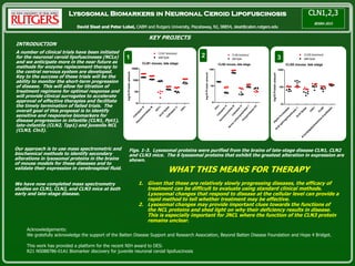
- 1. Lysosomal Biomarkers in Neuronal Ceroid Lipofuscinosis David Sleat and Peter Lobel, CABM and Rutgers University, Piscataway, NJ, 08854, sleat@cabm.rutgers.edu KEY PROJECTS WHAT THIS MEANS FOR THERAPY 1. Given that these are relatively slowly progressing diseases, the efficacy of treatment can be difficult to evaluate using standard clinical methods. Lysosomal changes that respond to disease at the cellular level can provide a rapid method to tell whether treatment may be effective. 2. Lysosomal changes may provide important clues towards the functions of the NCL proteins and shed light on why their deficiency results in disease. This is especially important for JNCL where the function of the CLN3 protein remains unclear. Acknowledgements: We gratefully acknowledge the support of the Batten Disease Support and Research Association, Beyond Batten Disease Foundation and Hope 4 Bridget. This work has provided a platform for the recent NIH award to DES: R21 NS088786-01A1 Biomarker discovery for juvenile neuronal ceroid lipofuscinosis INTRODUCTION A number of clinical trials have been initiated for the neuronal ceroid lipofuscinoses (NCLs) and we anticipate more in the near future as methods for enzyme replacement therapy to the central nervous system are developed. Key to the success of these trials will be the ability to monitor the short-term progression of disease. This will allow for titration of treatment regimens for optimal response and will provide clinical surrogates to accelerate approval of effective therapies and facilitate the timely termination of failed trials. The overall goal of this proposal is to identify sensitive and responsive biomarkers for disease progression in infantile (CLN1, Ppt1), late-infantile (CLN2, Tpp1) and juvenile NCL (CLN3, Cln3). 1 CLN1 mouse, late stage C athepsin D H exosam inidase B PPT1 A cid lipase C athepsin C TPP1 10 100 1000 10000 CLN1 knockout wild-type log10Proteinamount 2 CLN2 mouse, late stage TPP1Cathepsin S Hexosam inidase BCathepsin H Serine carboxypeptidase 1 Phospholipase D3 10 100 1000 CLN2 knockout wild-type log10Proteinamount 8 3 CLN3 mouse, late stage A lpha fucosidase A cid sphingom yelinase 1A cid lipase TPP1 C LN 5 N -acetylgalactosam ine 6-sulfatase 10 100 1000 CLN3 knockout wild-type log10Proteinamount Our approach is to use mass spectrometric and biochemical methods to identify secondary alterations in lysosomal proteins in the brains of mouse models for these diseases and to validate their expression in cerebrospinal fluid. We have now completed mass spectrometry studies on CLN1, CLN2, and CLN3 mice at both early and late-stage disease. Figs. 1-3. Lysosomal proteins were purified from the brains of late-stage disease CLN1, CLN2 and CLN3 mice. The 6 lysosomal proteins that exhibit the greatest alteration in expression are shown.