

Recomendados
Recomendados
Mais conteúdo relacionado
Mais procurados
Mais procurados (20)
Semelhante a Imitators of severe preeclampsia Sibai 2009
Semelhante a Imitators of severe preeclampsia Sibai 2009 (20)
Mais de Asha Reddy
Mais de Asha Reddy (20)
Imitators of severe preeclampsia Sibai 2009
- 1. Imitators of Severe Pre-eclampsia Baha M. Sibai, MD There are many obstetric, medial, and surgical disorders that share many of the clinical and laboratory findings of patients with severe pre-eclampsia– eclampsia. Imitators of severe pre-eclampsia– eclampsia are life-threatening emergencies that can develop during preg- nancy or in the postpartum period. These conditions are associated with high maternal and perinatal mortalities and morbidities, and survivors may face long-term sequelae. The pathophysiologic abnormalities in many of these disorders include vasospasm, platelet activation or destruction, microvascular thrombosis, endothelial cell dysfunction, and reduced tissue perfusion. Some of these disorders include acute fatty liver of pregnancy, thrombotic thrombocytopenic purpura, hemolytic uremic syndrome, acute exacerbation of systemic lupus erythematosus, and disseminated herpes simplex and sepsis syndromes. Differential diagnosis may be difficult due to the overlap of several clinical and laboratory findings of these syndrome. It is important that the clinician make the accurate diagnosis when possible because the management and complications from these syndromes may be different. Because of the rarity of these conditions during pregnancy and postpartum, the available literature includes only case reports and case series describing these syndromes. This review focuses on diagnosis, management, and counseling of women who develop these syndromes based on results of recent studies and my own clinical experience. Semin Perinatol 33:196-205 © 2009 Elsevier Inc. All rights reserved. KEYWORDS severe pre-eclampsia, acute fatty liver, TTP, HUS S everal microangiopathic disorders that are encountered during pregnancy provide physicians with a formidable, if not impossible, diagnostic challenge. Severe pre-eclampsia Acute Fatty Liver of Pregnancy Acute fatty liver of pregnancy (AFLP) is a rare but potentially with hemolysis, elevated liver enzymes, and low platelets fatal complication of the third trimester. The incidence of this (HELLP) syndrome and many other obstetric and medical or disorder ranges from 1 in 10,000 to 1 in 15,000 deliveries. surgical conditions produce similar clinical presentations The incidence is probably lower than that because the re- and laboratory study results to pre-eclampsia.1 In addition, ported rates are usually from large referral centers, which pre-eclampsia is not infrequently superimposed upon one of tend to overestimate the true incidence.1-11 It has been sug- these disorders, further confounding an already difficult dif- gested that AFLP is more common in nulliparous women as well as in those with multifetal gestation.1-7,9 The clinical onset ferential diagnosis. Because of the remarkably similar clinical of symptoms ranges from 27 to 40 weeks, with an average of 36 and laboratory findings of these disease processes, even the weeks’ gestation6; however, cases have been reported in the sec- most experienced physician will face a difficult diagnostic ond trimester.11,2 In some cases, the first onset of signs/symp- challenge.1 Therefore, an effort should be made to attempt to toms may be in the postpartum period.2,9 The patient typically identify an accurate diagnosis given the fact that management presents with a 1- to 2-week history of malaise, anorexia, nau- strategies and outcome may differ among these conditions. In sea, vomiting, midepigastric or right upper quadrant pain, head- this review, I will describe the pathogenesis, differential di- ache, or jaundice. The urine will have a bright yellow appear- agnosis, and management of the medical conditions de- ance (Fig. 1). Rarely, the patient may present with hepatic scribed in the box below. encephalopathy.13 Symptoms of preterm labor or lack of fetal movement may be the presenting complaint in some of these patients.2,3 In about 15-20%, the patient might not present with Department of Obstetrics and Gynecology, University of Cincinnati, College any of the above symptoms.2-5 of Medicine, Cincinnati, OH. Address reprint requests to Baha M. Sibai, MD, Department of Obstetrics and Physical examination reveals an ill-appearing patient with Gynecology, University of Cincinnati, 231 Albert Sabin Way, Cincinnati, jaundice. Some patients will have a low-grade fever. Other OH 45267. E-mail: baha.sibai@uc.edu findings may include hypertension and even proteinuria and 196 0146-0005/09/$-see front matter © 2009 Elsevier Inc. All rights reserved. doi:10.1053/j.semperi.2009.02.004
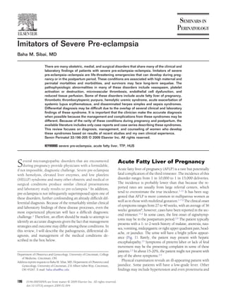
- 2. Imitators of severe pre-eclampsia 197 ascites and bleeding from severe coagulopathy. Because of Table 1 Imitators of Severe Pre-eclampsia/HELLP Syndrome these findings, the diagnosis may be initially confused with ● AFLP pre-eclampsia.1,2,6 Neurologic findings may range from nor- ● TTP mal to lethargy, agitation, confusion, and even coma.8,9,13,14 ● HUS ● Exacerbation of lupus erythematosus Laboratory Findings ● Catastrophic antiphospholipid syndrome The complete blood count usually reveals hemoconcentra- ● Systemic viral sepsis (disseminated herpes) ● Systemic inflammatory response syndrome/septic shock tion, elevated white blood count, and platelet count that is ● Other conditions (cholestasis of pregnancy, necrotizing initially normal but may be low.1-6 Coagulation findings re- pancreatitis, etc.) veal low fibrinogen, prolonged prothrombin time (PT), and low levels of antithrombin.1-10 These abnormalities are re- lated to reduced production of these factors by the liver and are consistent with disseminated intravascular coagulopathy is not possible to be performed once the tissue has been (DIC). In contrast, the DIC seen in severe pre-eclampsia and submitted to routine paraffin blocks. Liver biopsy should be abruptio placentae is due to abnormal consumption.15 Serum performed only after correction of coagulopathy. However, electrolytes will reveal evidence of metabolic acidosis with this is rarely used in clinical practice, and the diagnosis is elevated creatinine and uric acid values. Blood sugar may be usually made based on clinical and laboratory findings.1-9 normal but is usually low in the postpartum period.1-6,8,9,13 In addition to the other conditions listed in Box I, the Blood sugars may also be elevated in association with second- differential diagnosis of AFLP should include idiopathic cho- ary pancreatitis.1,8 Liver enzymes, such as AST, ALT, alkaline lestasis of pregnancy, Budd-Chiari syndrome, adult-onset phosphatase, and bilirubin, will be elevated. The increase in Reye’s syndrome, and drug-induced hepatic toxicity (acet- bilirubin is mainly of the conjugated form, with levels usually aminophen overdose, tetracycline-induced toxicity, anticon- exceeding 5 mg/dL. Ammonia levels are also increased, par- vulsant drugs hypersensitivity, and methyldopa hepati- ticularly in late stage of the disease. Amylase and lipase values tis).1,4,21 may be elevated in the presence of concomitant pancreati- tis.1,8 Hepatitis profile for A, B, and C will be negative. Ultrasonography of the liver may reveal the presence of Maternal and Perinatal Complications increased echogenicity in severe cases; however, it is less AFLP is associated with an increased risk of maternal mortal- sensitive than computed tomography (CT) and magnetic res- ity and morbidities. In the past, the rate of maternal death was onance imaging (MRI).16-20 In addition, quantitation of liver close to 70%; however, recent data indicate a mortality rate of density by ultrasound is subjective and operator-dependent. Ͻ10%, but maternal complications remain high. Table 1 descri- CT scan of the liver may show decrease or diffuse attenuation bes maternal complications in recent series of AFLP.1-11,14,22,23 in the liver. However, none of these techniques are suffi- Recently, Davidson and coworkers reported on three women ciently sensitive to exclude a diagnosis of AFLP.2,20 with triplet gestation who had AFLP proven by liver biopsy Liver biopsy is the gold standard for confirming the diag- who survived. They suggested that women with triplets are at nosis of AFLP. Histopathologic findings in AFLP reveal swol- increased risk for AFLP because of potential for increased len, pale hepatocytes with central nuclei.21 The diagnosis can production of fatty acid metabolites by three fetuses. It has be made only on a frozen section liver biopsy with special been suggested that the improved maternal survival in recent stains for fat, such as oiled red (O).1,4 Plans to perform this years was related to the supportive care and aggressive man- stain should be made before the biopsy procedure because it agement of serious maternal complications by a multidisci- plinary group of physicians from various specialties.2-9 Recently, Knight, and coworkers11 reported a prospective national study of AFLP in the UK. The participants included 57 women with AFLP diagnosed mostly by clinical criteria; 55 had abdominal ultrasound scan, but only 12 (27%) had either ascites and/or abnormal liver findings. Forty-two (74%) were diagnosed antepartum and 15 (26%) postpartum (within 4 days of delivery). In general, most of these patients had a biochemical diagnosis of AFLP (mild prolongation of PT, PTT, normal platelets), only 8 (16%) had acute renal failure, and 4 (7%) required ventilatory support. There was only 1 maternal death (1.8%) in a patient who received a liver transplant.14 Both perinatal mortality and morbidities are increased in patients of AFLP.2-9,20 The perinatal mortality among 160 births (142 pregnancies) in recent case series was 13.1%.9,22 Figure 1 Liver biopsy findings in herpes hepatitis. (Color version of In addition, neonatal morbidity remains high because of the figure is available online.) high rate of preterm delivery (74%) among reported cases.
- 3. 198 B.M. Sibai The average gestational age at delivery was 34 weeks (range, can be used to maintain blood sugars above 60 mg/dL. Ane- 25-42 weeks).2-9,22 mia and DIC should be treated as needed by a packed red In the study by Knight and coworkers,11 the perinatal mor- blood cells, platelets, and fresh frozen plasma. It is important tality was 10.4% among 67 infants (6 stillbirths and 1 neo- to aggressively treat maternal hypotension to avoid further natal death). injury to the liver, kidneys, and other organs. Invasive hemo- dynamic monitoring may be necessary in some patients to assess fully the intravascular volumes and maintain cardiac Management of AFLP output, and in patients who develop acute respiratory dis- The clinical course of women with AFLP is usually character- tress syndrome (ARDS). ized by progressive and sometimes sudden deterioration in Pancreatitis is a potentially lethal complication of AFLP, maternal and fetal conditions.9 Therefore, patients in whom and thus all patients with AFLP should undergo serial screen- AFLP is considered require hospitalization in a labor and ing of serum lipase and amylase for several days after the delivery unit. Fetal heart rate monitoring and/or performing onset of hepatic dysfunction.1,8 Abnormalities in these en- of a biophysical profile should be performed concurrently zymes typically appear after hepatic and renal dysfunction. with maternal evaluation. Evidence of fetal compromise may The development of pseudocysts with secondary infections be present even in those with stable maternal conditions. or hemorrhagic pancreatitis with resultant retroperitoneal Nonreassuring fetal testing may be secondary to maternal bleeding increases the risk for maternal death.8 acidosis and/or reduced uteroplacental blood flow.1-6 The In general, most patients with AFLP will start to improve presence of maternal acidosis may be reflected in reduced- 2-3 days after delivery. In some cases, however, deterioration to-absent fetal movement, absent fetal breathing, or tone dur- in liver function tests, renal function, mental status, and co- ing biophysical profile testing.1 agulopathy may continue for about 1 week. In such cases, The next step in management is to confirm or exclude the some authors suggest using plasmapheresis to improve ma- diagnosis of AFLP according to the clinical findings and re- ternal outcome. This method of treatment was recently re- sults of blood tests.1,23 The presence of bleeding and/or severe ported in six such cases with no maternal death. However, coagulopathy requires transfusion with fresh frozen plasma the value of such therapy remains unclear. In rare cases, a and other blood products as needed. The ultimate treatment patient will progress into fulminant hepatic failure, requiring for this condition is maternal stabilization and delivery be- liver transplantation.21 cause there are no reports of women recovering from AFLP spontaneously. The presence of AFLP is not an indication for delivery by cesarean section because of the risks of bleeding Counseling of Women with AFLP complications in the presence of coagulopathy.2-6,10 The de- Several case reports and case series22-26 have noted an asso- cision to perform cesarean delivery should be based on fetal ciation between the development of AFLP and/or HELLP gestational age, fetal condition, and presence of labor. Induc- syndrome and a deficiency of long-chain 3-hydroxyacyl-co- tion of labor with an attempt for vaginal delivery within 24 enzyme A dehydrogenase (LCHAD) in infants born to hours is a reasonable approach. Because of the associated women with the above complications. This disorder of mito- coagulopathy, most anesthesiologists will avoid epidural an- chondrial fatty acid oxidation might lead to significant in- algesia. Maternal analgesia during labor can be provided by crease in maternal fatty acid levels that are highly toxic to the intermittent use of small doses of systemic opioids. The use of liver. Based on these findings, some authors suggest that pudendal block should be avoided because of the risk of women with AFLP as well as their partners and children bleeding and hematoma formation into this area. Caution should undergo molecular testing for Glu 474 Gln mutation should be exercised to avoid vaginal trauma and lacerations in the LCHAD.22,24 Screening for this mutation would allow during vaginal delivery.1 early diagnosis and treatment in newborns of affected moth- In case of cesarean delivery, the patient should receive ers and would allow counseling about subsequent pregnan- general anesthesia. It is advisable to avoid incisions that re- cies.22,25 The risk of recurrence is increased in women who quire extensive dissection, such as the Pfannenstiel incision, are carriers for this mutation, particularly if the fetus is also and meticulous attention should be given to secure hemosta- affected during a subsequent pregnancy. There are few case sis. It is advisable to perform a midline incision, to use a reports describing recurrent AFLP in women without the subfascial drain, and to keep the skin incision open for at above mutation2,4; however, the risk of this recurrence re- least 48 hours to avoid hematoma formation.1,23 mains unknown because of the limited number of pregnan- In the postpartum period, the patient should be monitored cies reported following AFLP in these women. very closely with careful evaluation of vital signs, intake– output, and bleeding. Some of these patients may develop acute refractory hypotensive shock in the immediate postpar- Thrombotic Microangiopathies tum period. Serial measurements of hematologic, hepatic, Thrombotic thrombocytopenic purpura (TTP) and hemo- and renal function should be performed and recorded in an lytic uremic syndrome (HUS) are two microangiopathic dis- organized fashion. Blood sugars should be monitored every orders that are extremely rare during pregnancy/postpartum. few hours with a bedside glucometer because of the risk of They are usually reported as case reports or small case se- hypoglycemia in the postpartum period. Glucose infusions ries.1,26-35 Thus, their expected development during preg-