SlideShare uma empresa Scribd logo
1 de 106
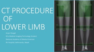
CT PROCEDURE
OF
LOWER LIMBAnjan Dangal
B.Sc.Medical Imaging Technology Student
National Academy of Medical Sciences
Bir Hospital, kathmandu, Nepal
Contents
• Indication
• Contraindication
• Anatomy of HIP Joint and Thigh + CT Procedure of Hip Joint
WHY Lower Extremity CT
Computed tomography (CT) is used for evaluation of tumors, metastatic
lesions, infection, fractures and other problems.
Magnetic resonance imaging (MRI) is the first-line choice for imaging of
many conditions, but CT may be used in these cases if MRI is
contraindicated or unable to be performed
• Evaluation of suspicious mass/ tumor (unconfirmed cancer
diagnosis
Initial evaluation of suspicious mass/tumor , which has been
nondiagnostic after x ray and ultrasound
Suspected tumor size increase or recurrence based on a sign,
symptom, imaging study or abnormal lab value
Evaluation of Known Cancer
Initial staging of known cancer in the lower extremity.
Follow-up of known cancer of patient undergoing active treatment within the past year.
Known cancer with suspected lower extremity metastasis based on a sign, symptom, imaging
study or abnormal lab value.Initial staging of known cancer in the lower extremity.
Follow-up of known cancer of patient undergoing active treatment within the past year.
Known cancer with suspected lower extremity metastasis based on a sign, symptom, imaging
study or abnormal lab value.
For evaluation of known or suspected fracture and/or injury:
Further evaluation of an abnormality or non-diagnostic findings on prior imaging.
Suspected fracture when imaging is negative or equivocal.
Determine position of known fracture fragments/dislocation.
For evaluation of persistent pain, initial imaging (e.g. x-ray) has been
performed and MRI is contraindicated or cannot be performed:
Chronic (lasting 3 months or greater) pain and/or persistent tendonitis
unresponsive to conservative treatment*, which include - medical
therapy (may include physical therapy or chiropractic treatments)
Pre-operative evaluation.
Post-operative/procedural evaluation:
When imaging, physical, or laboratory findings indicate joint infection,
delayed or non-healing, or other surgical/procedural complications.
A follow-up study may be needed to help evaluate a patient’s progress
after treatment, procedure, intervention, or surgery. Documentation
requires a medical reason that clearly indicates why additional imaging is
needed for the type and area(s) requested.
• For evaluation of known or suspected infection or inflammatory disease (e.g.
osteomyelitis)
• For evaluation of suspected (AVN) avascular necrosis (e.g., aseptic necrosis,
Legg-Calve-Perthes disease in children) and MRI is contraindicated or cannot be
performed
• For evaluation of suspected or known Auto Immune Disease, (e.g. Rheumatoid
arthritis)
• Abnormal bone scan and radiograph is non-diagnostic or requires further
evaluation.
• For evaluation of leg length discrepancy when physical deformities of the lower
extremities would prevent standard modalities such as x-rays or a Scanogram
from being performed.
• CT arthrogram and MRI is contraindicated or cannot be performed.
• To assess status of osteochondral abnormalities including osteochondral
fractures, osteochondritis dissecans, treated osteochondral defects where
physical or imaging findings suggest its presence and MRI is contraindicated or
cannot be performed.
Lower limb
Lower limb consists of :
Thigh
Leg
Ankle and Foot
HIP joint
Articulating Surface:
• Head of femur
• lunate surface of acetabulum:
Cup like cavity : Acetabulum
Ilium , Ischium and Pubis
Acetabular
fossaNon Articulating surface:
Loose connective tisssue,
mobile fat pad
not covered by hyaline
cartilage
ACETABULAR
LABRUM
Surrounds Bony rim of Acetabulum
Fovea
capitis femoris
ligament of head of femur connets
at fovea acetabular ligament
On Axial Section
Anterior column
Posterior Column
Ligaments
Outer ligaments
Inner ligaments
ILiofemoral ligament
Ischiofemoral ligament
pubofemoral ligament
ligament of head of femur
transverse Acetabular ligament
Head
Muscle of Hip and Thigh
Muscles of Hip and Thigh
• Anterior Hip Muscle
• Posterior Hip Muscle (Glueteal )
Superficial Gluteal Muscle
Deep Gluteal Muscle
• Anterior Compartment
• Medial compartment
• Posterior Compartment
Anterior Hip Muscles
Iliopsoas Muscle
Psoas Minor Muscle
Iliopsoas
Muscle
Psoas Muscle
Illiacus Muscle
Psoas
Minor
Muscle
• Posterior Hip Muscle (Glueteal )
Superficial Gluteal Muscle
Gluteus Maximus Muscle
Gluteal Medius Muscle
Gleteal Minimus Muscle
Tensor fasciae latae
Deep Gluteal Muscle
Superficial Gluteal Muscle
Gluteus Maximus Muscle
Gluteal Medius Muscle
Gleteal Minimus Muscle
Tensor fsciae latae
Gluteus Maximus Muscle
Gluteal
Medius
Muscle
Gluteus
Minimus
Muscle
Tensor
Fascia
Latae
Deep Gluteal Muscle
Piriformis muscle
Superior Gemellus Muscle
Obturator Internus Muscle
Inferior gemellus muscle
Quadratus femoris muscle
Piriformis
Muscle
Superior
Gemellus
Muscle
Obturator
Internus
Muscle
Inferior
Gemellus
Muscle
Quadratus
Femoris
Muscle
Muscles of Thigh
• Anterior Compartment
• Medial compartment
• Posterior Compartment
Muscle of thigh :
Ant Compartment
Sartorius Muscle
Quadriceps femoris Muscle
Sartorius
Muscle
Rectus
Femoris
Muscle
Blood Supply
major contributing set contains the medial and lateral
circumflex arteries that arise from the deep branch of
the femoral artery
Femoral
Artery
lateral
Circumflex
Femoral
Artery
Medial
Circumflex
Femoral
Artery
CT Procedure of Hip
Computed tomography is primarily used to evaluate acute trauma,
e.g., acetabular fracture or hip dislocation. It can detect
intraarticular fragments and associated articular surface fractures
and it is useful in surgical planning.
Additional Indications Specific to Hip CT
For any evaluation of patient with hip prosthesis or other implanted metallic
hardware where prosthetic loosening or dysfunction is suspected on physical
examination or imaging.
For evaluation of total hip arthroplasty patients with suspected loosening
and/or wear or osteolysis or assessment of bone stock is needed.
For evaluation of suspected slipped capital femoral epiphysis with non-
diagnostic or equivocal imaging and MRI is contraindicated or cannot be
performed.
Suspected labral tear of the hip with signs of clicking and pain with hip
motion especially with hip flexion, internal rotation and adduction which can
also be associated with locking and giving way sensations of the hip on
ambulation and MRI is contraindicated or cannot be performed.
Patient preperation
Remove any non-fixed metal prosthesis, jewelry or zippers that
might interfere with the region to be scanned.
- Discuss the procedure with the patient. The patient must not
move during any part of the scanning.
-
Patient Position
and Posture
• Patient laying supine with legs extended.
• Legs in natural alignment with neutral rotation.
• No un-natural tilt or lift of the pelvis.
• Arms folded upward away from the pelvis
• Position the patient to maximize comfort and minimize motion.
• Only true axial slices are allowed: no oblique or reformatted images and no gantry tilt
Hip Joint/ Proximal femurProtocol
Positioning patient supine, with feet first
Scouts AP and Lat
Scan Type Helical
Start Location Just Above SI Joint
End Location Approx 4cm below Lesser trochanter
DFOV ~ 30 cm ( Include Skin Surface )
Acqusition Detector Width * Number of detector in row= coverage
Reconstruction ( Slice Thickness/ Interval ) 1.25mm/ 0.625 mm
Pitch 0.5
Kvp 120
mA 200
Clinical indications that may necessitate IV contrast include infection
or tumor. When IV contrast is ordered, 80 mL of LOCM
is injected at 3 mL/s and scanning begins after 40 seconds.
Algorithm Bone
Window Width 2000
Window level 500
Algorithm Standard
Window width 350
Window level 50
A. Axial MPR can be programmed from an AP scout. B. Coronal MPR can be
programmed from an axial image and should follow the long axis of the femoral neck.
C. Sagittal MPR can be programmed from an axial image and should be perpendicular to the
coronal MPR plane.
MPR
1. Acetabulum (anterior column)
2. Acetabulum
3.Femoral head
4. Acetabulum posterior column
5. Hip Joint
6. Illiopsoas mUscle
7. Sarotrius M
8.Gluteus Minimus M
9. Gletues Medius M
10. Gluteal Maximus Muscle
11. Bladder
12. Rectus Femoris
1. Femoral head
2. Iliopsoas m.
3. Femoral neck
4. Rectus femoris m.
5. Tensor fascia lata m.
6. Greater trochanter
7. Ischium/Ischial tuberosity
8. Obturator internus m.
9. Pubis
10. Pectineus m.
11. Gluteus maximus m.
12. Sartorius m.
Adductor brevis m.
2. Rectus femoris m.
3. Vastus intermedius m.
4. Femur
5. Pubis, inferior ramus
6. Obturator externus m.
7. Iliopsoas m.
8. Femur, lesser trochanter
9. Gluteus maximus m.
10. Sartorius m.
11. Tensor fascia lata m.
12. Vastus lateralis m
VRT MPR : Coronal and
Sag Images
Knee Joint
Articulating Surface : Femur: lateral and medial condyles,
intercondylar groove, patellar surface
Tibia: tibial plateaus
Patella: posterior surface
Meniscus:Ameniscus(me-NIS-kus;a crescent;plural,menisci) isa pad
offibrocartilage betweenopposing boneswithinasynovial joint.
Meniscus:A meniscus (me-NIS-kus; a crescent; plural,
menisci) is a pad offibrocartilage between opposing
bones within a synovial joint.
Medial
Meniscus
Lateral
Meniscus
The transverse
ligament
connects
the menisci
anteriorly and
holds them in
place during knee
extension.
The anterior and
posterior
meniscofemoral
ligaments attach the
menisci to the femur
and the bases of the
menisci are attached
to the joint capsule.
Bursa: bursa is a small, thin, fluid-filled pocket that forms in
connective tissue outside of a joint capsule. It contains synovial
fluid and is lined by a synovial membrane.Bursae often form
where a tendon or ligament rubs against other tissues.
Suprapatellar
Infrapatellar
prepatellar
Suprapatellar
Infrapatellar
Prepatellar
Ligaments
Frontal ligamnets
Medial/lateral ligaments
dorsal
Cruciate ligament
Frontal ligament: The frontal ligamentous apparatus
holds the patella in place.
Patellar ligament
Retinaculum
Patellar
ligament
Lateral
patellar
Retinaculum
Medial
Patellar
Retinaculum
Medial/Lateral
ligament:The lateral and medial
ligaments secure thekneejoint,
preventexcessive sideways
movement.
Cruciate ligament:two cruciate ligaments cross in the
centre of the joint, preventing slippage of the femur on
the tibia
ACL
PCL
Flexors & Extensors of Knee
CT PROTOCOL OF KNEE
Additional indications specific for KNEE CT and MRI is
contraindicated or cannot be performed:
Accompanied by blood in the joint (hemarthrosis) demonstrated by aspiration.
Presence of a joint effusion.
Accompanied by physical findings of a meniscal injury determined by physical
examination tests (McMurray’s, Apley’s) or significant laxity on varus or valgus stress
tests.
Accompanied by physical findings of anterior cruciate ligament (ACL) or posterior
cruciate ligament (PCL) ligamental injury determined by the drawer test or the
Lachman test.
Patient preperation
Remove any non-fixed metal prosthesis, jewelry or zippers that
might interfere with the region to be scanned.
- Discuss the procedure with the patient. The patient must not
move during any part of the scanning.
-
Patient Position
and Posture
• Patient laying supine with legs extended.
• Legs in natural alignment with neutral rotation.
• No un-natural tilt or lift of the pelvis.
• Arms folded upward away from the pelvis
• Position the patient to maximize comfort and minimize motion.
• Only true axial slices are allowed: no oblique or reformatted images and no gantry tilt
KNEE/ TIBIAL PLATEAU
Positioning Patient Supine with feet first , Legs Flat on table
Scouts Ap and Lateral
Scan Type Helical
Start Location Just Above Patella
End Location Just below fibular Head
DFOV ~ 20cm ( adjust to include Skin Surface : affected knee only )
SFOV large Body
Acquisition Detector Width * Number of detector in row= coverage
Reconstruction ( Slice thickness/ interval ) 1.25mm/0.625 mm
Pitch 0.5
Kvp/ mA 120
Reconstruction 1
Algorithm Bone Plus
Window Width 2000
Window level 500
Reconstruction 2
Algorithm Standard
Window Width 350
Window level 50
MPR: Bone AlgorithmSlice Thickness/ Interval : 2mm/2mm
Planes: Axial, coronal, sagittal
A. Axial MPR can be programmed from an AP scout and should be parallel to the tibial plateau.
B. Coronal MPR can be programmed from an axial image and should be parallel to the femoral condyles.
C. Sagittal MPR can be programmed from an axial image and should be perpendicular to the coronal
MPR.
ANKLE JOINT
Articulation
Tibiotarsal joint: fibula, tibia, talus
Talotarsal joint: talus, calcaneus, navicular bone
LigamentsAnterior talofibular,
posterior talofibular,
Anterior Tibiofibular ligament
Posterior Tibiofibular ligament
calcaneofibular ligamnent
Anterior Tibiotalar ligamnet
Posterior Tibiotalar ligament
Tibionavicular ligamnet
Tibiotalar ligament
CT Protocol of Ankle/ Foot
Ankle/ Distal Tibia
Positioning Patient Supine with feet first , Legs Flat on table, use foot
holder
Scouts Ap and Lateral
Scan Type Helical
Start Location Just Above ankle joint
End Location Through calcaneus
DFOV ~ 16 cm ( adjust to include Skin Surface )
SFOV large Body
Acquisition Detector Width * Number of detector in row= coverage
Reconstruction ( Slice thickness/ interval ) 0.625mm/0.3 mm
Pitch 0.562
Kvp/ mA 120
Reconstruction 1
Algorithm Bone Plus
Window Width 2000
Window level 500
Reconstruction 2
Algorithm Standard
Window Width 350
Window level 50
MPR: Bone Algorithm
A. Axial MPR can be programmed from an AP scout parallel to the top of the talus.
B. Coronal MPR can be programmed from an axial image at the level of the distal tibia.
C. Sagittal MPR can be programmed from an axial image at the level of the distal tibia. They are perpendicular to the coronal MPR
plane.
Slice Thickness/ Interval : 2mm/2mm
Planes: Axial, coronal, sagittal
Thankyou

Mais conteúdo relacionado

Mais procurados

Ct image quality artifacts and it remedy
Ct image quality artifacts and it remedyCt image quality artifacts and it remedy
Ct image quality artifacts and it remedyYashawant Yadav
 
Difference Between Single Slice and Multi Slice CT Scanner
Difference Between Single Slice and Multi Slice CT ScannerDifference Between Single Slice and Multi Slice CT Scanner
Difference Between Single Slice and Multi Slice CT ScannerAbdul Rehman (R)(CT)(MR)
 
Post processing of computed tomography
Post processing of computed tomographyPost processing of computed tomography
Post processing of computed tomographyBeuniquewithNehaSing
 
Tissue harmonic imaging
Tissue harmonic imaging Tissue harmonic imaging
Tissue harmonic imaging MAMTA PANDA
 
CT Procedure OF Abdomen & Pelvis
CT Procedure OF Abdomen & Pelvis CT Procedure OF Abdomen & Pelvis
CT Procedure OF Abdomen & Pelvis Upakar Paudel
 
Arthrography of the Shoulder, Ankle and Wrist.pptx
Arthrography of the Shoulder, Ankle and Wrist.pptxArthrography of the Shoulder, Ankle and Wrist.pptx
Arthrography of the Shoulder, Ankle and Wrist.pptxDeneicer Guy
 
Portable ct mobile ct
Portable ct mobile ctPortable ct mobile ct
Portable ct mobile ctanilayyakutty
 
Computed Tomography Dose Index
Computed Tomography Dose IndexComputed Tomography Dose Index
Computed Tomography Dose IndexAnjan Dangal
 
Mammography physics and technique
Mammography  physics and techniqueMammography  physics and technique
Mammography physics and techniqueArchana Koshy
 
K Space in MRI
K Space in MRIK Space in MRI
K Space in MRIKajal Jha
 
Helical and Multislice CT
Helical and Multislice CTHelical and Multislice CT
Helical and Multislice CTManojzz Bhatta
 
CT ITS BASIC PHYSICS
CT ITS BASIC PHYSICSCT ITS BASIC PHYSICS
CT ITS BASIC PHYSICSDEEPAK
 
Perfusion and dynamic contrast enhanced mri
Perfusion and dynamic contrast enhanced mriPerfusion and dynamic contrast enhanced mri
Perfusion and dynamic contrast enhanced mrifahad shafi
 

Mais procurados (20)

Ct image quality artifacts and it remedy
Ct image quality artifacts and it remedyCt image quality artifacts and it remedy
Ct image quality artifacts and it remedy
 
CT artifact
CT artifact CT artifact
CT artifact
 
Ultrasound contrast agents
Ultrasound contrast agentsUltrasound contrast agents
Ultrasound contrast agents
 
CT History
CT HistoryCT History
CT History
 
Difference Between Single Slice and Multi Slice CT Scanner
Difference Between Single Slice and Multi Slice CT ScannerDifference Between Single Slice and Multi Slice CT Scanner
Difference Between Single Slice and Multi Slice CT Scanner
 
Post processing of computed tomography
Post processing of computed tomographyPost processing of computed tomography
Post processing of computed tomography
 
Tissue harmonic imaging
Tissue harmonic imaging Tissue harmonic imaging
Tissue harmonic imaging
 
CT Procedure OF Abdomen & Pelvis
CT Procedure OF Abdomen & Pelvis CT Procedure OF Abdomen & Pelvis
CT Procedure OF Abdomen & Pelvis
 
Arthrography of the Shoulder, Ankle and Wrist.pptx
Arthrography of the Shoulder, Ankle and Wrist.pptxArthrography of the Shoulder, Ankle and Wrist.pptx
Arthrography of the Shoulder, Ankle and Wrist.pptx
 
Portable ct mobile ct
Portable ct mobile ctPortable ct mobile ct
Portable ct mobile ct
 
MR Elastography
MR ElastographyMR Elastography
MR Elastography
 
Computed Tomography Dose Index
Computed Tomography Dose IndexComputed Tomography Dose Index
Computed Tomography Dose Index
 
Mammography physics and technique
Mammography  physics and techniqueMammography  physics and technique
Mammography physics and technique
 
K Space in MRI
K Space in MRIK Space in MRI
K Space in MRI
 
MRI protocol of Knee
MRI protocol of KneeMRI protocol of Knee
MRI protocol of Knee
 
Helical and Multislice CT
Helical and Multislice CTHelical and Multislice CT
Helical and Multislice CT
 
CT Angiography Lower Limb
CT Angiography Lower LimbCT Angiography Lower Limb
CT Angiography Lower Limb
 
Chapter 4 image display
Chapter 4 image displayChapter 4 image display
Chapter 4 image display
 
CT ITS BASIC PHYSICS
CT ITS BASIC PHYSICSCT ITS BASIC PHYSICS
CT ITS BASIC PHYSICS
 
Perfusion and dynamic contrast enhanced mri
Perfusion and dynamic contrast enhanced mriPerfusion and dynamic contrast enhanced mri
Perfusion and dynamic contrast enhanced mri
 

Semelhante a Computed Tomography procedure of lower limb

lect 1 Anthropometry -.pdf
lect 1 Anthropometry -.pdflect 1 Anthropometry -.pdf
lect 1 Anthropometry -.pdfssuser650c771
 
Lateral patellar compression syndrome
Lateral patellar compression syndromeLateral patellar compression syndrome
Lateral patellar compression syndromeAmanj Gardi
 
Transfemoral protheses
Transfemoral prothesesTransfemoral protheses
Transfemoral prothesesSoundar Rajan
 
Medial meniscus injury and physiotherapy treatment
Medial meniscus injury and physiotherapy treatmentMedial meniscus injury and physiotherapy treatment
Medial meniscus injury and physiotherapy treatmentMuskan Rastogi
 
Case Review #50: 29 year old woman presents with dislodged instrumentation fo...
Case Review #50: 29 year old woman presents with dislodged instrumentation fo...Case Review #50: 29 year old woman presents with dislodged instrumentation fo...
Case Review #50: 29 year old woman presents with dislodged instrumentation fo...Robert Pashman
 
NCCT OR CECT PELVIS BONE.pptx, INDICATIONS OF PELVIS BONE
NCCT OR CECT PELVIS BONE.pptx, INDICATIONS OF PELVIS BONENCCT OR CECT PELVIS BONE.pptx, INDICATIONS OF PELVIS BONE
NCCT OR CECT PELVIS BONE.pptx, INDICATIONS OF PELVIS BONERukamaneeYadav
 
MRI procedure of pelvis and hip suman duwal
MRI procedure of pelvis and hip suman duwalMRI procedure of pelvis and hip suman duwal
MRI procedure of pelvis and hip suman duwalsuman duwal
 
EVALUATION METHODS.presentation for evaluation
EVALUATION METHODS.presentation for evaluationEVALUATION METHODS.presentation for evaluation
EVALUATION METHODS.presentation for evaluationPranavTrehan2
 
Ankylosing spondilitis
Ankylosing spondilitisAnkylosing spondilitis
Ankylosing spondilitisRachita Hada
 
Hip Joint anatomy, surgical approches & AVN review
Hip Joint anatomy, surgical approches & AVN reviewHip Joint anatomy, surgical approches & AVN review
Hip Joint anatomy, surgical approches & AVN reviewdocortho Patel
 
Ankylosing spondylitis.pptx
Ankylosing spondylitis.pptxAnkylosing spondylitis.pptx
Ankylosing spondylitis.pptxrohanbirajdar2
 
Rotator Cuff Injuries present at Chirayu Medical College.pptx
Rotator Cuff Injuries present at Chirayu Medical College.pptxRotator Cuff Injuries present at Chirayu Medical College.pptx
Rotator Cuff Injuries present at Chirayu Medical College.pptxNamanSharda2
 
Conference ext.สิทธิกร ปภาวิน orthokorat 1
Conference ext.สิทธิกร ปภาวิน orthokorat 1Conference ext.สิทธิกร ปภาวิน orthokorat 1
Conference ext.สิทธิกร ปภาวิน orthokorat 1sittikornpaphawin
 
Advancement in back pain treatment
Advancement in back pain treatmentAdvancement in back pain treatment
Advancement in back pain treatmentManish Raj
 
Shoulder Biomechanics -.pptx
 Shoulder Biomechanics -.pptx Shoulder Biomechanics -.pptx
Shoulder Biomechanics -.pptxDmaarGroup
 
General examination ms 2020
General examination ms 2020General examination ms 2020
General examination ms 2020cardilogy
 

Semelhante a Computed Tomography procedure of lower limb (20)

lect 1 Anthropometry -.pdf
lect 1 Anthropometry -.pdflect 1 Anthropometry -.pdf
lect 1 Anthropometry -.pdf
 
Lateral patellar compression syndrome
Lateral patellar compression syndromeLateral patellar compression syndrome
Lateral patellar compression syndrome
 
Transfemoral protheses
Transfemoral prothesesTransfemoral protheses
Transfemoral protheses
 
Medial meniscus injury and physiotherapy treatment
Medial meniscus injury and physiotherapy treatmentMedial meniscus injury and physiotherapy treatment
Medial meniscus injury and physiotherapy treatment
 
Case Review #50: 29 year old woman presents with dislodged instrumentation fo...
Case Review #50: 29 year old woman presents with dislodged instrumentation fo...Case Review #50: 29 year old woman presents with dislodged instrumentation fo...
Case Review #50: 29 year old woman presents with dislodged instrumentation fo...
 
NCCT OR CECT PELVIS BONE.pptx, INDICATIONS OF PELVIS BONE
NCCT OR CECT PELVIS BONE.pptx, INDICATIONS OF PELVIS BONENCCT OR CECT PELVIS BONE.pptx, INDICATIONS OF PELVIS BONE
NCCT OR CECT PELVIS BONE.pptx, INDICATIONS OF PELVIS BONE
 
MRI procedure of pelvis and hip suman duwal
MRI procedure of pelvis and hip suman duwalMRI procedure of pelvis and hip suman duwal
MRI procedure of pelvis and hip suman duwal
 
EVALUATION METHODS.presentation for evaluation
EVALUATION METHODS.presentation for evaluationEVALUATION METHODS.presentation for evaluation
EVALUATION METHODS.presentation for evaluation
 
Ankylosing spondilitis
Ankylosing spondilitisAnkylosing spondilitis
Ankylosing spondilitis
 
Case discussion 10
Case discussion 10Case discussion 10
Case discussion 10
 
Hip anatomy, approaches & AVN
Hip anatomy, approaches & AVN Hip anatomy, approaches & AVN
Hip anatomy, approaches & AVN
 
Hip Joint anatomy, surgical approches & AVN review
Hip Joint anatomy, surgical approches & AVN reviewHip Joint anatomy, surgical approches & AVN review
Hip Joint anatomy, surgical approches & AVN review
 
Ankylosing spondylitis.pptx
Ankylosing spondylitis.pptxAnkylosing spondylitis.pptx
Ankylosing spondylitis.pptx
 
Rotator Cuff Injuries present at Chirayu Medical College.pptx
Rotator Cuff Injuries present at Chirayu Medical College.pptxRotator Cuff Injuries present at Chirayu Medical College.pptx
Rotator Cuff Injuries present at Chirayu Medical College.pptx
 
Conference ext.สิทธิกร ปภาวิน orthokorat 1
Conference ext.สิทธิกร ปภาวิน orthokorat 1Conference ext.สิทธิกร ปภาวิน orthokorat 1
Conference ext.สิทธิกร ปภาวิน orthokorat 1
 
Mri knee
Mri kneeMri knee
Mri knee
 
Hip impingement fai
Hip impingement faiHip impingement fai
Hip impingement fai
 
Advancement in back pain treatment
Advancement in back pain treatmentAdvancement in back pain treatment
Advancement in back pain treatment
 
Shoulder Biomechanics -.pptx
 Shoulder Biomechanics -.pptx Shoulder Biomechanics -.pptx
Shoulder Biomechanics -.pptx
 
General examination ms 2020
General examination ms 2020General examination ms 2020
General examination ms 2020
 

Mais de Anjan Dangal

Contrast agents used in radiology
Contrast agents used in radiologyContrast agents used in radiology
Contrast agents used in radiologyAnjan Dangal
 
Image Reconstruction in Computed Tomography
Image Reconstruction in Computed TomographyImage Reconstruction in Computed Tomography
Image Reconstruction in Computed TomographyAnjan Dangal
 
Radiography round on dacrocystography
Radiography round on dacrocystographyRadiography round on dacrocystography
Radiography round on dacrocystographyAnjan Dangal
 
MRI Parameters for Imaging
MRI Parameters for ImagingMRI Parameters for Imaging
MRI Parameters for ImagingAnjan Dangal
 
Magnetic resonance cholangiopancreatography ppt
Magnetic resonance cholangiopancreatography pptMagnetic resonance cholangiopancreatography ppt
Magnetic resonance cholangiopancreatography pptAnjan Dangal
 
Magnetic Resonance Angiography and Venography
Magnetic Resonance Angiography and VenographyMagnetic Resonance Angiography and Venography
Magnetic Resonance Angiography and VenographyAnjan Dangal
 
Tools in interventional radiology
Tools in interventional radiologyTools in interventional radiology
Tools in interventional radiologyAnjan Dangal
 
Computed Tomography Instrumentation and Detector Configuration
Computed Tomography Instrumentation and Detector ConfigurationComputed Tomography Instrumentation and Detector Configuration
Computed Tomography Instrumentation and Detector ConfigurationAnjan Dangal
 
Ultrasound imaging
Ultrasound imagingUltrasound imaging
Ultrasound imagingAnjan Dangal
 
Picture archiving and communication in medicines ( pacs
Picture archiving and communication in medicines ( pacsPicture archiving and communication in medicines ( pacs
Picture archiving and communication in medicines ( pacsAnjan Dangal
 
Computed radiography
Computed radiographyComputed radiography
Computed radiographyAnjan Dangal
 
X ray generator, circuits
X  ray generator, circuitsX  ray generator, circuits
X ray generator, circuitsAnjan Dangal
 
Radiation hazard and protection
Radiation hazard and protection  Radiation hazard and protection
Radiation hazard and protection Anjan Dangal
 
Interaction of x ray with matter
Interaction of x ray with matterInteraction of x ray with matter
Interaction of x ray with matterAnjan Dangal
 

Mais de Anjan Dangal (19)

Contrast agents used in radiology
Contrast agents used in radiologyContrast agents used in radiology
Contrast agents used in radiology
 
Radiobiology
RadiobiologyRadiobiology
Radiobiology
 
Image Reconstruction in Computed Tomography
Image Reconstruction in Computed TomographyImage Reconstruction in Computed Tomography
Image Reconstruction in Computed Tomography
 
Radiography round on dacrocystography
Radiography round on dacrocystographyRadiography round on dacrocystography
Radiography round on dacrocystography
 
MRI Artefacts
MRI ArtefactsMRI Artefacts
MRI Artefacts
 
MRI Parameters for Imaging
MRI Parameters for ImagingMRI Parameters for Imaging
MRI Parameters for Imaging
 
Magnetic resonance cholangiopancreatography ppt
Magnetic resonance cholangiopancreatography pptMagnetic resonance cholangiopancreatography ppt
Magnetic resonance cholangiopancreatography ppt
 
Barium Meal
Barium MealBarium Meal
Barium Meal
 
Magnetic Resonance Angiography and Venography
Magnetic Resonance Angiography and VenographyMagnetic Resonance Angiography and Venography
Magnetic Resonance Angiography and Venography
 
Tools in interventional radiology
Tools in interventional radiologyTools in interventional radiology
Tools in interventional radiology
 
Computed Tomography Instrumentation and Detector Configuration
Computed Tomography Instrumentation and Detector ConfigurationComputed Tomography Instrumentation and Detector Configuration
Computed Tomography Instrumentation and Detector Configuration
 
Ultrasound imaging
Ultrasound imagingUltrasound imaging
Ultrasound imaging
 
Picture archiving and communication in medicines ( pacs
Picture archiving and communication in medicines ( pacsPicture archiving and communication in medicines ( pacs
Picture archiving and communication in medicines ( pacs
 
Fluroscopy
FluroscopyFluroscopy
Fluroscopy
 
Ptc and pbd
Ptc and pbdPtc and pbd
Ptc and pbd
 
Computed radiography
Computed radiographyComputed radiography
Computed radiography
 
X ray generator, circuits
X  ray generator, circuitsX  ray generator, circuits
X ray generator, circuits
 
Radiation hazard and protection
Radiation hazard and protection  Radiation hazard and protection
Radiation hazard and protection
 
Interaction of x ray with matter
Interaction of x ray with matterInteraction of x ray with matter
Interaction of x ray with matter
 

Último

Coimbatore Call Girls in Thudiyalur : 7427069034 High Profile Model Escorts |...
Coimbatore Call Girls in Thudiyalur : 7427069034 High Profile Model Escorts |...Coimbatore Call Girls in Thudiyalur : 7427069034 High Profile Model Escorts |...
Coimbatore Call Girls in Thudiyalur : 7427069034 High Profile Model Escorts |...chennailover
 
Best Rate (Patna ) Call Girls Patna ⟟ 8617370543 ⟟ High Class Call Girl In 5 ...
Best Rate (Patna ) Call Girls Patna ⟟ 8617370543 ⟟ High Class Call Girl In 5 ...Best Rate (Patna ) Call Girls Patna ⟟ 8617370543 ⟟ High Class Call Girl In 5 ...
Best Rate (Patna ) Call Girls Patna ⟟ 8617370543 ⟟ High Class Call Girl In 5 ...Dipal Arora
 
Call Girls Madurai Just Call 9630942363 Top Class Call Girl Service Available
Call Girls Madurai Just Call 9630942363 Top Class Call Girl Service AvailableCall Girls Madurai Just Call 9630942363 Top Class Call Girl Service Available
Call Girls Madurai Just Call 9630942363 Top Class Call Girl Service AvailableGENUINE ESCORT AGENCY
 
Premium Bangalore Call Girls Jigani Dail 6378878445 Escort Service For Hot Ma...
Premium Bangalore Call Girls Jigani Dail 6378878445 Escort Service For Hot Ma...Premium Bangalore Call Girls Jigani Dail 6378878445 Escort Service For Hot Ma...
Premium Bangalore Call Girls Jigani Dail 6378878445 Escort Service For Hot Ma...tanya dube
 
Trichy Call Girls Book Now 9630942363 Top Class Trichy Escort Service Available
Trichy Call Girls Book Now 9630942363 Top Class Trichy Escort Service AvailableTrichy Call Girls Book Now 9630942363 Top Class Trichy Escort Service Available
Trichy Call Girls Book Now 9630942363 Top Class Trichy Escort Service AvailableGENUINE ESCORT AGENCY
 
Call Girls Ahmedabad Just Call 9630942363 Top Class Call Girl Service Available
Call Girls Ahmedabad Just Call 9630942363 Top Class Call Girl Service AvailableCall Girls Ahmedabad Just Call 9630942363 Top Class Call Girl Service Available
Call Girls Ahmedabad Just Call 9630942363 Top Class Call Girl Service AvailableGENUINE ESCORT AGENCY
 
Top Rated Hyderabad Call Girls Erragadda ⟟ 9332606886 ⟟ Call Me For Genuine ...
Top Rated  Hyderabad Call Girls Erragadda ⟟ 9332606886 ⟟ Call Me For Genuine ...Top Rated  Hyderabad Call Girls Erragadda ⟟ 9332606886 ⟟ Call Me For Genuine ...
Top Rated Hyderabad Call Girls Erragadda ⟟ 9332606886 ⟟ Call Me For Genuine ...chandars293
 
Independent Call Girls In Jaipur { 8445551418 } ✔ ANIKA MEHTA ✔ Get High Prof...
Independent Call Girls In Jaipur { 8445551418 } ✔ ANIKA MEHTA ✔ Get High Prof...Independent Call Girls In Jaipur { 8445551418 } ✔ ANIKA MEHTA ✔ Get High Prof...
Independent Call Girls In Jaipur { 8445551418 } ✔ ANIKA MEHTA ✔ Get High Prof...parulsinha
 
💕SONAM KUMAR💕Premium Call Girls Jaipur ↘️9257276172 ↙️One Night Stand With Lo...
💕SONAM KUMAR💕Premium Call Girls Jaipur ↘️9257276172 ↙️One Night Stand With Lo...💕SONAM KUMAR💕Premium Call Girls Jaipur ↘️9257276172 ↙️One Night Stand With Lo...
💕SONAM KUMAR💕Premium Call Girls Jaipur ↘️9257276172 ↙️One Night Stand With Lo...khalifaescort01
 
Call Girls Service Jaipur {9521753030 } ❤️VVIP BHAWNA Call Girl in Jaipur Raj...
Call Girls Service Jaipur {9521753030 } ❤️VVIP BHAWNA Call Girl in Jaipur Raj...Call Girls Service Jaipur {9521753030 } ❤️VVIP BHAWNA Call Girl in Jaipur Raj...
Call Girls Service Jaipur {9521753030 } ❤️VVIP BHAWNA Call Girl in Jaipur Raj...khalifaescort01
 
Call Girls Raipur Just Call 9630942363 Top Class Call Girl Service Available
Call Girls Raipur Just Call 9630942363 Top Class Call Girl Service AvailableCall Girls Raipur Just Call 9630942363 Top Class Call Girl Service Available
Call Girls Raipur Just Call 9630942363 Top Class Call Girl Service AvailableGENUINE ESCORT AGENCY
 
(Low Rate RASHMI ) Rate Of Call Girls Jaipur ❣ 8445551418 ❣ Elite Models & Ce...
(Low Rate RASHMI ) Rate Of Call Girls Jaipur ❣ 8445551418 ❣ Elite Models & Ce...(Low Rate RASHMI ) Rate Of Call Girls Jaipur ❣ 8445551418 ❣ Elite Models & Ce...
(Low Rate RASHMI ) Rate Of Call Girls Jaipur ❣ 8445551418 ❣ Elite Models & Ce...parulsinha
 
Jogeshwari ! Call Girls Service Mumbai - 450+ Call Girl Cash Payment 90042684...
Jogeshwari ! Call Girls Service Mumbai - 450+ Call Girl Cash Payment 90042684...Jogeshwari ! Call Girls Service Mumbai - 450+ Call Girl Cash Payment 90042684...
Jogeshwari ! Call Girls Service Mumbai - 450+ Call Girl Cash Payment 90042684...Anamika Rawat
 
Call Girls Kolkata Kalikapur 💯Call Us 🔝 8005736733 🔝 💃 Top Class Call Girl Se...
Call Girls Kolkata Kalikapur 💯Call Us 🔝 8005736733 🔝 💃 Top Class Call Girl Se...Call Girls Kolkata Kalikapur 💯Call Us 🔝 8005736733 🔝 💃 Top Class Call Girl Se...
Call Girls Kolkata Kalikapur 💯Call Us 🔝 8005736733 🔝 💃 Top Class Call Girl Se...Namrata Singh
 
Coimbatore Call Girls in Coimbatore 7427069034 genuine Escort Service Girl 10...
Coimbatore Call Girls in Coimbatore 7427069034 genuine Escort Service Girl 10...Coimbatore Call Girls in Coimbatore 7427069034 genuine Escort Service Girl 10...
Coimbatore Call Girls in Coimbatore 7427069034 genuine Escort Service Girl 10...chennailover
 
Call Girl In Pune 👉 Just CALL ME: 9352988975 💋 Call Out Call Both With High p...
Call Girl In Pune 👉 Just CALL ME: 9352988975 💋 Call Out Call Both With High p...Call Girl In Pune 👉 Just CALL ME: 9352988975 💋 Call Out Call Both With High p...
Call Girl In Pune 👉 Just CALL ME: 9352988975 💋 Call Out Call Both With High p...chetankumar9855
 
Call Girls Service Jaipur {9521753030} ❤️VVIP RIDDHI Call Girl in Jaipur Raja...
Call Girls Service Jaipur {9521753030} ❤️VVIP RIDDHI Call Girl in Jaipur Raja...Call Girls Service Jaipur {9521753030} ❤️VVIP RIDDHI Call Girl in Jaipur Raja...
Call Girls Service Jaipur {9521753030} ❤️VVIP RIDDHI Call Girl in Jaipur Raja...Sheetaleventcompany
 
Call Girls Vasai Virar Just Call 9630942363 Top Class Call Girl Service Avail...
Call Girls Vasai Virar Just Call 9630942363 Top Class Call Girl Service Avail...Call Girls Vasai Virar Just Call 9630942363 Top Class Call Girl Service Avail...
Call Girls Vasai Virar Just Call 9630942363 Top Class Call Girl Service Avail...GENUINE ESCORT AGENCY
 

Último (20)

Coimbatore Call Girls in Thudiyalur : 7427069034 High Profile Model Escorts |...
Coimbatore Call Girls in Thudiyalur : 7427069034 High Profile Model Escorts |...Coimbatore Call Girls in Thudiyalur : 7427069034 High Profile Model Escorts |...
Coimbatore Call Girls in Thudiyalur : 7427069034 High Profile Model Escorts |...
 
Best Rate (Patna ) Call Girls Patna ⟟ 8617370543 ⟟ High Class Call Girl In 5 ...
Best Rate (Patna ) Call Girls Patna ⟟ 8617370543 ⟟ High Class Call Girl In 5 ...Best Rate (Patna ) Call Girls Patna ⟟ 8617370543 ⟟ High Class Call Girl In 5 ...
Best Rate (Patna ) Call Girls Patna ⟟ 8617370543 ⟟ High Class Call Girl In 5 ...
 
Call Girls Madurai Just Call 9630942363 Top Class Call Girl Service Available
Call Girls Madurai Just Call 9630942363 Top Class Call Girl Service AvailableCall Girls Madurai Just Call 9630942363 Top Class Call Girl Service Available
Call Girls Madurai Just Call 9630942363 Top Class Call Girl Service Available
 
🌹Attapur⬅️ Vip Call Girls Hyderabad 📱9352852248 Book Well Trand Call Girls In...
🌹Attapur⬅️ Vip Call Girls Hyderabad 📱9352852248 Book Well Trand Call Girls In...🌹Attapur⬅️ Vip Call Girls Hyderabad 📱9352852248 Book Well Trand Call Girls In...
🌹Attapur⬅️ Vip Call Girls Hyderabad 📱9352852248 Book Well Trand Call Girls In...
 
Premium Bangalore Call Girls Jigani Dail 6378878445 Escort Service For Hot Ma...
Premium Bangalore Call Girls Jigani Dail 6378878445 Escort Service For Hot Ma...Premium Bangalore Call Girls Jigani Dail 6378878445 Escort Service For Hot Ma...
Premium Bangalore Call Girls Jigani Dail 6378878445 Escort Service For Hot Ma...
 
Trichy Call Girls Book Now 9630942363 Top Class Trichy Escort Service Available
Trichy Call Girls Book Now 9630942363 Top Class Trichy Escort Service AvailableTrichy Call Girls Book Now 9630942363 Top Class Trichy Escort Service Available
Trichy Call Girls Book Now 9630942363 Top Class Trichy Escort Service Available
 
Call Girls Ahmedabad Just Call 9630942363 Top Class Call Girl Service Available
Call Girls Ahmedabad Just Call 9630942363 Top Class Call Girl Service AvailableCall Girls Ahmedabad Just Call 9630942363 Top Class Call Girl Service Available
Call Girls Ahmedabad Just Call 9630942363 Top Class Call Girl Service Available
 
Call Girls in Gagan Vihar (delhi) call me [🔝 9953056974 🔝] escort service 24X7
Call Girls in Gagan Vihar (delhi) call me [🔝  9953056974 🔝] escort service 24X7Call Girls in Gagan Vihar (delhi) call me [🔝  9953056974 🔝] escort service 24X7
Call Girls in Gagan Vihar (delhi) call me [🔝 9953056974 🔝] escort service 24X7
 
Top Rated Hyderabad Call Girls Erragadda ⟟ 9332606886 ⟟ Call Me For Genuine ...
Top Rated  Hyderabad Call Girls Erragadda ⟟ 9332606886 ⟟ Call Me For Genuine ...Top Rated  Hyderabad Call Girls Erragadda ⟟ 9332606886 ⟟ Call Me For Genuine ...
Top Rated Hyderabad Call Girls Erragadda ⟟ 9332606886 ⟟ Call Me For Genuine ...
 
Independent Call Girls In Jaipur { 8445551418 } ✔ ANIKA MEHTA ✔ Get High Prof...
Independent Call Girls In Jaipur { 8445551418 } ✔ ANIKA MEHTA ✔ Get High Prof...Independent Call Girls In Jaipur { 8445551418 } ✔ ANIKA MEHTA ✔ Get High Prof...
Independent Call Girls In Jaipur { 8445551418 } ✔ ANIKA MEHTA ✔ Get High Prof...
 
💕SONAM KUMAR💕Premium Call Girls Jaipur ↘️9257276172 ↙️One Night Stand With Lo...
💕SONAM KUMAR💕Premium Call Girls Jaipur ↘️9257276172 ↙️One Night Stand With Lo...💕SONAM KUMAR💕Premium Call Girls Jaipur ↘️9257276172 ↙️One Night Stand With Lo...
💕SONAM KUMAR💕Premium Call Girls Jaipur ↘️9257276172 ↙️One Night Stand With Lo...
 
Call Girls Service Jaipur {9521753030 } ❤️VVIP BHAWNA Call Girl in Jaipur Raj...
Call Girls Service Jaipur {9521753030 } ❤️VVIP BHAWNA Call Girl in Jaipur Raj...Call Girls Service Jaipur {9521753030 } ❤️VVIP BHAWNA Call Girl in Jaipur Raj...
Call Girls Service Jaipur {9521753030 } ❤️VVIP BHAWNA Call Girl in Jaipur Raj...
 
Call Girls Raipur Just Call 9630942363 Top Class Call Girl Service Available
Call Girls Raipur Just Call 9630942363 Top Class Call Girl Service AvailableCall Girls Raipur Just Call 9630942363 Top Class Call Girl Service Available
Call Girls Raipur Just Call 9630942363 Top Class Call Girl Service Available
 
(Low Rate RASHMI ) Rate Of Call Girls Jaipur ❣ 8445551418 ❣ Elite Models & Ce...
(Low Rate RASHMI ) Rate Of Call Girls Jaipur ❣ 8445551418 ❣ Elite Models & Ce...(Low Rate RASHMI ) Rate Of Call Girls Jaipur ❣ 8445551418 ❣ Elite Models & Ce...
(Low Rate RASHMI ) Rate Of Call Girls Jaipur ❣ 8445551418 ❣ Elite Models & Ce...
 
Jogeshwari ! Call Girls Service Mumbai - 450+ Call Girl Cash Payment 90042684...
Jogeshwari ! Call Girls Service Mumbai - 450+ Call Girl Cash Payment 90042684...Jogeshwari ! Call Girls Service Mumbai - 450+ Call Girl Cash Payment 90042684...
Jogeshwari ! Call Girls Service Mumbai - 450+ Call Girl Cash Payment 90042684...
 
Call Girls Kolkata Kalikapur 💯Call Us 🔝 8005736733 🔝 💃 Top Class Call Girl Se...
Call Girls Kolkata Kalikapur 💯Call Us 🔝 8005736733 🔝 💃 Top Class Call Girl Se...Call Girls Kolkata Kalikapur 💯Call Us 🔝 8005736733 🔝 💃 Top Class Call Girl Se...
Call Girls Kolkata Kalikapur 💯Call Us 🔝 8005736733 🔝 💃 Top Class Call Girl Se...
 
Coimbatore Call Girls in Coimbatore 7427069034 genuine Escort Service Girl 10...
Coimbatore Call Girls in Coimbatore 7427069034 genuine Escort Service Girl 10...Coimbatore Call Girls in Coimbatore 7427069034 genuine Escort Service Girl 10...
Coimbatore Call Girls in Coimbatore 7427069034 genuine Escort Service Girl 10...
 
Call Girl In Pune 👉 Just CALL ME: 9352988975 💋 Call Out Call Both With High p...
Call Girl In Pune 👉 Just CALL ME: 9352988975 💋 Call Out Call Both With High p...Call Girl In Pune 👉 Just CALL ME: 9352988975 💋 Call Out Call Both With High p...
Call Girl In Pune 👉 Just CALL ME: 9352988975 💋 Call Out Call Both With High p...
 
Call Girls Service Jaipur {9521753030} ❤️VVIP RIDDHI Call Girl in Jaipur Raja...
Call Girls Service Jaipur {9521753030} ❤️VVIP RIDDHI Call Girl in Jaipur Raja...Call Girls Service Jaipur {9521753030} ❤️VVIP RIDDHI Call Girl in Jaipur Raja...
Call Girls Service Jaipur {9521753030} ❤️VVIP RIDDHI Call Girl in Jaipur Raja...
 
Call Girls Vasai Virar Just Call 9630942363 Top Class Call Girl Service Avail...
Call Girls Vasai Virar Just Call 9630942363 Top Class Call Girl Service Avail...Call Girls Vasai Virar Just Call 9630942363 Top Class Call Girl Service Avail...
Call Girls Vasai Virar Just Call 9630942363 Top Class Call Girl Service Avail...
 

Computed Tomography procedure of lower limb

  • 1. CT PROCEDURE OF LOWER LIMBAnjan Dangal B.Sc.Medical Imaging Technology Student National Academy of Medical Sciences Bir Hospital, kathmandu, Nepal
  • 2. Contents • Indication • Contraindication • Anatomy of HIP Joint and Thigh + CT Procedure of Hip Joint
  • 3. WHY Lower Extremity CT Computed tomography (CT) is used for evaluation of tumors, metastatic lesions, infection, fractures and other problems. Magnetic resonance imaging (MRI) is the first-line choice for imaging of many conditions, but CT may be used in these cases if MRI is contraindicated or unable to be performed
  • 4. • Evaluation of suspicious mass/ tumor (unconfirmed cancer diagnosis Initial evaluation of suspicious mass/tumor , which has been nondiagnostic after x ray and ultrasound Suspected tumor size increase or recurrence based on a sign, symptom, imaging study or abnormal lab value
  • 5. Evaluation of Known Cancer Initial staging of known cancer in the lower extremity. Follow-up of known cancer of patient undergoing active treatment within the past year. Known cancer with suspected lower extremity metastasis based on a sign, symptom, imaging study or abnormal lab value.Initial staging of known cancer in the lower extremity. Follow-up of known cancer of patient undergoing active treatment within the past year. Known cancer with suspected lower extremity metastasis based on a sign, symptom, imaging study or abnormal lab value.
  • 6. For evaluation of known or suspected fracture and/or injury: Further evaluation of an abnormality or non-diagnostic findings on prior imaging. Suspected fracture when imaging is negative or equivocal. Determine position of known fracture fragments/dislocation.
  • 7. For evaluation of persistent pain, initial imaging (e.g. x-ray) has been performed and MRI is contraindicated or cannot be performed: Chronic (lasting 3 months or greater) pain and/or persistent tendonitis unresponsive to conservative treatment*, which include - medical therapy (may include physical therapy or chiropractic treatments)
  • 8. Pre-operative evaluation. Post-operative/procedural evaluation: When imaging, physical, or laboratory findings indicate joint infection, delayed or non-healing, or other surgical/procedural complications. A follow-up study may be needed to help evaluate a patient’s progress after treatment, procedure, intervention, or surgery. Documentation requires a medical reason that clearly indicates why additional imaging is needed for the type and area(s) requested.
  • 9. • For evaluation of known or suspected infection or inflammatory disease (e.g. osteomyelitis) • For evaluation of suspected (AVN) avascular necrosis (e.g., aseptic necrosis, Legg-Calve-Perthes disease in children) and MRI is contraindicated or cannot be performed • For evaluation of suspected or known Auto Immune Disease, (e.g. Rheumatoid arthritis) • Abnormal bone scan and radiograph is non-diagnostic or requires further evaluation. • For evaluation of leg length discrepancy when physical deformities of the lower extremities would prevent standard modalities such as x-rays or a Scanogram from being performed. • CT arthrogram and MRI is contraindicated or cannot be performed. • To assess status of osteochondral abnormalities including osteochondral fractures, osteochondritis dissecans, treated osteochondral defects where physical or imaging findings suggest its presence and MRI is contraindicated or cannot be performed.
  • 10. Lower limb Lower limb consists of : Thigh Leg Ankle and Foot
  • 12. Articulating Surface: • Head of femur • lunate surface of acetabulum:
  • 13. Cup like cavity : Acetabulum Ilium , Ischium and Pubis
  • 14. Acetabular fossaNon Articulating surface: Loose connective tisssue, mobile fat pad not covered by hyaline cartilage
  • 16. Fovea capitis femoris ligament of head of femur connets at fovea acetabular ligament
  • 17. On Axial Section Anterior column Posterior Column
  • 20.
  • 21.
  • 22. ligament of head of femur transverse Acetabular ligament
  • 23.
  • 24.
  • 25. Head
  • 26. Muscle of Hip and Thigh
  • 27. Muscles of Hip and Thigh • Anterior Hip Muscle • Posterior Hip Muscle (Glueteal ) Superficial Gluteal Muscle Deep Gluteal Muscle • Anterior Compartment • Medial compartment • Posterior Compartment
  • 28. Anterior Hip Muscles Iliopsoas Muscle Psoas Minor Muscle
  • 31. • Posterior Hip Muscle (Glueteal ) Superficial Gluteal Muscle Gluteus Maximus Muscle Gluteal Medius Muscle Gleteal Minimus Muscle Tensor fasciae latae Deep Gluteal Muscle
  • 32. Superficial Gluteal Muscle Gluteus Maximus Muscle Gluteal Medius Muscle Gleteal Minimus Muscle Tensor fsciae latae
  • 37. Deep Gluteal Muscle Piriformis muscle Superior Gemellus Muscle Obturator Internus Muscle Inferior gemellus muscle Quadratus femoris muscle
  • 43. Muscles of Thigh • Anterior Compartment • Medial compartment • Posterior Compartment
  • 44. Muscle of thigh : Ant Compartment Sartorius Muscle Quadriceps femoris Muscle
  • 47. Blood Supply major contributing set contains the medial and lateral circumflex arteries that arise from the deep branch of the femoral artery
  • 51. CT Procedure of Hip Computed tomography is primarily used to evaluate acute trauma, e.g., acetabular fracture or hip dislocation. It can detect intraarticular fragments and associated articular surface fractures and it is useful in surgical planning.
  • 52. Additional Indications Specific to Hip CT For any evaluation of patient with hip prosthesis or other implanted metallic hardware where prosthetic loosening or dysfunction is suspected on physical examination or imaging. For evaluation of total hip arthroplasty patients with suspected loosening and/or wear or osteolysis or assessment of bone stock is needed. For evaluation of suspected slipped capital femoral epiphysis with non- diagnostic or equivocal imaging and MRI is contraindicated or cannot be performed. Suspected labral tear of the hip with signs of clicking and pain with hip motion especially with hip flexion, internal rotation and adduction which can also be associated with locking and giving way sensations of the hip on ambulation and MRI is contraindicated or cannot be performed.
  • 53. Patient preperation Remove any non-fixed metal prosthesis, jewelry or zippers that might interfere with the region to be scanned. - Discuss the procedure with the patient. The patient must not move during any part of the scanning. -
  • 54. Patient Position and Posture • Patient laying supine with legs extended. • Legs in natural alignment with neutral rotation. • No un-natural tilt or lift of the pelvis. • Arms folded upward away from the pelvis • Position the patient to maximize comfort and minimize motion. • Only true axial slices are allowed: no oblique or reformatted images and no gantry tilt
  • 55. Hip Joint/ Proximal femurProtocol Positioning patient supine, with feet first Scouts AP and Lat Scan Type Helical Start Location Just Above SI Joint End Location Approx 4cm below Lesser trochanter DFOV ~ 30 cm ( Include Skin Surface ) Acqusition Detector Width * Number of detector in row= coverage Reconstruction ( Slice Thickness/ Interval ) 1.25mm/ 0.625 mm Pitch 0.5 Kvp 120 mA 200
  • 56. Clinical indications that may necessitate IV contrast include infection or tumor. When IV contrast is ordered, 80 mL of LOCM is injected at 3 mL/s and scanning begins after 40 seconds. Algorithm Bone Window Width 2000 Window level 500 Algorithm Standard Window width 350 Window level 50
  • 57. A. Axial MPR can be programmed from an AP scout. B. Coronal MPR can be programmed from an axial image and should follow the long axis of the femoral neck. C. Sagittal MPR can be programmed from an axial image and should be perpendicular to the coronal MPR plane.
  • 58. MPR
  • 59. 1. Acetabulum (anterior column) 2. Acetabulum 3.Femoral head 4. Acetabulum posterior column 5. Hip Joint 6. Illiopsoas mUscle 7. Sarotrius M 8.Gluteus Minimus M 9. Gletues Medius M 10. Gluteal Maximus Muscle 11. Bladder 12. Rectus Femoris
  • 60. 1. Femoral head 2. Iliopsoas m. 3. Femoral neck 4. Rectus femoris m. 5. Tensor fascia lata m. 6. Greater trochanter 7. Ischium/Ischial tuberosity 8. Obturator internus m. 9. Pubis 10. Pectineus m. 11. Gluteus maximus m. 12. Sartorius m.
  • 61. Adductor brevis m. 2. Rectus femoris m. 3. Vastus intermedius m. 4. Femur 5. Pubis, inferior ramus 6. Obturator externus m. 7. Iliopsoas m. 8. Femur, lesser trochanter 9. Gluteus maximus m. 10. Sartorius m. 11. Tensor fascia lata m. 12. Vastus lateralis m
  • 62. VRT MPR : Coronal and Sag Images
  • 64. Articulating Surface : Femur: lateral and medial condyles, intercondylar groove, patellar surface Tibia: tibial plateaus Patella: posterior surface
  • 65. Meniscus:Ameniscus(me-NIS-kus;a crescent;plural,menisci) isa pad offibrocartilage betweenopposing boneswithinasynovial joint. Meniscus:A meniscus (me-NIS-kus; a crescent; plural, menisci) is a pad offibrocartilage between opposing bones within a synovial joint.
  • 68. The transverse ligament connects the menisci anteriorly and holds them in place during knee extension.
  • 69. The anterior and posterior meniscofemoral ligaments attach the menisci to the femur and the bases of the menisci are attached to the joint capsule.
  • 70. Bursa: bursa is a small, thin, fluid-filled pocket that forms in connective tissue outside of a joint capsule. It contains synovial fluid and is lined by a synovial membrane.Bursae often form where a tendon or ligament rubs against other tissues. Suprapatellar Infrapatellar prepatellar
  • 75. Frontal ligament: The frontal ligamentous apparatus holds the patella in place. Patellar ligament Retinaculum
  • 77.
  • 80. Medial/Lateral ligament:The lateral and medial ligaments secure thekneejoint, preventexcessive sideways movement.
  • 81.
  • 82. Cruciate ligament:two cruciate ligaments cross in the centre of the joint, preventing slippage of the femur on the tibia
  • 83. ACL
  • 84. PCL
  • 87. Additional indications specific for KNEE CT and MRI is contraindicated or cannot be performed: Accompanied by blood in the joint (hemarthrosis) demonstrated by aspiration. Presence of a joint effusion. Accompanied by physical findings of a meniscal injury determined by physical examination tests (McMurray’s, Apley’s) or significant laxity on varus or valgus stress tests. Accompanied by physical findings of anterior cruciate ligament (ACL) or posterior cruciate ligament (PCL) ligamental injury determined by the drawer test or the Lachman test.
  • 88. Patient preperation Remove any non-fixed metal prosthesis, jewelry or zippers that might interfere with the region to be scanned. - Discuss the procedure with the patient. The patient must not move during any part of the scanning. -
  • 89. Patient Position and Posture • Patient laying supine with legs extended. • Legs in natural alignment with neutral rotation. • No un-natural tilt or lift of the pelvis. • Arms folded upward away from the pelvis • Position the patient to maximize comfort and minimize motion. • Only true axial slices are allowed: no oblique or reformatted images and no gantry tilt
  • 90. KNEE/ TIBIAL PLATEAU Positioning Patient Supine with feet first , Legs Flat on table Scouts Ap and Lateral Scan Type Helical Start Location Just Above Patella End Location Just below fibular Head DFOV ~ 20cm ( adjust to include Skin Surface : affected knee only ) SFOV large Body Acquisition Detector Width * Number of detector in row= coverage Reconstruction ( Slice thickness/ interval ) 1.25mm/0.625 mm Pitch 0.5 Kvp/ mA 120
  • 91. Reconstruction 1 Algorithm Bone Plus Window Width 2000 Window level 500 Reconstruction 2 Algorithm Standard Window Width 350 Window level 50
  • 92. MPR: Bone AlgorithmSlice Thickness/ Interval : 2mm/2mm Planes: Axial, coronal, sagittal A. Axial MPR can be programmed from an AP scout and should be parallel to the tibial plateau. B. Coronal MPR can be programmed from an axial image and should be parallel to the femoral condyles. C. Sagittal MPR can be programmed from an axial image and should be perpendicular to the coronal MPR.
  • 93.
  • 95. Articulation Tibiotarsal joint: fibula, tibia, talus Talotarsal joint: talus, calcaneus, navicular bone
  • 96. LigamentsAnterior talofibular, posterior talofibular, Anterior Tibiofibular ligament Posterior Tibiofibular ligament calcaneofibular ligamnent Anterior Tibiotalar ligamnet Posterior Tibiotalar ligament Tibionavicular ligamnet Tibiotalar ligament
  • 97.
  • 98.
  • 99.
  • 100.
  • 101. CT Protocol of Ankle/ Foot
  • 102.
  • 103. Ankle/ Distal Tibia Positioning Patient Supine with feet first , Legs Flat on table, use foot holder Scouts Ap and Lateral Scan Type Helical Start Location Just Above ankle joint End Location Through calcaneus DFOV ~ 16 cm ( adjust to include Skin Surface ) SFOV large Body Acquisition Detector Width * Number of detector in row= coverage Reconstruction ( Slice thickness/ interval ) 0.625mm/0.3 mm Pitch 0.562 Kvp/ mA 120
  • 104. Reconstruction 1 Algorithm Bone Plus Window Width 2000 Window level 500 Reconstruction 2 Algorithm Standard Window Width 350 Window level 50
  • 105. MPR: Bone Algorithm A. Axial MPR can be programmed from an AP scout parallel to the top of the talus. B. Coronal MPR can be programmed from an axial image at the level of the distal tibia. C. Sagittal MPR can be programmed from an axial image at the level of the distal tibia. They are perpendicular to the coronal MPR plane. Slice Thickness/ Interval : 2mm/2mm Planes: Axial, coronal, sagittal

Notas do Editor

  1. cdskcndsjkjs
  2. hhhjhjhj