SlideShare uma empresa Scribd logo
1 de 38
Muscular Physiology 1
‫م‬‫ي‬‫ح‬‫الر‬‫من‬‫ح‬‫الر‬‫هللا‬‫م‬‫س‬‫ب‬
Muscle Tissues
• Definition: muscle is a tissue specialized for contraction.
• Always associated with C.T
• Almost always associated with nervous tissue.
• Functions of Muscle
1. Produce movement
2. Maintain posture
3. Stabilize joints
4. Generate heat
2
The muscular Tissue
The cells that constitute this tissue are referred to as muscle fibers .
Classification of muscle tissue:
Three types of muscles are usually distinguished according to striations into
striated (skeletal and cardiac)and unstriated (smooth).
1. The most familiar is the skeletal ,voluntary .This type is named skeletal muscle
because it is connected to the skeleton and is concerned with body movements.
2. The second type forms the contractile portion of the heart and is therefore called
cardiac muscle .It is striated but involuntary in action. They contract
spontaneously.
3. The third type is also involuntary and because it does not exhibit striations, it is
known as smooth muscle. It is found as part of the walls of the viscera.
The muscle type
4
The muscular Tissue
 Each type of muscle has its own particular characteristics, but all types of
muscles exhibit common features which are:
 Muscle cells are elongated in shape.
1. The plasma membrane of the muscle fiber is called sarcolemma and its
cytoplasm is called sarcoplasm.
2. Organoids and inclusions are present in the cytoplasm (mitochondria and
smooth ER )which is called sarcoplasmic reticulum. Glycogen is the main
muscle inclusions.
3. Sarcoplasmic reticulum (SR) associated with the initiation and cessation of
muscle contraction .
4. Muscle fibers are mesodermal in origin.
5
The Skeletal Muscular Tissues
 Attached to and covers bony skeleton and Responsible for overall body mobility.
 The entire muscle is enclosed by a sheath of dense CT called epimysium .
 Thin fibrous septa ,that constitute the perimysium exetnd the epimysium and divide the
muscle into bundles of muscles fibers called fascicles .
 The epimysium sends delicate CT sheets to surround individual muscle fibers in each
fascicle. These connective tissue sheets constitute the endomysium.
 Blood vessels and nerves penetrate the epimysium to the perimysium and endomysium
but Lymphatic vessels are only present in the epimysium and perimysium.
6
Skeletal muscle fibers
 Long cylindrical cells, enclosed by the sarcolemma.
 The fibers are multinucleated ,pale oval nuclei that lie under its sarcolemma
(peripheral situation).
 The sarcoplasm is filled with numerous myofibrils.
 The myofibrils, The thick filament (myosin) and the thin myofilaments (actin).
Muscle contraction involves the sliding of filaments past each other, stimulated
by nerve impulses.
7
Skeletal Muscle Tissue
8
Muscle belly
Tendon
All movement is the result of contraction of skeletal muscle
across a movable joint
9
The muscle fiber and the basis of striations
(The myofibrils)
Two principal types of myofilaments have been identified ;thick and thin ones .
1. The thick filament is composed of a protein called myosin.
2. The thin myofilaments are attached to the Z line ,and are composed of a protein called
actin together with two other associated protein called tropomyosin and troponin.
The above arrangement of thin and thick myofilaments shows that:
• The I-band is formed of thin myofilaments (actin filaments).
• The A-band is formed of thick filaments (myosin) and actin.
• The H-band has myosin filament only
• The M line is a site where fine transverse filaments appear to interconnect the thick myosin
filaments.
Transvese tubules (T-tubules) :
These are tubular invaginations of the sarcolemma that penetrate deep into the interior of
the muscle fiber 10
Skeletalmuscleorganization
11
12
13
14
Bipolar aggregation of two different myosin molecules
15
Portion of a thin filament
16
Muscle contraction mechanism
1. Muscle fiber is stimulated to contract.
2. sarcolemma depolarizes (a wave of depolarization passes to the T tubules)
3. In the sarcoplasm, sarcoplasm reticulum (SR) undergoes a sudden
permeability changes that release the stored calcium ions to sarcoplasm.
4. Calcium ions bind to one kind of troponin, causing a change in the
configuration of the troponin complex.
5. Which causes a change in position of the tropomysin which lead to expose
the myosin interacting sites on the actin molecule .
6. This activation induces the myosin heads (levers)to exert a pull on the
actin filament causing it undergo a sliding movement.
17
(Relaxed)
(Contracting)
18
Actomyosin System
19
The sliding of action
along the myosin
molecule results in
physical shortening
of the sarcomere
20
Skeletal Muscle Action Potential
21
Excitation-Contraction Coupling
• Skeletal muscles contraction controlled by the nervous system.
• The transmission of electrical excitation to the actomyosin system is called
Excitation-Contraction Coupling.
• Converting an electrical stimulus (action potential ) to a mechanical response
(contraction).
22
23
• At the neuromuscular
junction, the
neurotransmitter released
from the motor neuron is
acetylcholine (ACh).
• The ACh is received by a
nicotinic cholinergic
receptor.
• The nicotinic receptor is a
non-selective cation
channel, that allows Na+ to
enter the cell; Na+ entry
causes depolarization.
Neuromuscular Transmission
24
Neuromuscular Transmission
Fig. 11.7
• This depolarization, called
an end plate potential, is
an example of an
excitatory postsynaptic
potential (EPSP).
• If the EPSP causes the
area next to the motor
end plate to depolarize to
threshold, an action
potential is generated.
25
The action potentials on the sarcolemma spread to the
interior of the muscle cell along the transverse tubules 26
• T-tubules are tube-shaped invaginations of
the sarcolemma (skeletal muscle plasma
membrane) that penetrate throughout the
muscle fiber.
• The action potential in the muscle fiber is
conducted into the interior of the muscle cell
along the T-tubules.
• The lumen of the T-tubule is continuous with
the extracellular fluid, and the membrane
depolarization during an action potential occurs
across the T-tubule membrane.
27
How does depolarization in the T-tubule
membrane open a Ca++ channel in the
sarcoplasmic reticulum membrane?
Dihydropyridine receptor
++++
28
The muscle action potential activates the T-tubules (voltage sensors) the
dihydropyridine (DHP) receptors.
• DHP is a prototypical calcium channel blocker.
• In cardiac and smooth muscle, the DHP receptor is a functional voltage-
gated Ca++ channel.
• In skeletal muscle, the DHP receptor does not function as a Ca++ channel.
It is only a voltage sensor.
The activated DHP receptors cause the calcium release channels (ryanodine
receptors) of the SR to open.
• direct coupling?
• the favored model for skeletal muscle
• via a second messenger (e.g., calcium-induced calcium release)?
• the favored model for cardiac muscle
Calcium enters the cytosol.
29
30
End of Excitation
• The ACh lasts only a short time because it is broken down by an
acetylcholinesterase.
• The ACh-esterase is anchored to the postsynaptic membrane.
Fig. 11.10 31
2. Smooth muscle
• Individual smooth appears as spindle shaped cell has a single elongated rod
nucleus, centrally located
• Sarcoplasm is abundant, eosinophilic .
• Does not have clearly defined striations visible on the cells. This is because
smooth muscle cells are organized in a different way than other muscle cells.
• The actin and myosin fibers are arranged an angles to each other as they run
through the cell.
• The actin filaments in smooth muscle run from one side of the cell to the
other, connecting at dense bodies and at the cell membrane.
• In skeletal and cardiac muscle, the actin filaments are attached to Z plates,
which hold many actin filaments and show up as dark bands under the
microscope.
32
33
Deference between smooth muscle and skeletal muscle
contraction
• In skeletal muscle, a signal from the somatic nervous system traverses to
the muscle, where it stimulates organelles in the muscle cell to release
calcium. Since there was always available ATP, the myosin uses it to quickly
contract the cell.
• In smooth muscle, the contraction is not controlled voluntarily by the
somatic nervous system, but by signals from the autonomous nervous
system, such as nerve impulses, hormones, and other chemicals released
by specialized organs.
• Smooth muscle is specialized to contract persistently, unlike skeletal muscle
which much contract and release quickly.
34
Smooth muscle location and function
• Like all muscle tissue, the function of smooth muscle is to contract.
• In the circulatory system, smooth muscle plays a vital role in maintaining
and controlling the blood pressure and flow of oxygen throughout the
body. While the majority of the pressure is applied by the heart, every
vein and artery is lined with smooth muscle.
• lines the majority of the digestive system, such in swallowing process. The
cells on that side contract in reaction, a wave begins to propagate itself
down your digestive tract. This phenomena is known as peristalsis, which
responsible for moving food.
• Smooth muscle is also responsible for contracting the irises, raising the
small hairs on your arm, contracting the many sphincters in your body,
and even moving fluids through organs by applying pressure to them.
35
3. Cardiac muscle
• Each muscle fiber is enclosed with a sarcolemma.
• Each muscle fibers composed of several short columnar cellular units join
end to end by special surface specializations, intercalated discs, While
skeletal muscle cells can have multiple nuclei, cardiac muscle cells typically
only have one nucleus (large oval ,central position).
• Sarcoplasm of a cardiac muscle fiber is more abundant and its
mitochondria are much more numerous.
• They branch and faintly striated.
Purkinje muscle fibers:
modified cardiac muscle fibers present in the interventricular septum.
They specialized to conduct the cardiac impulse four and half times faster.
• Contraction of cardiac muscles is spontaneous.
36
Cont.
• Intercalated discs: are small connections that join cardiac muscle cells
(cardiomyocytes) to each other.
• Gap junction: are part of the intercalated discs. When one cardiac
muscle cell is stimulated to contract, a gap junction transfers the
stimulation to the next cardiac cell. This allows the muscle to contract in
a coordinated way.
• Desmosomes: Like gap junctions, desmosomes are also found within
intercalated discs. They help hold the cardiac muscle fibers together
during a contraction.
37
38

Mais conteúdo relacionado

Mais procurados

Musculoskeletal system
Musculoskeletal systemMusculoskeletal system
Musculoskeletal system
Kerrie O'Bryan
 
Muscles classification
Muscles classificationMuscles classification
Muscles classification
Rajesh Goit
 

Mais procurados (20)

Muscular system
Muscular systemMuscular system
Muscular system
 
Muscle Ppt
Muscle PptMuscle Ppt
Muscle Ppt
 
Myology
MyologyMyology
Myology
 
Unit Three - Excitable Tissues (Muscle).ppt
Unit Three  - Excitable Tissues (Muscle).pptUnit Three  - Excitable Tissues (Muscle).ppt
Unit Three - Excitable Tissues (Muscle).ppt
 
Muscle metabolism
Muscle metabolismMuscle metabolism
Muscle metabolism
 
Bone (basic)
Bone (basic)Bone (basic)
Bone (basic)
 
Muscle
MuscleMuscle
Muscle
 
LT5.2 Muscle Anatomy (Micro & Macro)
LT5.2 Muscle Anatomy (Micro & Macro)LT5.2 Muscle Anatomy (Micro & Macro)
LT5.2 Muscle Anatomy (Micro & Macro)
 
Muscular system
Muscular  systemMuscular  system
Muscular system
 
Muscular tissue
Muscular tissueMuscular tissue
Muscular tissue
 
Human skeletal system - Movement and Locomotion
Human skeletal system - Movement and LocomotionHuman skeletal system - Movement and Locomotion
Human skeletal system - Movement and Locomotion
 
Myology: The Study of Muscles (Anatomy: 1st Semester Lecture 5)
Myology: The Study of Muscles (Anatomy: 1st Semester Lecture 5)Myology: The Study of Muscles (Anatomy: 1st Semester Lecture 5)
Myology: The Study of Muscles (Anatomy: 1st Semester Lecture 5)
 
Membranes
MembranesMembranes
Membranes
 
The Muscular system
The Muscular systemThe Muscular system
The Muscular system
 
Musculoskeletal system
Musculoskeletal systemMusculoskeletal system
Musculoskeletal system
 
11.2 muscle contraction
11.2  muscle contraction11.2  muscle contraction
11.2 muscle contraction
 
Muscles classification
Muscles classificationMuscles classification
Muscles classification
 
The muscular system
The muscular systemThe muscular system
The muscular system
 
Muscle Tissue Types
Muscle Tissue TypesMuscle Tissue Types
Muscle Tissue Types
 
Molecular basis of Skeletal Muscle Contraction
Molecular basis of Skeletal Muscle ContractionMolecular basis of Skeletal Muscle Contraction
Molecular basis of Skeletal Muscle Contraction
 

Semelhante a Muscular system

Lect. 8 muscular tissues
Lect. 8   muscular tissuesLect. 8   muscular tissues
Lect. 8 muscular tissues
Hara O.
 
Med Muscle physiology merged.ppt HAWASSA
Med Muscle  physiology merged.ppt HAWASSAMed Muscle  physiology merged.ppt HAWASSA
Med Muscle physiology merged.ppt HAWASSA
EYOSIASABIY
 
Muscle funccellularlevel animal systems
Muscle funccellularlevel animal systemsMuscle funccellularlevel animal systems
Muscle funccellularlevel animal systems
Yukti Sharma
 
skeletal, cardiac & smooth Muscles by Thiru Murugan.pptx
skeletal, cardiac & smooth Muscles by Thiru Murugan.pptxskeletal, cardiac & smooth Muscles by Thiru Murugan.pptx
skeletal, cardiac & smooth Muscles by Thiru Murugan.pptx
thiru murugan
 

Semelhante a Muscular system (20)

10 muscular tissue
10 muscular tissue10 muscular tissue
10 muscular tissue
 
Lecture 6.dr hameed alsarraf
Lecture 6.dr hameed alsarrafLecture 6.dr hameed alsarraf
Lecture 6.dr hameed alsarraf
 
Muscle Tissue
Muscle TissueMuscle Tissue
Muscle Tissue
 
luctur muscular tissue physiology .pptx
luctur muscular tissue physiology  .pptxluctur muscular tissue physiology  .pptx
luctur muscular tissue physiology .pptx
 
Lect. 8 muscular tissues
Lect. 8   muscular tissuesLect. 8   muscular tissues
Lect. 8 muscular tissues
 
Med Muscle physiology merged.ppt HAWASSA
Med Muscle  physiology merged.ppt HAWASSAMed Muscle  physiology merged.ppt HAWASSA
Med Muscle physiology merged.ppt HAWASSA
 
A&P Chapter 09
A&P Chapter 09A&P Chapter 09
A&P Chapter 09
 
Muscle physiology and muscles of facial expressions & mastication
Muscle physiology and muscles of facial expressions & masticationMuscle physiology and muscles of facial expressions & mastication
Muscle physiology and muscles of facial expressions & mastication
 
skeletal musclemicroemulsions microemulsions microemulsions microemulsions mi...
skeletal musclemicroemulsions microemulsions microemulsions microemulsions mi...skeletal musclemicroemulsions microemulsions microemulsions microemulsions mi...
skeletal musclemicroemulsions microemulsions microemulsions microemulsions mi...
 
Muscle Tissue.pptx
Muscle Tissue.pptxMuscle Tissue.pptx
Muscle Tissue.pptx
 
MUSCLE physiology assignment and good notes for exam
MUSCLE physiology assignment and good notes for examMUSCLE physiology assignment and good notes for exam
MUSCLE physiology assignment and good notes for exam
 
MUSCLES (THREE TYPES OF MUSCLES IN THE BODY)
MUSCLES (THREE TYPES OF MUSCLES IN THE BODY) MUSCLES (THREE TYPES OF MUSCLES IN THE BODY)
MUSCLES (THREE TYPES OF MUSCLES IN THE BODY)
 
Muscle funccellularlevel animal systems
Muscle funccellularlevel animal systemsMuscle funccellularlevel animal systems
Muscle funccellularlevel animal systems
 
The Muscular System
The Muscular SystemThe Muscular System
The Muscular System
 
7.-The-Histology-and-Physiology-of-Muscles.ppt
7.-The-Histology-and-Physiology-of-Muscles.ppt7.-The-Histology-and-Physiology-of-Muscles.ppt
7.-The-Histology-and-Physiology-of-Muscles.ppt
 
muscle-2.pptx
muscle-2.pptxmuscle-2.pptx
muscle-2.pptx
 
M5 Muscular System.pdf
M5 Muscular System.pdfM5 Muscular System.pdf
M5 Muscular System.pdf
 
skeletal, cardiac & smooth Muscles by Thiru Murugan.pptx
skeletal, cardiac & smooth Muscles by Thiru Murugan.pptxskeletal, cardiac & smooth Muscles by Thiru Murugan.pptx
skeletal, cardiac & smooth Muscles by Thiru Murugan.pptx
 
150 ch9 muscle
150 ch9 muscle150 ch9 muscle
150 ch9 muscle
 
T - Types of Muscle
T - Types of MuscleT - Types of Muscle
T - Types of Muscle
 

Mais de Amani Riyadh

Marine and freshwater ecology understanding the complexities of food chains...
Marine and freshwater ecology  understanding the complexities of  food chains...Marine and freshwater ecology  understanding the complexities of  food chains...
Marine and freshwater ecology understanding the complexities of food chains...
Amani Riyadh
 
Implantation, Embryonic membrane and Twinning
Implantation, Embryonic membrane and TwinningImplantation, Embryonic membrane and Twinning
Implantation, Embryonic membrane and Twinning
Amani Riyadh
 

Mais de Amani Riyadh (20)

Flow cytometry in cell biology
Flow cytometry in cell biologyFlow cytometry in cell biology
Flow cytometry in cell biology
 
Isosopora belli
Isosopora belliIsosopora belli
Isosopora belli
 
Impact of air pollution on sperm parameter
Impact of air pollution on sperm parameterImpact of air pollution on sperm parameter
Impact of air pollution on sperm parameter
 
Marine and freshwater ecology understanding the complexities of food chains...
Marine and freshwater ecology  understanding the complexities of  food chains...Marine and freshwater ecology  understanding the complexities of  food chains...
Marine and freshwater ecology understanding the complexities of food chains...
 
Effect of Psychotropic Drugs on Male fertility
Effect of Psychotropic Drugs  on Male fertilityEffect of Psychotropic Drugs  on Male fertility
Effect of Psychotropic Drugs on Male fertility
 
Regulation of gonadal function
Regulation of gonadal function Regulation of gonadal function
Regulation of gonadal function
 
Review
ReviewReview
Review
 
Spermatogensis
SpermatogensisSpermatogensis
Spermatogensis
 
Fertilization
FertilizationFertilization
Fertilization
 
Sperm and eeg transport
Sperm and eeg transportSperm and eeg transport
Sperm and eeg transport
 
Metabolism and energy supply
Metabolism and energy supplyMetabolism and energy supply
Metabolism and energy supply
 
Hormones and chemical
Hormones and chemicalHormones and chemical
Hormones and chemical
 
Enzyme
EnzymeEnzyme
Enzyme
 
Cell death
Cell deathCell death
Cell death
 
Cleavage
CleavageCleavage
Cleavage
 
Implantation, Embryonic membrane and Twinning
Implantation, Embryonic membrane and TwinningImplantation, Embryonic membrane and Twinning
Implantation, Embryonic membrane and Twinning
 
Fertilization
FertilizationFertilization
Fertilization
 
Gastrulation
GastrulationGastrulation
Gastrulation
 
Gametogensis
GametogensisGametogensis
Gametogensis
 
Respiratory system
 Respiratory system Respiratory system
Respiratory system
 

Último

GUIDELINES ON SIMILAR BIOLOGICS Regulatory Requirements for Marketing Authori...
GUIDELINES ON SIMILAR BIOLOGICS Regulatory Requirements for Marketing Authori...GUIDELINES ON SIMILAR BIOLOGICS Regulatory Requirements for Marketing Authori...
GUIDELINES ON SIMILAR BIOLOGICS Regulatory Requirements for Marketing Authori...
Lokesh Kothari
 
Conjugation, transduction and transformation
Conjugation, transduction and transformationConjugation, transduction and transformation
Conjugation, transduction and transformation
Areesha Ahmad
 
Presentation Vikram Lander by Vedansh Gupta.pptx
Presentation Vikram Lander by Vedansh Gupta.pptxPresentation Vikram Lander by Vedansh Gupta.pptx
Presentation Vikram Lander by Vedansh Gupta.pptx
gindu3009
 
Discovery of an Accretion Streamer and a Slow Wide-angle Outflow around FUOri...
Discovery of an Accretion Streamer and a Slow Wide-angle Outflow around FUOri...Discovery of an Accretion Streamer and a Slow Wide-angle Outflow around FUOri...
Discovery of an Accretion Streamer and a Slow Wide-angle Outflow around FUOri...
Sérgio Sacani
 
Labelling Requirements and Label Claims for Dietary Supplements and Recommend...
Labelling Requirements and Label Claims for Dietary Supplements and Recommend...Labelling Requirements and Label Claims for Dietary Supplements and Recommend...
Labelling Requirements and Label Claims for Dietary Supplements and Recommend...
Lokesh Kothari
 
Pests of mustard_Identification_Management_Dr.UPR.pdf
Pests of mustard_Identification_Management_Dr.UPR.pdfPests of mustard_Identification_Management_Dr.UPR.pdf
Pests of mustard_Identification_Management_Dr.UPR.pdf
PirithiRaju
 

Último (20)

SAMASTIPUR CALL GIRL 7857803690 LOW PRICE ESCORT SERVICE
SAMASTIPUR CALL GIRL 7857803690  LOW PRICE  ESCORT SERVICESAMASTIPUR CALL GIRL 7857803690  LOW PRICE  ESCORT SERVICE
SAMASTIPUR CALL GIRL 7857803690 LOW PRICE ESCORT SERVICE
 
Call Girls Alandi Call Me 7737669865 Budget Friendly No Advance Booking
Call Girls Alandi Call Me 7737669865 Budget Friendly No Advance BookingCall Girls Alandi Call Me 7737669865 Budget Friendly No Advance Booking
Call Girls Alandi Call Me 7737669865 Budget Friendly No Advance Booking
 
GBSN - Microbiology (Unit 1)
GBSN - Microbiology (Unit 1)GBSN - Microbiology (Unit 1)
GBSN - Microbiology (Unit 1)
 
Proteomics: types, protein profiling steps etc.
Proteomics: types, protein profiling steps etc.Proteomics: types, protein profiling steps etc.
Proteomics: types, protein profiling steps etc.
 
GUIDELINES ON SIMILAR BIOLOGICS Regulatory Requirements for Marketing Authori...
GUIDELINES ON SIMILAR BIOLOGICS Regulatory Requirements for Marketing Authori...GUIDELINES ON SIMILAR BIOLOGICS Regulatory Requirements for Marketing Authori...
GUIDELINES ON SIMILAR BIOLOGICS Regulatory Requirements for Marketing Authori...
 
Conjugation, transduction and transformation
Conjugation, transduction and transformationConjugation, transduction and transformation
Conjugation, transduction and transformation
 
GBSN - Microbiology (Unit 3)
GBSN - Microbiology (Unit 3)GBSN - Microbiology (Unit 3)
GBSN - Microbiology (Unit 3)
 
Presentation Vikram Lander by Vedansh Gupta.pptx
Presentation Vikram Lander by Vedansh Gupta.pptxPresentation Vikram Lander by Vedansh Gupta.pptx
Presentation Vikram Lander by Vedansh Gupta.pptx
 
Discovery of an Accretion Streamer and a Slow Wide-angle Outflow around FUOri...
Discovery of an Accretion Streamer and a Slow Wide-angle Outflow around FUOri...Discovery of an Accretion Streamer and a Slow Wide-angle Outflow around FUOri...
Discovery of an Accretion Streamer and a Slow Wide-angle Outflow around FUOri...
 
Botany 4th semester file By Sumit Kumar yadav.pdf
Botany 4th semester file By Sumit Kumar yadav.pdfBotany 4th semester file By Sumit Kumar yadav.pdf
Botany 4th semester file By Sumit Kumar yadav.pdf
 
9654467111 Call Girls In Raj Nagar Delhi Short 1500 Night 6000
9654467111 Call Girls In Raj Nagar Delhi Short 1500 Night 60009654467111 Call Girls In Raj Nagar Delhi Short 1500 Night 6000
9654467111 Call Girls In Raj Nagar Delhi Short 1500 Night 6000
 
Labelling Requirements and Label Claims for Dietary Supplements and Recommend...
Labelling Requirements and Label Claims for Dietary Supplements and Recommend...Labelling Requirements and Label Claims for Dietary Supplements and Recommend...
Labelling Requirements and Label Claims for Dietary Supplements and Recommend...
 
module for grade 9 for distance learning
module for grade 9 for distance learningmodule for grade 9 for distance learning
module for grade 9 for distance learning
 
Kochi ❤CALL GIRL 84099*07087 ❤CALL GIRLS IN Kochi ESCORT SERVICE❤CALL GIRL
Kochi ❤CALL GIRL 84099*07087 ❤CALL GIRLS IN Kochi ESCORT SERVICE❤CALL GIRLKochi ❤CALL GIRL 84099*07087 ❤CALL GIRLS IN Kochi ESCORT SERVICE❤CALL GIRL
Kochi ❤CALL GIRL 84099*07087 ❤CALL GIRLS IN Kochi ESCORT SERVICE❤CALL GIRL
 
American Type Culture Collection (ATCC).pptx
American Type Culture Collection (ATCC).pptxAmerican Type Culture Collection (ATCC).pptx
American Type Culture Collection (ATCC).pptx
 
High Profile 🔝 8250077686 📞 Call Girls Service in GTB Nagar🍑
High Profile 🔝 8250077686 📞 Call Girls Service in GTB Nagar🍑High Profile 🔝 8250077686 📞 Call Girls Service in GTB Nagar🍑
High Profile 🔝 8250077686 📞 Call Girls Service in GTB Nagar🍑
 
Connaught Place, Delhi Call girls :8448380779 Model Escorts | 100% verified
Connaught Place, Delhi Call girls :8448380779 Model Escorts | 100% verifiedConnaught Place, Delhi Call girls :8448380779 Model Escorts | 100% verified
Connaught Place, Delhi Call girls :8448380779 Model Escorts | 100% verified
 
Pests of mustard_Identification_Management_Dr.UPR.pdf
Pests of mustard_Identification_Management_Dr.UPR.pdfPests of mustard_Identification_Management_Dr.UPR.pdf
Pests of mustard_Identification_Management_Dr.UPR.pdf
 
TEST BANK For Radiologic Science for Technologists, 12th Edition by Stewart C...
TEST BANK For Radiologic Science for Technologists, 12th Edition by Stewart C...TEST BANK For Radiologic Science for Technologists, 12th Edition by Stewart C...
TEST BANK For Radiologic Science for Technologists, 12th Edition by Stewart C...
 
GBSN - Biochemistry (Unit 1)
GBSN - Biochemistry (Unit 1)GBSN - Biochemistry (Unit 1)
GBSN - Biochemistry (Unit 1)
 

Muscular system

  • 2. Muscle Tissues • Definition: muscle is a tissue specialized for contraction. • Always associated with C.T • Almost always associated with nervous tissue. • Functions of Muscle 1. Produce movement 2. Maintain posture 3. Stabilize joints 4. Generate heat 2
  • 3. The muscular Tissue The cells that constitute this tissue are referred to as muscle fibers . Classification of muscle tissue: Three types of muscles are usually distinguished according to striations into striated (skeletal and cardiac)and unstriated (smooth). 1. The most familiar is the skeletal ,voluntary .This type is named skeletal muscle because it is connected to the skeleton and is concerned with body movements. 2. The second type forms the contractile portion of the heart and is therefore called cardiac muscle .It is striated but involuntary in action. They contract spontaneously. 3. The third type is also involuntary and because it does not exhibit striations, it is known as smooth muscle. It is found as part of the walls of the viscera.
  • 5. The muscular Tissue  Each type of muscle has its own particular characteristics, but all types of muscles exhibit common features which are:  Muscle cells are elongated in shape. 1. The plasma membrane of the muscle fiber is called sarcolemma and its cytoplasm is called sarcoplasm. 2. Organoids and inclusions are present in the cytoplasm (mitochondria and smooth ER )which is called sarcoplasmic reticulum. Glycogen is the main muscle inclusions. 3. Sarcoplasmic reticulum (SR) associated with the initiation and cessation of muscle contraction . 4. Muscle fibers are mesodermal in origin. 5
  • 6. The Skeletal Muscular Tissues  Attached to and covers bony skeleton and Responsible for overall body mobility.  The entire muscle is enclosed by a sheath of dense CT called epimysium .  Thin fibrous septa ,that constitute the perimysium exetnd the epimysium and divide the muscle into bundles of muscles fibers called fascicles .  The epimysium sends delicate CT sheets to surround individual muscle fibers in each fascicle. These connective tissue sheets constitute the endomysium.  Blood vessels and nerves penetrate the epimysium to the perimysium and endomysium but Lymphatic vessels are only present in the epimysium and perimysium. 6
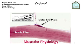
  • 7. Skeletal muscle fibers  Long cylindrical cells, enclosed by the sarcolemma.  The fibers are multinucleated ,pale oval nuclei that lie under its sarcolemma (peripheral situation).  The sarcoplasm is filled with numerous myofibrils.  The myofibrils, The thick filament (myosin) and the thin myofilaments (actin). Muscle contraction involves the sliding of filaments past each other, stimulated by nerve impulses. 7
  • 9. Muscle belly Tendon All movement is the result of contraction of skeletal muscle across a movable joint 9
  • 10. The muscle fiber and the basis of striations (The myofibrils) Two principal types of myofilaments have been identified ;thick and thin ones . 1. The thick filament is composed of a protein called myosin. 2. The thin myofilaments are attached to the Z line ,and are composed of a protein called actin together with two other associated protein called tropomyosin and troponin. The above arrangement of thin and thick myofilaments shows that: • The I-band is formed of thin myofilaments (actin filaments). • The A-band is formed of thick filaments (myosin) and actin. • The H-band has myosin filament only • The M line is a site where fine transverse filaments appear to interconnect the thick myosin filaments. Transvese tubules (T-tubules) : These are tubular invaginations of the sarcolemma that penetrate deep into the interior of the muscle fiber 10
  • 12. 12
  • 13. 13
  • 14. 14
  • 15. Bipolar aggregation of two different myosin molecules 15
  • 16. Portion of a thin filament 16
  • 17. Muscle contraction mechanism 1. Muscle fiber is stimulated to contract. 2. sarcolemma depolarizes (a wave of depolarization passes to the T tubules) 3. In the sarcoplasm, sarcoplasm reticulum (SR) undergoes a sudden permeability changes that release the stored calcium ions to sarcoplasm. 4. Calcium ions bind to one kind of troponin, causing a change in the configuration of the troponin complex. 5. Which causes a change in position of the tropomysin which lead to expose the myosin interacting sites on the actin molecule . 6. This activation induces the myosin heads (levers)to exert a pull on the actin filament causing it undergo a sliding movement. 17
  • 20. The sliding of action along the myosin molecule results in physical shortening of the sarcomere 20
  • 21. Skeletal Muscle Action Potential 21
  • 22. Excitation-Contraction Coupling • Skeletal muscles contraction controlled by the nervous system. • The transmission of electrical excitation to the actomyosin system is called Excitation-Contraction Coupling. • Converting an electrical stimulus (action potential ) to a mechanical response (contraction). 22
  • 23. 23
  • 24. • At the neuromuscular junction, the neurotransmitter released from the motor neuron is acetylcholine (ACh). • The ACh is received by a nicotinic cholinergic receptor. • The nicotinic receptor is a non-selective cation channel, that allows Na+ to enter the cell; Na+ entry causes depolarization. Neuromuscular Transmission 24
  • 25. Neuromuscular Transmission Fig. 11.7 • This depolarization, called an end plate potential, is an example of an excitatory postsynaptic potential (EPSP). • If the EPSP causes the area next to the motor end plate to depolarize to threshold, an action potential is generated. 25
  • 26. The action potentials on the sarcolemma spread to the interior of the muscle cell along the transverse tubules 26
  • 27. • T-tubules are tube-shaped invaginations of the sarcolemma (skeletal muscle plasma membrane) that penetrate throughout the muscle fiber. • The action potential in the muscle fiber is conducted into the interior of the muscle cell along the T-tubules. • The lumen of the T-tubule is continuous with the extracellular fluid, and the membrane depolarization during an action potential occurs across the T-tubule membrane. 27
  • 28. How does depolarization in the T-tubule membrane open a Ca++ channel in the sarcoplasmic reticulum membrane? Dihydropyridine receptor ++++ 28
  • 29. The muscle action potential activates the T-tubules (voltage sensors) the dihydropyridine (DHP) receptors. • DHP is a prototypical calcium channel blocker. • In cardiac and smooth muscle, the DHP receptor is a functional voltage- gated Ca++ channel. • In skeletal muscle, the DHP receptor does not function as a Ca++ channel. It is only a voltage sensor. The activated DHP receptors cause the calcium release channels (ryanodine receptors) of the SR to open. • direct coupling? • the favored model for skeletal muscle • via a second messenger (e.g., calcium-induced calcium release)? • the favored model for cardiac muscle Calcium enters the cytosol. 29
  • 30. 30
  • 31. End of Excitation • The ACh lasts only a short time because it is broken down by an acetylcholinesterase. • The ACh-esterase is anchored to the postsynaptic membrane. Fig. 11.10 31
  • 32. 2. Smooth muscle • Individual smooth appears as spindle shaped cell has a single elongated rod nucleus, centrally located • Sarcoplasm is abundant, eosinophilic . • Does not have clearly defined striations visible on the cells. This is because smooth muscle cells are organized in a different way than other muscle cells. • The actin and myosin fibers are arranged an angles to each other as they run through the cell. • The actin filaments in smooth muscle run from one side of the cell to the other, connecting at dense bodies and at the cell membrane. • In skeletal and cardiac muscle, the actin filaments are attached to Z plates, which hold many actin filaments and show up as dark bands under the microscope. 32
  • 33. 33
  • 34. Deference between smooth muscle and skeletal muscle contraction • In skeletal muscle, a signal from the somatic nervous system traverses to the muscle, where it stimulates organelles in the muscle cell to release calcium. Since there was always available ATP, the myosin uses it to quickly contract the cell. • In smooth muscle, the contraction is not controlled voluntarily by the somatic nervous system, but by signals from the autonomous nervous system, such as nerve impulses, hormones, and other chemicals released by specialized organs. • Smooth muscle is specialized to contract persistently, unlike skeletal muscle which much contract and release quickly. 34
  • 35. Smooth muscle location and function • Like all muscle tissue, the function of smooth muscle is to contract. • In the circulatory system, smooth muscle plays a vital role in maintaining and controlling the blood pressure and flow of oxygen throughout the body. While the majority of the pressure is applied by the heart, every vein and artery is lined with smooth muscle. • lines the majority of the digestive system, such in swallowing process. The cells on that side contract in reaction, a wave begins to propagate itself down your digestive tract. This phenomena is known as peristalsis, which responsible for moving food. • Smooth muscle is also responsible for contracting the irises, raising the small hairs on your arm, contracting the many sphincters in your body, and even moving fluids through organs by applying pressure to them. 35
  • 36. 3. Cardiac muscle • Each muscle fiber is enclosed with a sarcolemma. • Each muscle fibers composed of several short columnar cellular units join end to end by special surface specializations, intercalated discs, While skeletal muscle cells can have multiple nuclei, cardiac muscle cells typically only have one nucleus (large oval ,central position). • Sarcoplasm of a cardiac muscle fiber is more abundant and its mitochondria are much more numerous. • They branch and faintly striated. Purkinje muscle fibers: modified cardiac muscle fibers present in the interventricular septum. They specialized to conduct the cardiac impulse four and half times faster. • Contraction of cardiac muscles is spontaneous. 36
  • 37. Cont. • Intercalated discs: are small connections that join cardiac muscle cells (cardiomyocytes) to each other. • Gap junction: are part of the intercalated discs. When one cardiac muscle cell is stimulated to contract, a gap junction transfers the stimulation to the next cardiac cell. This allows the muscle to contract in a coordinated way. • Desmosomes: Like gap junctions, desmosomes are also found within intercalated discs. They help hold the cardiac muscle fibers together during a contraction. 37
  • 38. 38