intermediate filaments
•
4 gostaram•2,412 visualizações

a summaty about the intermediate filaments

Recomendados
Mais conteúdo relacionado
Mais procurados
Mais procurados (20)
Destaque
Destaque (10)
Semelhante a intermediate filaments
Semelhante a intermediate filaments (20)
Mais de Aly Barakat
Último
Último (20)
intermediate filaments
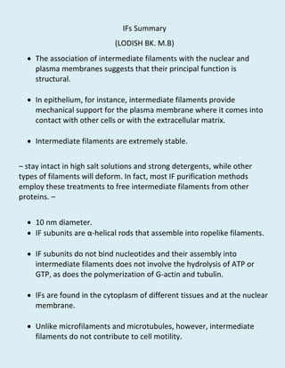
- 1. IFs Summary (LODISH BK. M.B) The association of intermediate filaments with the nuclear and plasma membranes suggests that their principal function is structural. In epithelium, for instance, intermediate filaments provide mechanical support for the plasma membrane where it comes into contact with other cells or with the extracellular matrix. Intermediate filaments are extremely stable. – stay intact in high salt solutions and strong detergents, while other types of filaments will deform. In fact, most IF purification methods employ these treatments to free intermediate filaments from other proteins. – 10 nm diameter. IF subunits are α-helical rods that assemble into ropelike filaments. IF subunits do not bind nucleotides and their assembly into intermediate filaments does not involve the hydrolysis of ATP or GTP, as does the polymerization of G-actin and tubulin. IFs are found in the cytoplasm of different tissues and at the nuclear membrane. Unlike microfilaments and microtubules, however, intermediate filaments do not contribute to cell motility.
- 2. The most ubiquitous group of IFs are the lamins. Lamins are found exclusively in the nucleus. Epithelial cells express acidic and basic keratins. They associate in a 1:1 ratio to form heterodimers, which assemble into heteropolymeric keratin filaments; neither type alone can assemble into a keratin filament. There are large number of keratin isoforms . Four proteins are classified as type III IF proteins. Unlike the keratins, the type III proteins can form both homo- and heteropolymeric IF filaments. The most widely distributed of all IF proteins is vimentin, which is typically expressed in leukocytes, blood vessel endothelial cells, some epithelial cells, and mesenchymal cells such as fibroblasts. Vimentin filaments help support cellular membranes. Vimentin networks may also help keep the nucleus and other organelles in a defined place within the cell. Vimentin is frequently associated with microtubules. The other type III IF proteins is Desmin –limitedly distributed–Desmin filaments in muscle cells are responsible for stabilizing sarcomeres in contracting muscle. Glial fibrillary acidic protein forms filaments in the glial cells that surround neurons and in astrocytes.
- 3. Peripherin is found in neurons of the peripheral nervous system, but little is known about it. The core of neuronal axons is filled with neurofilaments (NFs), each a heteropolymer composed of three polypeptides— NF-L, NF-M, and NF-H—which differ greatly in molecular weight. Neurofilaments are responsible for the radial growth of an axon and determine axonal diameter, (directly related to the speed of impulse conduction). ………………………………………………………………………………………………………………… All IF subunit proteins have a common domain structure: a central α-helical core flanked by globular N- and C-terminal domains. The core helical domain, which is conserved among all IF proteins, consists of four long α-helices separated by three nonhelical, “spacer” regions. The α-helical segments pair to form a coiled-coil dimer. An IF-protein dimer appears as a rodlike molecule with globular domains at the ends; two dimers associate laterally into a tetramer. The next steps in assembly include the end-to-end association of tetramers to form long protofilaments, which aggregate laterally into a loose bundle of protofibrils. Compaction of a protofibril yields
- 4. a mature 10-nm-diameter filament with the N- and C-terminal globular domains of the tetramers. In a mature filament, consisting of four protofibrils, the globular domains form beaded clusters on the surface. Interestingly, because the tetramer is symmetric, an intermediate filament may not have a polarity as does an actin filament or a microtubule. Although the α-helical core is common to all IF proteins, the N- and C-terminal domains of different types of IF proteins vary greatly in molecular weight and sequence. The true monomer of the IFs is still unknown. Some IF proteins form homopolymeric filaments; others form only heteropolymeric filaments with other proteins in their class; and some can form both homo- and heteropolymeric filaments. Some IF proteins, but not the keratins, can form heteropolymers with IF proteins in another class. NF-L self-associates to form a homopolymer, but NF-H and NF-M commonly co-assemble with the NF-L backbone, and so most neurofilaments contain all three proteins. The relative stability of intermediate filaments presents special problems in mitotic cells, which must reorganize all three cytoskeletal networks in the course of the cell cycle.
- 5. In particular, breakdown of the nuclear envelope early in mitosis depends on the disassembly of the lamin filaments that form a meshwork supporting the membrane. The phosphorylation of nuclear lamins by Cdc2, a cyclin-dependent kinase that becomes active early in mitosis (prophase), induces the disassembly of intact filaments and prevents their reassembly. Later in mitosis (telophase), removal of these phosphates by specific phosphatases promotes lamin reassembly, which is critical to re- formation of a nuclear envelope around the daughter chromosomes. Intermediate filament–associated proteins (IFAPs) cross-link intermediate filaments with one another, forming a bundle or a network, and with other cell structures, including the plasma membrane. IFAPs integrate the IF cytoskeleton with both the microfilament and the microtubule cytoskeletons, and attaching the IF cytoskeleton to the nuclear membrane and plasma membrane, especially at cell junctions. One family of IFAPs, the plakins, is responsible for linking IFs with both microtubules and microfilaments. One plakin family member is plectin, a 500,000-MW protein that has been shown to cross-link intermediate filaments with microtubules and actin filaments in vitro. Plectin also interacts with other cytoskeletal proteins, including spectrin, microtubule- associated proteins, and lamin B.
- 6. Immunoelectron microscopy reveals gold-labeled antibodies to plectin decorating short, thin connections between microtubules and vimentin, indicating the presence of plectin in these cross-links. A network of intermediate filaments is often found as a laminating layer adjacent to a cellular membrane, where it provides mechanical support. The best example is the nuclear lamina along the inner surface of the nuclear membrane. This supporting network is composed of lamin A and lamin C filaments cross-linked into an orthogonal lattice, which is attached by lamin B to the inner nuclear membrane through interactions with a lamin B receptor, an IFAP, in the membrane. Like the membrane skeleton of the plasma membrane, the lamin nuclear skeleton not only supports the inner nuclear membrane but also provides sites where nuclear pores and interphase chromosomes attach. Thus, the nuclear lamins organize the nuclear contents from the outside in.
- 7. In addition to forming the nuclear lamina, intermediate filaments are typically organized in the cytosol as an extended system that stretches from the nuclear envelope to the plasma membrane. Some intermediate filaments run parallel to the cell surface, whereas others traverse the cytosol; together they form an internal framework that helps support the shape and resilience of the cell. In muscle, a lattice composed of a band of desmin filaments surrounds the sarcomere. Longitudinal desmin filaments cross to neighboring Z disks within the myofibril, and connections between desmin filaments around Z disks in adjacent myofibrils serve to cross-link myofibrils into bundles within a muscle cell. The keratin filaments in one cell are indirectly connected to those in a neighboring cell by desmosomes (through E-cadherin) or to the extracellular matrix by hemidesmosomes (through integrin). The general role of keratin filaments appears to be to maintain the structural integrity of epithelial tissues by mechanically reinforcing the connections between cells. REFERENCE// MOLECULAR CELL BIOLOGY (5TH EDITION) –LODISH – BERK – MATISUDAIRA – KAISER – KRIEGER – SCOTT – ZIPURSKY – DARNELL ………………………………………………………………………………………………………........ DONE BY: ALY AHMED BARAKAT (MD STUDENT AT OMC)