
Recomendados
Mais conteúdo relacionado
Mais procurados
Mais procurados (20)
Semelhante a Nasopharyngeal Carcinoma
Semelhante a Nasopharyngeal Carcinoma (20)
Mais de Ali Azher
Último
Último (20)
Nasopharyngeal Carcinoma
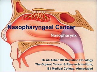
- 1. Nasopharyngeal Cancer Dr.Ali Azher MD Radiation Oncology The Gujarat Cancer & Research Institute, BJ Medical College, Ahmadabad
- 2. Introduction • Cuboidal chamber – below skull base • 4-5.5cm transverse, 2.5 to 3.5 AP diameter, 4cm height • Anteriorly nasal cavity via posterior choana • Inferiorly oropharynx • Roof sphenoid & occipital bones • Floor soft palate • Lateral walls Eustachian Tube • Lateral walls & fossa of rosenmuller common sites for malignancies
- 3. • Foramen lacerum – spread to middle cranial fossa • Close to cavernous sinus, CNs 2,3,4
- 4. Anatomy Lateral Eustachian tube, torus tubarius, fossa of Rosenmuller, Superior pharyngeal Constrictors medial pterygoid plate Anterior Posterior nasal septum/choanae Posterior Clivus and C1–2 Superior Sphenoid bone/ sinus Inferior Roof of soft palate
- 5. Foramen rotundum - Maxillary division of trigeminal (V2) nerve Foramen ovale - Mandibular division of trigeminal (V3) Nerve, Accessory meningeal artery, Lesser superficial petrosal nerve Foramen lacerum - Internal carotid, sympathetic carotid plexus; vidian nerve, meningeal branch of ascending pharyngeal artery, emissary vein Foramen spinosum - Recurrent branch of V3 nerve Middle meningeal artery and vein
- 6. Lymphatic Drainage • Rich lymphatic plexus – roof, posterior, lateral walls • Three lymph node collecting stations 1. RP node of Rouviere 2. Jugular chain – upper lateral JD nodes 3. Upper deep cervical node at the confluence of Spinal accessory • Lymphatic capillary plexus – lymphiod aggregates in the pharyngeal tonsil & tubal tonsil
- 7. • RP nodes – lateral parapharyngeal space • RP nodes – proximity to 9,10,11,12 • Classic NP node – 3-4 high lateral jugular nodes that lie deep into SCM and lateral to IJV
- 8. • Lymph collectors • Lateral side of the pharyngeal wall • The lateral lymph collector empties into multiple first-tier nodes (lateral pharyngeal node, the jugulodigastric/subdigastric nodes, 3,4,5 RP group • The posterior lymph collector empties into the first node (node of Rouviere) of RP
- 9. CT anatomy
- 13. Epidemiology • Highest in china 3/100,000 • Bimodal age distribution 15-25 yrs & 50-59yrs • Causation is multifactorial involving environmental, genetic and familial and viral factors • Early lymphatic spread and notorious predilection for distant metastases • Anatomical proximity to critical structures makes surgical extirpation difficult without morbidity. • RADIOSENSITIVE
- 14. Etiology • Genetic • Environmental Salted fish in southern china Dimethyl nitrosamine Alcohol, cigarette, dust etc • Viral – EBV NPC non keratinising type Tumorigenic potential – LMP 1, 2A, 2B Nuclear antigens – EBNA1 & 2
- 15. Natural history • Local extension Anterior – nasal fossa → lateral walls of nasal fossa → destruction of pterygoid plates • Ethmoid, maxillary sinus less common • Advanced disease orbital invasion • Superior & Posterior Base of skull, sphenoid sinus,clivus Cavernous sinus, • Inferiorly – tonsillar pillar, tonsillar fossa, oropharyngeal wall
- 16. Border Spread Significance Lateral Parapharyngeal Space Fossa of Rosenmuller is the most common site for NPC. Retroparotidian syndrome = involvement of CN IX–XII and cervical sympathetics Masticator space Trismus anterior Pterygopalatie fossa (PPF) via sphenopalatine foramen from nasal cavity Tumors can extend proximally along V2 from PPF to cavernous sinus Posterior Retropharyngeal (RP) nodes and prevertebral space >75% patients - cN+, 90% - subclinical nodes, 40–50% b/l nodes. Level 2 & lateral RP nodes are the first echelon Superior Skull base Foramen ovale (CN V3) and foramen lacerum commonly involved. True intracranial extension is uncommon (<10%). Petrosphenoidal syndrome = extension through foramen lacerum to cavernous sinus Inferior Hard palate (oropharynx) Infrequent
- 20. Lymphatic Spread • Vast avalvular lymph nodes • Ipsilateral 85-90% • Bilateral 50%
- 22. Hematogenous Spread • DM in 3-6% • Higher in advanced neck node mets • Bone > Lung > Liver
- 23. Signs & Symptoms • Neck masses – upper neck (upper posterior neck, palpable beneath SCM) • Nasopharynx Tumor Mass – epistaxis, nasal obstruction discharge • Skull base erosion – palsy of cranial nerves (headache, diplopia, facial pain, numbness) • Neck mass> nasal & aural symptoms • CN – V& VI frequently involved
- 24. Neurological symptoms • Advanced disease • Petrosphenoidal syndrome of Jacob – unilateral trigeminal neuralgia, unilateral ptosis, complete ophthalmoplegia, and amaurosis – CN II-VI by IC extension
- 25. Villaret Syndrome • Retroparotidian syndrome • dysphagia, alterted taste • problems in salivation • paralysis of trapezius & SCM • Weakness of tongue & soft palate • CN IX-XII involvement by RP node mets
- 26. Horners Syndrome • Enophthalmos • Ptosis • Miosis
- 27. Work up & Evaluation • Complete head & neck physical exam Node – size, mobility, extent of enlarged node Vision, hearing Exclusion of gross signs of DM • Nasopharyngoscopy + Biopsy of Primary / FNAC • Labs – CBC, LFT , EBV titers (IgA anti VCA, IgG anti EA) • Chest X ray • CT brain + PNS + Neck • MRI with contrast include skull base, nasopharynx, and neck to clavicles
- 28. • MRI better sensitivity than CT • Detection rates of MRI and CT Scan compared • IC Extension 57 % vs. 36 % • Skull base involvement 60 % vs. 40 % • Retropharyngeal node 58 % vs. 21 % • Prevertebral muscle infiltration 51 % vs. 22 % • MRI detected bone erosion in all cases, as seen on CT • Upstage of T stage in 22%, downstage in 4 %
- 29. • Metastatic work up PET-CT (especially for non-keratinizing histology, endemic phenotype, N2 or N3, and stage III –IV) CT chest & abdomen Bone scan
- 30. Consultations • Medical Oncology • Dental Evaluation • ENT – audiology • Nutrition – swallowing
- 31. Differentials • If small nasopharyngeal mass and confined to the mucosa: Prominent, but normal adenoidal tissue Nasopharyngeal lymphoma Early primary nasopharyngeal malignancy
- 32. • If larger nasopharyngeal mass +/-involvement of the skull base: Primary nasopharyngeal malignancy Adenoid cystic carcinoma Papillary adenocarcinoma Melanoma Plasmacytoma Lymphoma Chordoma/Chondrosarcoma Meningioma Rhabdomyosarcoma/other sarcoma
- 33. Histopathology • Poorly differentiated carcinoma • Pleomorphic epithelioid cells in a background of lymphocytes • Presence of keratin – adverse prognostic factor
- 34. WHO classification • Keratinizing Squamous cell carcinoma 20% • Nonkeratinizing carcinoma - Most Common Differentiated subtype 30-40% Undifferentiated subtype 50-60% - most favorable prognosis, EBV • Basaloid Squamous cell carcinoma <0.2% - agrressive clinical course – poor survival
- 35. AJCC 8th Edition T Criteria Tx Primary Tumor cannot be assessed T0 Tumor identified, EBV + CERVICAL NODES T1 Tumor Confined to NP or extension to oropharynx/nasal cavity WITHOUT PARAPHARYNGEAL INVOLVEMENT T2 Tumour with extension to PARAPHARYNGEAL SPACE and/or adjacent soft tissue involvement (medial pterygoid, lateral pterygoid, prevertebral muscles) T3 Tumour invades BONY STRUCTURES of skull base cervical vertebra, pterygoid structures, and/or PARANASAL SINUSES T4 Tumour with INTRACRANIAL EXTENSION, involvement of cranial nerves, hypopharynx, orbit, PAROTID GLAND and/or extensive soft tissue infiltration beyond the lateral surface of the lateral pterygoid muscle
- 36. N Criteria Nx Regional LN cannot be assessed N0 No regional LN N1 UNILATERAL metastasis, in cervical lymph node(s), and/or unilateral or bilateral metastasis in RETROPHARYNGEAL LN, 6 cm or less ,above the caudal border of cricoid cartilage N2 BILATERAL metastasis in cervical lymph node(s), 6 cm or less ABOVE the caudal border of cricoid cartilage N3 Metastasis in cervical lymph node(s) GREATER THAN 6 CM in dimension and/or extension BELOW the caudal border of cricoid cartilage
- 37. Staging T1 T2 T3 T4 N0 I II III IVA N1 II II III IVA N2 III III III IVA N3 IVA IVA IVA IVA M1 IVB IVB IVB IVB
- 38. Prognostic factors Younger age & female sex – better prognosis Histology – non keratinizing & undifferentiated – more radiosensitive – better prognosis Advanced T stage – worse LC Advanced N stage – increased risk of DM & worse survival EBV DNA load – higher recurrence EGFR, VEGF over expression – poor
- 39. Treatment strategy • Surgery difficult ; only role in: Biopsy for pathologic diagnosis Salvage for persistent / recurrent disease • RT ALONE / IN COMBINATION WITH CT
- 40. Treatment Algorithm Stage Treatment options T1 N0 Definitive RT T1 N+ T2-4 N0 T2-4 N+ Concurrent CRT +/– Adj Chemo Induction Chemo ► Concurrent CRT M1 Chemotherapy Concurrent CRT
- 41. Diagnosis of NPC Staging of NPC Stage IVA T4 or N3 M0 Stage IVB M1 Stage I T1N0M0 Stage II/III T1-3 N0-2, M0 Definitive IMRT CRT IMRT + Cisplatin Adj CT Cis+ 5FU NACT Cisplatin CRT IMRT + Cisplatin Adj CT Cis+ 5FU CT Cisplatin +/- Pall RT
- 42. Chemo radiotherapy ARM 3Yr SURVIVAL 5Yr SURVIVAL DM RATE RT 56.38% 41.09% 38.71% CRT 68.74% 51.91% 26.19% The result demonstrated that CRT increased overall survival by 12% at 3 years, and 11% at 5 years. After CRT, the rate of distant metastasis was reduced by 12%. Meta-analysis included 18 trials enrolling a total of 1993 patients from China. Yank AK,Liu TR, Guo x, QI gl, Chen FJ.
- 43. Dose Dose T stage Local Tumor Control Rate(%) >70Gy T1 T2 100 66-70 Gy T1 T2 80 >70GY T3 T4 <55 60 Gy T1 T2 76 TOTAL DOSE IMPORTANT DOSE FRACTION NOT 70Gy/35#/7weeks to GTV 50-60 Gy for potential risk sites FRACTIONAL DOSE OF > 2Gy SHOULD BE AVOIDED
- 44. Pediatric • Per COG ARAR 0331 protocol • Stage I: RT alone (61.2/1.8 Gy for Stage I; 66.6/1.8 Gy for StageII) • Stage ≥ II: Cisplatin/5-FU × 3c → RT (CR/PR to chemo 61.2/1.8 Gy, SD to chemo 70.2/1.8 Gy) and concurrent cisplatin • 36–46/2–3 Gy to unresectable metastases
- 45. Radiation Planning • Conventional • Conformal
- 46. Conventional set up • Direct marking open field • Simulator • 3 fields; 2 lateral opposed for primary and upper neck, matched to 1 anterior field for lower neck.
- 47. Portals B/L parallel opposed portals for primary & upper neck • Superior : 2 to 2.5 cm above the zygomatic arch and splits the pituitary fossa. In case of base of skull involvement or intracranial extension it is taken 4.0 to 5.0 cm above the zygomatic arch or 1 cm above the pituitary fossa. • Inferior : at the thyroid notch • Anterior : encompasses posterior ½ of nasal cavity or moved forward to cover the extensions if any. • Posterior : kept open to cover the posterior triangle
- 48. • Single anterior portal for lower neck Superior : matched to the inferior border of the lateral fields. Inferior : extend below to cover the lower edge of clavicles. Lateral : cover medial 2/3rd of the clavicle.
- 51. Dose prescription • Phase I : 40 to 44 GY in 20 to 22 fractions @ 2GY/#. • Phase II : fields are shrinked to avoid the spinal cord. • Primary tumor is boosted to an additional 20 to 25 GY. • T1 & T2 tumor : 60 to 65 GY • T3 & T4 tumor : 70 to 75 GY. • Dose to neck nodes : 45 to 50 GY to N0 neck.
- 52. Morbidity from RT Acute Toxicity Mucositis Dermatitis Pharyngitis Otitis Dose-limiting organs – Brain stem – Spinal cord – Pituitary-hypothalamic axis – Temporal lobes – Eyes – Middle/inner ears – Parotid glands Chronic Toxicity Xerostomia sub cutaneous fibrosis radiation myelitis cranial neuropathy endocrine dysfunction temporal lobe necrosis hearing loss otitis media
- 53. Limitations of Conventional RT • rectangular-shaped - suboptimal target coverage and inclusion of large volumes of normal tissues • high rate of xerostomia • Carotid stenosis • Cranial nerve palsy • Dose escalation difficult
- 54. Conformal • Immobilisation device orfit making • Neck extended, shoulder down • Fiducial markers • Laser matching • Topogram • IV contrast • Upto the level of carina
- 55. • IMRT is preferred
- 56. Rationale for IMRT • Anatomically complex H&N region • Treat target volumes adjacent to critical or sensitive normal tissues • Lack of organ motion in the H&N region • Allows for dose escalation , allows for concomitant boost.
- 57. Better normal tissue sparing Better conformality Dose escalation
- 58. Inverse Planning • Desired dosimetric & clinical objectives are stated mathematically • Appropriate specifications for normal tissue dose constraints • Overstringent dose constraints to normal tisues – ensure adequate dose cover to target
- 59. Forward Planning • From field definition to dose distribution • T/t parameters • Dose calculation • Dose distribution • Dose delivery with uniform radiation intensity
- 60. Inverse Planning • From dose distribution to field definition • Dose delivery with nonuniform radiation Intensity • leaf sequence generation • optimization • Treatment goals ( objective function )
- 61. Steps of IMRT Clinical evaluation & assessment Simulation Planning CT/MRI/PET-CT scan Target volume Delineation: Gross target volume,Clinical target volume, Planning target volume. Dose prescription : PTV dose and Organ at risk (OAR) constraint IMRT Planning, Dose Volume Histogram Quality Assurance Execution of IMRT
- 62. Import of Images • Monaco system • OAR contouring • Dose constraints
- 63. OAR • Critical normal structures • Could suffer significant morbidities • Might influence treatment planning & dose prescription • All non target tissues • Depend on the location of ctv
- 64. Rhyme respecting cord & brainstem • Brainstem – lower part of lateral ventricle to odontiod apical • Cord starts from odontoid apical upto 5cm below PTV • Check for missing slice – DON’T INTERPOLATE • Serial organs – maximum dose minimum • Brain window • PRV extra degree of freedom
- 65. • Optic nerve – delineating orbital part only NOT IDEAL • Draw till chiasm through optic canal
- 66. PTV • Geometric concept for treatment planning • Tool to shape absorbed dose distributions • Ensure the delivery of dose to all parts of ctv • Despite organ motion & set up variations
- 67. Contouring guide lines • GTV 70 = gross disease • GTV N = all nodes ≥10mm
- 68. LN Metastasis • Ellipsoid • Size ≥ 10mm (5mm RP) • Central necrosis • Extra capsular spread • Clustered nodes 3 or more
- 69. CTV70 • GTV 70 + 5 mm • Around the brainstem and spinal cord, 1-mm margin • entire nasopharynx, sphenoid sinus, cavernous sinus, base of skull, posterior ½ of nasal cavity, posterior 1/3 of maxillary sinuses, post. Ethmoid sinus, pterygoid fossa, lateral & posterior pharyngeal wall, retropharyngeal nodes, & b/L cervical nodes including level V & SCF.
- 70. SCOPE • S – Sinus (Sphenoid, Max post 1/3), skull base, soft palate • C- Clivus, cavity nasal post 1/3, cavernous sinus, cave meckels • O – ovale foramen • P – Pterygopalatine fossa, pterygoid fossa, parapharyngeal space, proper nasopharynx, • E- ethmoid posterior
- 71. PTV70 • CTV 70 + 3–5 mm • Around the brainstem and spinal cord 1-mm
- 72. PTV2 (59.4 Gy) High risk subclinical disease: Sphenoid sinus Cavernous sinus Skull base Clivus Posterior 1/3 of maxillary sinus Posterior 1/3 of nasal cavity Pterygopalatine fossae Parapharyngeal space Retropharyngeal space Soft palate
- 73. High risk nodal levels • Include all bilaterally • Upper deep jugular • Level I submandibular • Level II JD • Level III mid jugular • Level IV Low jugular & supraclavicular • Level V Posterior cervical • Retropharyngeal
- 75. PTV3 (54 Gy) Elective nodal coverage: Low risk sub clinical disease Retrostyloidspace Bilateral IB –V (can omit IB, if N0)
- 77. Two different approaches • Extended whole field EWF Total target volume in the IMRT plan • Split field SF Target volume superior to vocal cord – IMRT Lower neck – conventional anterior neck field Dose to vocal cords minimum
- 78. Dose prescription • PTV High Risk 70Gy • PTV Intermediate risk 59.4Gy • PTV Low risk 54Gy
- 79. OAR dose constraints • Critical normal structures (higher priority): • Brain stem D Max <54 • Optic Nerves <54 • Optic Chiasma <54 • Spinal Cord <45 • Mandible & TMJ < 70 • Temporal Lobes < 60 • Pituitary Dmax < 50Gy
- 80. • Other normal structures (lower priority): • Parotids • Dmean < 26 (atleast one gland) • at least 20 cc of the combined volume of both parotid glands to receive < 20 Gy, • at least 50% of one gland to receive < 30 Gy • TongueMax < 55 Gy • Inner/middle ears Mean < 50 Gy • (especially with concurrent Cisplatin) • EyesMean < 35 Gy • GlotticlarynxMean < 45 Gy • Lens < 6
- 81. Delineating Parotid as OAR • Identify the gland • Mid aspect of the gland • Then proceed cranially and caudally • Boundaries Cranial – EAC & Mastoid Caudal – submandbular space Anterior – masseteric muscle Posterior - SCM
- 82. Simultaneous integrated boost SIB • Simultaneously treat two or more volumes • Different fraction sizes • Use of same plan – less error • Dose painting
- 83. Plan Evaluation • DVH • 95% isodose to 100% target volume • Dose constraints to OAR
- 84. Treatment delivery • IMRT • Exact position • Fiducial matching • Isocentre • SSD
- 85. Chemotherapy • Induction • Concurrent • Adjuvant
- 86. NACT • Ongoing trail • Excellent outcomes & tolerability • NCCN Category 3
- 87. 480 patients, Stage III, IV Docetaxel, cisplatin, and fluorouracil (TPF)+CRT Vs CRT
- 88. Arm Distatnt FFS Locoregional FFS NACT+CRT 90 92 CRT 83 89
- 89. Trial No Description DFS International NPC study 339 RT vs NACT+RT 54 vs 40 Me et al 456 RT vs NACT+RT Cis+bleo+5FU 63 vs56 AOCOA 334 RT vs NACT +RT Cis 48 vs 42
- 90. CRT • CDDP (Cis diammine dichlorido platinum) 100mg/m2 • Weekly monitoring of CBC, RFT • Adequate hydration of patient • Weekly oxaliplatin 70mg/m2 • Carboplatin • Cetuximab EGFR target • Bevacizumab VEGF
- 91. CDDP • Pencillin of cancer • Peyrone’s chloride • Use with caution in altered RFT • Hydrated before, after and post chemo • Higher the dose more aggressive hydration (2L) • Furosemide diuresis every 2L • anti emetis (ondan, dexona, aprepitant) • Nephrotoxic, ototoxic, neurotoxic • Azoopermia, alopecia
- 92. CRT – key trials Trial No Method DFS LRC Toxicity NPC 9901 348 Conc Cis+RT No Adj CT 72 vs 62 92 vs 82 > CRT arm SQNP01 221 Al Sarraf regimen 72 vs 53 > CRT Taiwan 284 Cis/5Fu+RT vs RT 72 vs 53 96.8 vs 92.1
- 93. Al Sarraf Trial • 193 patients Trial 5 yr PFS 5 yr OS RT 29 37 Chemo RT + Adj RT 58 67 Adjuvant CT Cisplatin / 5FU Concurrent CRT (Cisplatin)
- 94. MAC-NPC meta-analysis (Blanchard, Lancet Oncol 2015) • 19 trials with 4806 pts. • 5-yr OS benefit for concurrent and adjuvant chemo (12.4%) or concurrent chemo alone (9.4%)
- 95. NPC 9901
- 96. Dose escalation • Improvement in local tumor control rates • External Beam RT • Brachytherapy boost • Stereotactic RT boost
- 97. Brachytherapy • Suitable Candidate Tumors restricted to Nasopharynx as BOOST Thickness of CTV <10 mm - well circumscribed, superficial local recurrences. • Local control upto 90 – 95 % for T1-T2 tumours with acceptable late toxicities • Dose delivered through brachytherapy is adequate only for superficial non-bulky tumours • Outcome depends on accurate placement of the catheters, which largely depends on patient anatomy and clinician’s skill
- 98. • Boost minimal, local,residual local disease • Salvage therapy well circumscribed superficial local recurrences
- 99. Techniques • Customised mould • Rotterdam nasopharyngeal applicator • Massachusetts general hospital technique • Transnasal permanent interstitial implants
- 100. RN Apllicator • Made of soft silicone • Endocavitary • Well tolerated by patient • The two silicone tubes, can accommodate standard 6 French afterloading catheters. • The legs of the applicator sort through the nostrils and are fixed with a silicone bridge, pushed against the nasal septum
- 104. Nasopharynx Implant points • Tumor tissue points Node of Rouviere (R) Nasopharynx (Na) • Normal tissue points Spinal cord, soft palate, base of skull, pituitary gland, optic chiasm, and retina
- 105. Nasopharynx RPN
- 106. Dose Prescription • 5-10mm from mucosal surface • Doses of 18Gy/6# [EBRT 60 Gy/30#]—T1 • 12Gy/4#[EBRT 70Gy/35#]—T2-T4 lesions
- 107. Boost with electrons • Lateral field • 9MeV • 90% Isodose line • 6x6cm field
- 108. Endemic NPC • known to occur in China, Hong Kong, South Eastern Asia, Greenland • Associated with EBV virus infection • In India similar pathology seen in Kashmiris. • Present a decade younger. • Not associated with smoking or alcohol consumption
- 109. • Associated with undifferentiated carcinoma ( WHO II and III) • Associated with more advanced disease at presentation • Nodal stage also more advanced and more frequently involved. • Both chemo and radio sensitive • Histologically more vascularized (Better Rx response) • Greater % of cell in the growth fraction. • Better loco regional control and survival than sporadic variants. Several markers for predicting biological behavior
- 110. Complications • Radiation induced • Mucositis • Xerostomia • Chemo induced
- 111. Complications • Temporal lobe necrosis • Cranial neuropathy • Oral complications • Aural toxicity • Carotid stenosis • Endocrine dysfunction • Second malignancies
- 112. Temporal Lobe Necrosis • Sequelae of treatment • Conventional 2D RT • 65% • > 2 Gy • Hallucination, absence seizures, déjà vu (31%) • Headache, convulsions, confusion, hemiparesis (14%) • Asymptomatic (16%) • IMRT 3-4%
- 113. Cranial neuropathy • IX, XII • Slurring of speech, twitching of neck muscles, dysphagia – fatal aspiration pneumonia • VI
- 114. Oral complications • Xerostomia • Salivary flow dropped by half 7.2Gy • Nadir after 36Gy • For 2 years no recovery • Parotid sparing IMRT • Osteoradionecrosis – maxilla (2.7%), mandible (1.7%)
- 115. Aural toxicity • Hearing loss - SNHL • Cisplatin increased the risk
- 116. Carotid stenosis • High risk patients - >60yrs, smoking, HTN, HCH, CVA
- 117. Endocrine dysfunction • Hypothalamic pituitary dysfunction • Amenorrhea, galactorrhea – hyperprolactinemia • Hypothyroidism • Hypoadrenalism
- 118. Second malignancies • Rare (0.04%) • Latency period > 10yr • Maxillary osteosarcoma • Soft tissue sarcome
- 119. Follow up H&P + complete H&N physical exam +/- mirror/fiberopticexam q1-3 months for year 1 q2-6 months for year 2 q4-8 months for years 3 –5 Yearly for years > 5 Imaging for signs/symptoms TSH yearly (if irradiated) Speech/swallowing/dental/hearing evaluations Consider EBV-DNA monitoring
- 120. Clinical Pearls • Complex anatomy, radiosensitive & curable • IMRT with SIB • Dose 70Gy • Concurrent cisplatin-based chemo + RT has been shown to improve OS • Brachytherapy for boost