2015.surgical treatment of colon cancer
•Transferir como PPTX, PDF•
0 gostou•62 visualizações

dr. Amer Odobašić

Recomendados
Mais conteúdo relacionado
Mais procurados
Mais procurados (20)
Semelhante a 2015.surgical treatment of colon cancer
Semelhante a 2015.surgical treatment of colon cancer (20)
Mais de Aleksandar Aničić
Mais de Aleksandar Aničić (11)
Último
Último (20)
2015.surgical treatment of colon cancer
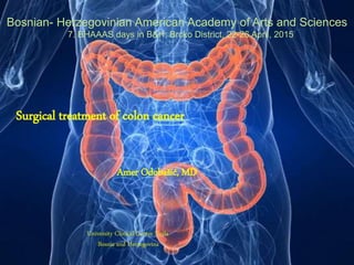
- 1. Surgical treatment of colon cancer Amer Odobašić, MD University Clinical Center Tuzla Bosnia and Herzegovina Bosnian- Herzegovinian American Academy of Arts and Sciences 7. BHAAAS days in B&H; Brcko District, 22-26 April, 2015
- 3. Facts • 2nd leading cancer killer in the U.S. – Every 9.3 minutes, a person in the U.S. dies of colon cancer • Survival depends on early detection – 90% five-year survival rate in early detected cases
- 4. • Estimated new cases and deaths from colon cancer in the United States in 2015: New cases: 93,090 (colon cancer only). Deaths: 49,700 (colon and rectal cancers combined) • American Cancer Society: Cancer Facts and Figures 2015. Atlanta, Ga: American Cancer Society, 2015. Available online. Last accessed January 7, 2015.
- 5. Republic of Korea had the highest rate of colorectal cancer, followed by Slovakia and Hungary. About 54 per cent of colorectal cancer cases occurred in more developed countries. The highest incidence of colorectal cancer was in Oceania and Europe and the lowest incidence in Africa and Asia. Source: Ferlay J, Soerjomataram I, Ervik M, Dikshit R, Eser S, Mathers C, Rebelo M, Parkin DM, Forman D, Bray, F. GLOBOCAN 2012 v1.1, Cancer Incidence and Mortality Worldwide: IARC CancerBase No. 11 [Internet]. Lyon, France: International Agency for Research on Cancer; 2014. Available from: http://globocan.iarc.fr, accessed on 16/01/2015.
- 6. How Can Reduce Risk For Colon Cancer?
- 7. • Know risk • Maintain a healthy weight throughout life • Be physically active • Eat a healthy diet • Limit consumption of alcoholic beverages • Do not use tobacco products
- 8. Groups at an increased risk: – Those age 50 and older – Individuals with a personal or family history of colon cancer, non-cancerous olon ps, or Irritable Bowel Syndrome (IBS)
- 9. Hereditary Colorectal Cancer • Familial adenomatous polyposis – FAP account for <1% of all colorectal cancers – Due to mutation of the adenomatosis polyposis coli (APC) gene – Numerous adenomas appear as early as childhood and virtually 100% have colorectal cancer by age 50 if untreated • Hereditary non-polyposis colorectal cancer / Lynch syndrome – More common than FAP and account for ~1-5% of all colonic adenocarcinomas – Due to a mutation in one of the mismatch repair genes – Earlier age onset of colorectal cancer and predominantly involve the right colon – HNPCC also increases the risk of • Endometrial, ovarian, breast ca • Stomach, small bowel, hepatobiliary ca – Renal pelvis or ureter ca
- 10. The best way to reduce risk is by getting screened.
- 11. • Common Screening Options: – Colonoscopy – Virtual colonoscopy (computerized tomographic colonography or CTC) – Sigmoidoscopy – Fecal occult blood test (FOBT) – Fecal immunochemical test (FIT) Screening Options
- 12. A colonoscopy is the most effective screening method. It can reduce the average person's risk of dying from colon cancer by 90%.
- 13. What is the better ?
- 14. Clinical Presentation Depends on location of cancer • Locations – ⅔ in descending colon and rectum – ½ in sigmoid colon and rectum (i.e. within reach of flexible sigmoidoscope) • Caecal and right sided cancer – Iron deficiency anaemia (most common) – Distal ileum obstruction (late) – Palpable mass (late)
- 15. Clinical Presentation • Left sided and sigmoid carcinoma Change of bowel habit • Alternating constipation + diarrhoea • Tenesmus • Thin stool PR bleeding, mucus • Bowel obstruction • Metastasis o Liver (hepatic pain, jaundice) o Lung (cough) o Bone (leucoerythroblastic anaemia) o Regional lymph nodes o Peritoneum o Others
- 16. Three elements: • T = Tumor – How large is the tumor? • N = Node – Are cancer cells in the lymph nodes? • M = Metastases – Has the cancer spread to other organs? Four stages: • Stage 0 • Stage I – Spread to the middle layers of the colon or rectum • Stage II • Stage III • Stage IV – Advanced disease, spread to other organs STAGING OF COLON CANCER
- 17. STAGE GROUPING STAGE T N M DUKES MAC 0 TIS NO MO - - I T1 NO MO A A T2 NO MO A B1 IIA T3 NO MO B B2 IIB T4 NO MO B B3 IIIA TI-T2 NI MO C CI IIIB T3-T4 NI MO C C2/C3 IIIC ANY T N2 MO C C1/C2/C3 IV ANY T ANY N MI D
- 18. STAGING OF COLON CANCER PRIMARY TUMOR REGIONAL NODES DISTANT METASTASES TX-CANNOT ASSESS NX-CANNOT ASSESS MX CANNOT ASSESS TO- NO PRIMARY TUMOR NO-NO METS RN MO- NO DISTANT METS TIS- TUMOR IN SITU N1-METS 1-3 RN M1- DISTANT METS T1- INVADES SUBMUCOSA N2- METS >3 RN T2- INVADES MUSCULARIS T3- INVADES THROUGH MUSCULARIS PROPIA INTO SUBSEROSA OR ONTO NON- PERITONIALISED PERICOLIC OR PERIRECTAL TISSUES T4-DIRECTLY INVADES OTHER ORGANS OR STRUCTURES AND/OR PERFORATES VISCERAL PERITONEUM
- 19. Colon anatomy
- 21. Stage 0 Colon Cancer Treatment Stage 0 colon cancer is the most superficial of all the lesions and is limited to the mucosa without invasion of the lamina propria. Because of its superficial nature, the surgical procedure may be limited. Standard treatment options for stage 0 colon cancer include the following: • Local excision or simple polypectomy with clear margins. • Colon resection for larger lesions not amenable to local excision.
- 22. Stage I Colon Cancer Treatment Because of its localized nature, stage I colon cancer has a high cure rate. Standard treatment options for stage I colon cancer include the following: • Wide surgical resection and anastomosis. The role of laparoscopic techniques [1] [2] [3] [4] in the treatment of colon cancer was examined in a multicenter, prospective, randomized trial (NCCTG-934653, now closed) comparing laparoscopic-assisted colectomy (LAC) with open colectomy.
- 23. Stage II Colon Cancer Treatment Standard treatment options for stage II colon cancer include the following: • Wide surgical resection and anastomosis. Evidence (laparoscopic techniques): The role of laparoscopic techniques [1] [2] [3] [4] in the treatment of colon cancer was examined in a multicenter, prospective, randomized trial (NCCTG-934653, now closed) comparing laparoscopic-assisted colectomy (LAC) to open colectomy.
- 24. Stage III Colon Cancer Treatment Stage III colon cancer denotes lymph node involvement. Studies have indicated that the number of lymph nodes involved affects prognosis; patients with one to three involved nodes have a significantly better survival than those with four or more involved nodes. Standard treatment options for stage III colon cancer include the following: • Surgery - wide surgical resection and anastomosis. • Adjuvant chemotherapy.
- 25. Stage IV and Recurrent Colon Cancer Treatment Stage IV colon cancer denotes distant metastatic disease. Treatment of recurrent colon cancer depends on the sites of recurrent disease demonstrable by physical examination and/or radiographic studies. Such approaches have not led to improvements in long-term outcome measures such as survival.
- 26. 1. Surgical resection of locally recurrent cancer. 2. Surgical resection and anastomosis or bypass of obstructing or bleeding primary lesions in selected metastatic cases. 3. Resection of liver metastases in selected metastatic patients (5-year cure rate for resection of solitary or combination metastases exceeds 20%) or ablation in selected patients. [2] [3] [4] [5] [6] [7] [8] [9] [10] [11] 4. Resection of isolated pulmonary or ovarian metastases in selected patients. [12] 5. Palliative radiation therapy. 6. Palliative chemotherapy. 7. Targeted therapy. 8. Clinical trials evaluating new drugs and biological therapy. 9. Clinical trials comparing various chemotherapy regimens or biological therapy, alone or in combination.
- 27. Survival rates for colon cancer, by stage: Stage 5-year Relative Survival Rate I 92% IIA 87% IIB 63%* IIIA 89%* IIIB 69% IIIC 53% IV 11% *These numbers are correct : patients with stage IIIA or IIIB cancers have better survival than those with stage IIB cancers. These statistics are based on a previous version of the staging system. In that version, there was no stage IIC (those cancers were grouped considered stage IIB). Also, some cancers that are now considered stage IIIC were classified as stage IIIB, while some other cancers that are now considered stage IIIB were classified as stage IIIC.
- 28. Malignant large bowel obstruction
- 29. • Malignant bowel obstruction needs: – Individualised approach – Team work (oncology, surgery, radiology, specialist palliative care team and other health care professionals) • Communication: – Treatment options, expectations & limitations, discharge plan and preferred place of care….the earlier you discuss with patient and family, the better coping and the less of unnecessary anxiety and fear of uncertainty
- 30. • Advantages: – Alternative option for patients unfit for surgery or do not want to have surgery – A quick fix while waiting for surgery – High success rate for left sided colonic obstruction – Quicker recovery & shorter hospital stay • Less successful: – Rapidly progressive cancers – Multifocal bowel obstruction – Diffuse carcinomatosis Colonic stenting
- 31. • What surgery – Resection/debulking….primary anastomosis – Bypass surgery – Defunctioning colostomy/ileostomy
- 32. OBSTRUCTING CANCERS ON THE RIGHT SIDE. RESECTION, ANASTOMOSIS ... STOMA ON THE LEFT SIDE DIVERTING STOMA, RESECTION, OR INTRA-OP COLONIC LAVAGE WITH ANASTOMOSIS PERFORATING CANCERS CAN BE FROM EROSION OR PERFORATION SECONDARY TO OBSTRUCTION. WASHOUT, RESECTION AND ANASTOMOSIS IF FEASIBLE. STOMA
- 33. • Safe • Return of early bowel function • Less postoperative pain • Better pulmonary postoperative function • Shorter hospital stay • Smaller incision LAP Surgery for Colon Cancer
- 34. Skilled surgical team • With experience in colorectal and laparoscopic surgery • Laparoscopic specialized equipment • Laparoscopic resection is feasible and safe
- 35. Robotic surgery for Colon Cancer The robotic system provides excellent ergonomics, tremor stabilization, enhanced ambidextrous capability, motion scaling, and instruments capable of moving with multiple degrees of freedom.
- 36. While robotic surgery for colon and rectal cancer appears feasible and safe, in the absence of long-term oncologic outcome studies, no clear recommendation can be made.
- 38. Surgical principles of D1, D2, and D3 resection for both (A) right-sided colon cancers and (B) left-sided colon cancers. Nicholas P. West et al. JCO 2012;30:1763-1769 ©2012 by American Society of Clinical Oncology Complete Mesocolic Excision / Central Vascular Ligation
- 39. D3 LN dissection for colon cancer. Hideki Ueno et al. Jpn. J. Clin. Oncol. 2014;44:547-555 © The Author 2014. Published by Oxford University Press. All rights reserved. For Permissions, please email: journals.permissions@oup.com
- 40. D3 resection in Japan. Kentaro Nakajima et al. Jpn. J. Clin. Oncol. 2014;44:799- 806 © The Author 2014. Published by Oxford University Press. All rights reserved. For Permissions, please email: journals.permissions@oup.com
- 42. Current possibilities Questions to be answered: • Radicality • Benefits • Security
- 43. Thanks for Your attention
Notas do Editor
- Surgical principles of D1, D2, and D3 resection for both (A) right-sided colon cancers and (B) left-sided colon cancers. Lymph nodes are classified according to their position: D1 (pericolic) nodes are situated close to the bowel wall, D2 (intermediate) nodes lie along the feeding arteries, and D3 (main) nodes are located at the origin of the feeding artery. The black heavy bars indicate transection points for the vessels. In Erlangen during complete mesocolic excision with central vascular ligation surgery, the ileocolic and right colic arteries (if present) are ligated at their origin from the superior mesenteric artery for right-sided tumors. If the right colic artery is not a distinct vessel, the right branch of the middle colic artery is ligated instead. Transverse tumors undergo central ligation of the middle colic artery. For left-sided tumors, the inferior mesenteric artery is ligated at its origin from the aorta.
- D3 LN dissection for colon cancer. D3 dissection is defined as systematic lymphadenectomy for pericolic, intermediate and central LNs. Pericolic LNs are located within 10 cm from the proximal and the distal margins of the primary tumor (LNs within 10 cm from the proximal margin of the tumor and those within 6 cm from the tumor for rectosigmoid cancer). Green, pericolic area; blue, intermediate area; red, central area.