Uneak White's Personal Brand Exploration Presentation
4 u1.0-b978-1-4160-4224-2..50038-7..docpdf
1. Chapter 35
Pregnancy-Related Hypertension
James M. Roberts, MD, and Edmund F. Funai, MD
mination. Because of the discrepancy between random protein deter-
minations and 24-hour urine protein values in women with
Classification of preeclampsia (which can be higher or lower),7-9 the diagnosis should
Hypertensive Disorders be based on a 24-hour urine specimen or on a timed collection
corrected for creatinine excretion if a 24-hour collection is not
Interpreting epidemiologic studies of the hypertensive disorders of feasible.3
pregnancy is difficult because the terminology is inconsistent.1 Several Preeclampsia occurs as a spectrum but is arbitrarily divided into
systems of nomenclature are in use around the world. The system mild and severe forms. This terminology is useful for descriptive pur-
prepared by the National Institutes of Health (NIH) Working Group poses but does not indicate specific diseases, nor should it indicate
on Hypertension in Pregnancy,2 although imperfect, has the advantage arbitrary cutoff points for therapy. The diagnosis of severe preeclamp-
of clarity and is available in published form for investigators through- sia is confirmed when any of the following criteria are met10:
out the world. The NIH system has four main classes: chronic hyper-
tension, preeclampsia and eclampsia, preeclampsia superimposed on Blood pressure of 160 mm Hg systolic or higher or 110 mm Hg
chronic hypertension, and gestational hypertension. diastolic or higher on two occasions at least 6 hours apart
while the patient is on bed rest
Proteinuria of 5 g or higher in a 24-hour urine specimen or 3+
Chronic Hypertension or greater on two random urine samples collected at least 4
Chronic hypertension is defined as hypertension that is observable hours apart
before pregnancy or that is diagnosed before the 20th week of gesta- Oliguria of less than 500 mL in 24 hours
tion. Hypertension is defined as a persistent blood pressure greater Cerebral or visual disturbances
than 140/90 mm Hg. Hypertension for which a diagnosis is confirmed Pulmonary edema or cyanosis
for the first time during pregnancy and that persists beyond the 84th Epigastric or right upper quadrant pain
day after delivery is also classified as chronic hypertension. Impaired liver function
Thrombocytopenia
Fetal growth restriction
Preeclampsia and Eclampsia
The diagnosis of preeclampsia is determined by increased blood pres- Eclampsia is the occurrence of seizures that cannot be attributed to
sure accompanied by proteinuria. The diagnosis requires a systolic other causes in a woman with preeclampsia.
pressure of 140 mm Hg or higher or a diastolic pressure of 90 mm Hg Edema occurs in too many normal pregnant women to be discrimi-
or higher. Diastolic blood pressure is defined as the Korotkoff phase V nant and has been abandoned as a marker in preeclampsia by the
value (i.e., disappearance of sounds). Gestational blood pressure eleva- National High Blood Pressure Education Program and by other clas-
tion should be determined by at least two measurements, with the sification schemes.11,12 Edema of the hands and face occurs in 10% to
repeat blood pressure performed in a manner that reduces the likeli- 15% of women whose blood pressure remains normal throughout
hood of artifact and patient anxiety.3 Absent from the diagnostic cri- pregnancy.13 Edema can be massive in women with severe preeclamp-
teria is the former inclusion of an increment of 30 mm Hg in systolic sia, rendering the patient virtually unrecognizable (Fig. 35-1).
or 15 mm Hg in diastolic blood pressure, even when absolute values
are below 140/90 mm Hg. This definition was excluded because avail-
able evidence shows that women in this group are not likely to suffer
Preeclampsia Superimposed on
increased adverse outcomes.4,5 Nonetheless, women who have an Chronic Hypertension
increase of 30 mm Hg in systolic or 15 mm Hg in diastolic blood pres- There is ample evidence that preeclampsia can occur in women who
sure warrant close observation, especially if proteinuria and hyperuri- are already hypertensive and that the prognosis for mother and fetus
cemia (i.e., uric acid ≥ 5.5 mg/dL)6 are also present.3 is much worse with both conditions than with either alone. Distin-
Proteinuria is defined as the urinary excretion of at least 300 mg of guishing superimposed preeclampsia from worsening chronic hyper-
protein in a 24-hour specimen. This usually correlates with 30 mg/dL tension tests the skills of the clinician. For clinical management, the
of protein (i.e., 1+ dipstick reading) or more in a random urine deter- principles of high sensitivity and unavoidable overdiagnosis are appro-
2. 652 CHAPTER 35 Pregnancy-Related Hypertension
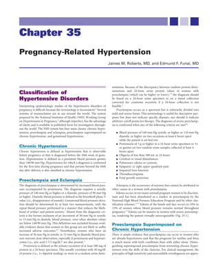
A B
FIGURE 35-1 Facial edema in severe preeclampsia. Markedly edematous facies of this severely
preeclamptic woman (A) is especially evident when compared with her appearance 6 weeks after delivery
(B).
priate, especially with advancing gestational age. The suspicion of be assigned. If blood pressure elevation persists, the diagnosis is chronic
superimposed preeclampsia mandates close observation, with delivery hypertension. The diagnosis of gestational hypertension is used during
indicated by the overall assessment of maternal and fetal well-being pregnancy only until a more specific diagnosis can be assigned after
rather than by any fixed end point. The diagnosis of superimposed delivery.3
preeclampsia is highly likely with the following findings:
1. In women with documented hypertension and no proteinuria Problems with Classification
before 20 weeks’ gestation The degree of blood pressure elevation that constitutes gestational
New-onset proteinuria, defined as the urinary excretion of 0.3 g hypertension is controversial. Because average blood pressure in
of protein or more in a 24-hour specimen women younger than 30 years is 120/60 mm Hg, the standard defini-
2. In women with hypertension and proteinuria before 20 weeks’ tion of hypertension (i.e., blood pressure >140/90 mm Hg) is judged
gestation by some to be too high,14 resulting in the suggestion that women with
A sudden increase in proteinuria blood pressure increases greater than 30 mm Hg systolic or 15 mm Hg
A sudden increase in blood pressure in a woman whose blood diastolic should be observed closely even if absolute blood pressure has
pressure has previously been well controlled not exceeded 140/90 mm Hg.3
Objective evidence of involvement of multiple organ systems, Blood pressures measured in early pregnancy to diagnose chronic
such as thrombocytopenia (platelet count < 100,000/mm3), an hypertension are problematic. Blood pressure usually decreases early
increase in liver transaminases to abnormal levels,3 or sudden in pregnancy, reaching its nadir at about the time women often present
worsening of renal function for obstetric care (Fig. 35-2). The decrease averages 7 mm Hg for dia-
stolic and systolic readings. In some women, blood pressure may
decline by more than 7 mm Hg; in others, the early decline and sub-
Gestational Hypertension sequent return of blood pressure to pre-pregnant levels in late gesta-
A woman who has no proteinuria and a blood pressure elevation tion may satisfy criteria for a diagnosis of preeclampsia. Women with
detected for the first time during pregnancy is classified as having ges- hypertension before pregnancy have a greater decrease in blood pres-
tational hypertension. This is a provisional diagnosis that includes sure in early pregnancy than do normotensive women,15 and they are
women with preeclampsia who have not yet manifested proteinuria more likely to be misdiagnosed as preeclamptic according to blood
and women who do not have preeclampsia. The hypertension may be pressure criteria.
accompanied by other concerning signs or symptoms that can influ- The diagnosis of chronic hypertension based on the failure of blood
ence management. A final determination that the woman does not pressure to return to normal by 84 days after delivery can be in error.
have preeclampsia can be made only after delivery. If preeclampsia has In a long-range, prospective study by Chesley,16 many women who
not developed and blood pressure has returned to normal by 12 weeks remained hypertensive 6 weeks after delivery were normotensive at
after delivery, the diagnosis of transient hypertension of pregnancy can long-term follow-up. Neither proteinuria nor hypertension is specific
3. CHAPTER 35 Pregnancy-Related Hypertension 653
gresses at various rates. In most cases, progression is slow, and the
mm Hg
disorder may remain mild. In others, the disease can progress rapidly,
125 changing from mild to severe over days to weeks or, in fulminant cases,
Systolic progressing in days or hours.
120
PARA 0 In a series of eclamptic women analyzed by Chesley,18 25% had
evidence of only mild preeclampsia in the days preceding convulsions.
115 PARA 1+ For purposes of clinical management, overdiagnosis must be accepted
because prevention of the serious complications of preeclampsia and
110 eclampsia requires increased sensitivity and early treatment, primarily
through the timing of delivery. For this reason, studies of preeclampsia
75 are necessarily confounded by inclusion of women diagnosed as pre-
Diastolic eclamptic who have another cardiovascular or renal disorder.
70 PARA 0
65 PARA 1+
HELLP Syndrome
The pathophysiologic changes of preeclampsia can occur in the absence
60 of hypertension and proteinuria. This is not surprising, because the
16 20 24 28 32 36 40 traditional diagnostic criteria have more historical than pathophysio-
Gestational age (weeks) logic relevance.18 This situation presents a challenge to clinicians and
demands that they remain alert to the possibility of preeclampsia in
FIGURE 35-2 Blood pressure correlated with gestational age. The pregnant women with signs and symptoms that may be explained by
mean blood pressure was plotted against gestational age for 6000 reduced organ perfusion. One clear setting in which this occurs is the
white women between the ages of 25 and 34 years who delivered HELLP syndrome (hemolysis, elevated liver enzymes, and low plate-
singleton term infants. (From Christianson R, Page EW: Studies on
lets), a combination of findings that defines a reasonably consistent
blood pressure during pregnancy: Influence of parity and age. Am J
syndrome.19
Obstet Gynecol 125:509, 1976. Courtesy of the American College of
Obstetricians and Gynecologists.) For management purposes, it is appropriate to consider HELLP as
a variant of preeclampsia, but they may be different entities. Women
with HELLP are more often older, white, and multiparous than
TABLE 35-1 RENAL BIOPSY FINDINGS IN preeclamptic women. Not all women with HELLP have hypertension.20
PATIENTS WITH A CLINICAL From a pathophysiologic perspective, changes in the renin-angiotensin
DIAGNOSIS OF PREECLAMPSIA system characteristic of preeclampsia are not present in HELLP.21
Nonetheless, progression of the disease and its termination with deliv-
Primigravidas Multigravidas ery argue for an observation and management strategy similar to that
Biopsy Findings (n = 62) (n = 152) for preeclampsia.
Glomeruloendotheliosis with 70% 14%
or without nephrosclerosis
Normal histology
Chronic renal disease, chronic
5%
25%
53%
21%
Preeclampsia and Eclampsia
glomerulonephritis, or
chronic pyelonephritis
Epidemiology of Preeclampsia
Arteriolar nephrosclerosis 0% 12% and Eclampsia
Despite the difficulties in clinical diagnosis, there exists a disorder
Modified from McCartney CP: Pathological anatomy of acute
hypertension of pregnancy. Circulation 30(Suppl II):37, 1964; by unique to pregnancy characterized by poor perfusion of many vital
permission of the American Heart Association, Inc. organs (including the fetoplacental unit) that is completely rever-
sible with the termination of pregnancy. Pathologic, pathophysiologic,
and prognostic findings indicate that preeclampsia is not merely
to preeclampsia, and their presence in pregnancy can have other an unmasking of preexisting, underlying hypertension. Although the
explanations. unique nature of preeclampsia has been well documented for many
Renal biopsy specimens from women with preeclampsia demon- years, controversies in therapy persist because of management strate-
strate these diagnostic difficulties (Table 35-1).17 Of 62 women with a gies based on principles used to treat hypertension in nonpregnant
diagnosis of preeclampsia in their first pregnancies, 70% had a glo- individuals. The successful management of preeclampsia requires an
merular lesion believed to be characteristic of the disorder, but 24% understanding of the pathophysiologic changes in this condition and
had evidence of chronic renal disease that was not previously sus- recognition that the signs of preeclampsia (i.e., increased blood pres-
pected. Renal biopsy specimens of multiparous women with a clinical sure and proteinuria) are only signs and do not cause the other features
diagnosis of superimposed preeclampsia also demonstrate the uncer- of preeclampsia.
tainty of diagnosis. Of 152 subjects, only 3% had the characteristic
glomerular lesion, but 43% had evidence of preexisting renal or vas- Women at Risk
cular disease. Preeclampsia occurs in about 4% of pregnancies that continue past
Preeclampsia has a clinical spectrum ranging from mild to severe the first trimester. Nulliparity is the most common feature of women
forms. The illness in affected women does not begin with eclampsia or who develop preeclampsia. At least two thirds of cases occur in the
the severe manifestations of preeclampsia. Rather, the disease pro- first pregnancy that progresses beyond the first trimester. Other risk
4. 654 CHAPTER 35 Pregnancy-Related Hypertension
factors for preeclampsia are similar in nulliparous and parous Obesity is a risk factor for preeclampsia.28,51 In the National Insti-
women.22 tute of Child Health and Human Development (NICHD) study of
Although preeclampsia was thought to be more common among aspirin to prevent preeclampsia in low-risk pregnancies,31 the inci-
women of lower socioeconomic status, this impression may be a con- dence of preeclampsia increased with maternal body mass index. Even
sequence of the associations of preeclampsia with age, race, and parity. in women of normal weight, there is a linear relationship between
Studies of pregnant women in Scotland23 from Aberdeen,24 Finland,25 pre-pregnancy body mass index and the frequency of preeclampsia.52
and Israel26 found that preeclampsia was not related to socioeconomic The mechanism may be related to increased insulin resistance, because
status. Eclampsia, in contrast, is clearly more common in women of preeclampsia is more common in another setting of increased insulin
lower socioeconomic status,23,25,26 related to the lack of availability of resistance: gestational diabetes.53 With a threefold increased risk for
quality obstetric care for indigent women. Remarkably, preeclampsia obese women and with 35% to 50% of women of reproductive age in
and eclampsia were once thought to occur more frequently in women the United States being obese, obesity has become a major attributable
of higher socioeconomic status.18 risk factor for preeclampsia, which is associated with more than one
There is a relationship between the extremes of childbearing age third of cases of preeclampsia.
and the incidence of eclampsia and preeclampsia. Because most first Certain conditions of pregnancy increase the risk of preeclampsia.
pregnancies occur in young women, most cases of preeclampsia and The incidence is increased among parous and nulliparous women with
eclampsia occur in this age group, but the association with young multiple gestations, although to a larger degree in the latter.36,54 In a
maternal age is lost when parity is considered. In the studies cited,23,25,26 study of 34,374 pregnancies with singleton, twin, triplet, or quadruplet
a higher incidence of preeclampsia was found in older women inde- pregnancies, the incidence of preeclampsia increased with each addi-
pendent of parity. tional fetus. The incidences were 6.7%, 12.7%, 20.0%, and 19.6%,
The relationship of preeclampsia and eclampsia to race is compli- respectively.55 The disease process may be initiated earlier and may be
cated by the higher prevalence of chronic hypertension in African more severe in these cases.54
Americans and the difficulty in differentiating preeclampsia from Preeclampsia affects 70% of women with large, rapidly growing
unrecognized preexisting chronic hypertension. Some studies indicate hydatidiform moles and occurs earlier than usual during gestation.56
a relationship.26,27 In a small case-control study of carefully defined In cases of preeclampsia occurring before 24 weeks’ gestation, hyda-
preeclampsia, black race was a significant risk factor only in nullipa- tidiform mole should be suspected and sought.
rous women (odds ratio [OR] = 12.3; 95% confidence interval [CI], An interesting variant of preeclampsia is the mirror syndrome, in
1.6 to 100.8).28 Other studies support a more modest increased risk in which the mother’s peripheral edema mirrors the fetal hydrops. It
African-American women.29,30 Studies that include the more severe occurs with fetal hydrops, although not with erythroblastosis uncom-
forms of preeclampsia more often suggest an increased incidence plicated by hydrops. The incidence approaches 50% of pregnancies
among African-American women.28 complicated by hydrops. The mirror syndrome is not confined to
In contrast, the incidence of rigorously defined preeclampsia did hydrops resulting from isoimmunization. In one series, mirror syn-
not differ by race after other risk factors were controlled in two large, drome occurred in 9 of 11 pregnancies with hydropic infants of non-
prospective trials of medical prophylaxis that enrolled 294731 and immune origin.57 This condition can manifest early in pregnancy with
431432 nulliparous women. Maternal nonwhite race appears to be severe signs and symptoms of preeclampsia, and it has resolved with
related more to the severity than the incidence of disease. treatment of the underlying process.58-60 Proteinuria is massive, and
A diverse array of medical disorders that often coexist with preg- blood pressure elevation and edema are marked. Eclampsia occurs
nancy, including diabetes, chronic hypertension, chronic renal disor- rarely (see Chapter 26).
ders, and rheumatologic conditions, have been associated with
preeclampsia. The existence and severity of diabetes have been
associated with an increased risk for preeclampsia, and diabetic Short-Term Prognosis for Preeclampsia
microvascular disease further increases this risk. This relationship PERINATAL MORTALITY
has been found in Sweden33 and in the United States.34 Both The perinatal mortality rate is increased in infants of preeclamptic
studies33,34 demonstrated that the risk of preeclampsia was approxi- women.61-63 In a study that examined 10,614,679 singleton pregnancies
mately 20% and 21% in 491 and 462 pregnancies, respectively. This in the United States from 1995 to 1997 after 24 weeks’ gestation, the
estimate is far more modest than the 50% incidence reported in his- relative risk for fetal death was 1.4 for infants born to women with any
torical cohorts.18 The preeclampsia risk increased according to the of the gestational hypertensive disorders and 2.7 for those born to
severity of disease, with an 11% to 12% risk among women with class women with chronic hypertensive disorders compared with low-risk
B diabetes and 21% to 23% with class C and D diabetes. Microvascular controls. Causes of perinatal death are placental insufficiency and
disease increased this risk to 36% to 54% in diabetics with class F or abruptio placentae,64 which cause intrauterine death before or during
R disease.33,34 labor, and prematurity. Predictably, the mortality rate is higher for
Chronic renal insufficiency and hypertension are well-recognized infants of women with more severe forms of the disorder. At any level
risk factors. Of women with hypertension antedating pregnancy, 25% of disease severity, the perinatal mortality rate is greatest for women
develop preeclampsia.35,36 Renal insufficiency with33,37 and without dia- with preeclampsia superimposed on preexisting vascular disease.
betes38-40 also is an important risk factor.38,40 The stillbirth rate attributable to preeclampsia has declined dra-
Connective tissue disorders such as systemic lupus erythe- matically in the past 35 years. However, infants born of preeclamptic
matosus41,42 and antiphospholipid antibody syndrome43-45 have been pregnancies continue to have an approximately twofold increased risk
reported as risk factors for preeclampsia. With lupus, the risk is par- for neonatal death.65 Although neonatal survival rates have improved
ticularly elevated with hypertension or nephropathy.46,47 However, dramatically, delivery before 34 weeks’ gestation continues to be associ-
data concerning an association between isolated antiphospholipid ated with an increased risk of long-range neurologic disability (see
antibodies and preeclampsia have been conflicting, with some Chapter 58).
studies demonstrating no relationship48,49 and others confirming the Growth restriction is more common in infants born to preeclamp-
association.44,50 tic women (see Chapter 34) and more pronounced with increasing
5. CHAPTER 35 Pregnancy-Related Hypertension 655
severity and earlier diagnosis.66 As with perinatal mortality, intrauter- To determine the subsequent pregnancy outcomes of women who
ine growth restriction (IUGR) is more common in infants of chroni- clearly had preeclampsia, Chesley and colleagues74 followed 270 women
cally hypertensive women with superimposed preeclampsia.67 with eclampsia for more than 40 years; only two were lost to follow-up.
The dramatic decrease in perinatal mortality rate among infants of Among 187 women who had eclampsia in the first pregnancy, 33% had
preeclamptic women is the result in part of improved medical and a hypertensive disorder in any subsequent pregnancy. In most, the
obstetric management, including improved assessment of fetal well- condition was not severe, but 5% had recurrent eclampsia. Twenty
being in the antepartum and intrapartum periods. The primary effect women with eclampsia as multiparas had recurrent hypertension in
on the perinatal mortality rate, however, has come from improvements 50% of subsequent pregnancies.
in neonatal care. Women with a clinical diagnosis of preeclampsia have increased
risk for hypertensive disorders in subsequent pregnancies. The chances
MATERNAL MORTALITY of recurrence decrease as the likelihood of true preeclampsia increases.
Maternal death associated with preeclampsia predominantly re- If the condition does recur, it will usually not be worse, and if pre-
sults from complications of abruptio placentae, hepatic rupture, and eclampsia truly arose de novo, it probably will be less severe in subse-
eclampsia. Historically, the mortality rate of eclamptic women was quent pregnancies. Some women, however, are normotensive between
most effectively reduced by avoiding iatrogenic complications related pregnancies but have recurrent preeclampsia. The risk of such recur-
to overmedication and overzealous attempts at vaginal delivery. In rence is increased when preeclampsia occurs in the late second or
series from the late 19th century, when immediate delivery was the early third trimester.73 The recurrence of severe preeclampsia or
practice, the mortality rate of eclamptic women was 20% to 30%. eclampsia in one pregnancy predicts its likely recurrence in subsequent
Expectant management with profound maternal sedation with narcot- pregnancies.
ics and hypnotics in the early 20th century was associated with a 10%
to 15% mortality rate. The change to magnesium as the exclusive agent Preeclampsia and Cardiovascular Disease
in the 1920s and 1930s resulted in a maternal mortality rate of 5%. in Later Life
Although magnesium was undoubtedly helpful, the primary factor Evidence that preeclampsia is associated with long-term maternal
responsible for improved mortality was decreased maternal sedation.18 health consequences is based on the work of Chesley and coworkers,74
The currently used combination of magnesium sulfate (MgSO4) and who followed a cohort of white women with eclampsia in their first
antihypertensive drugs as sole pharmacologic agents, followed by pregnancy and reported no increased risk of subsequent chronic
timely delivery, has produced a maternal mortality rate of almost hypertension. However, mortality was twofold to fivefold higher over
zero68,69 because of an appreciation of the profound pathophysiologic the next 35 years among women with eclampsia in any pregnancy after
abnormalities of preeclampsia, careful cardiopulmonary monitoring, the first (Fig. 35-3). The findings of Chesley and colleagues74 led to
and limitation of unproven interventions. speculation that multiparous women with preeclampsia or eclampsia
were more likely to have had unrecognized underlying chronic hyper-
RECURRENCE IN SUBSEQUENT PREGNANCIES tension and that this, not preeclampsia, caused the subsequent increase
Data from classic series indicate that the likelihood of recurrent in mortality. Sibai and associates71 also found that women with recur-
preeclampsia is influenced by the certainty of the clinical diagnosis in rent preeclampsia were more likely to develop chronic hypertension.
the first pregnancy. Of 225 women with hypertension during preg- These studies are the basis for a statement by The National High Blood
nancy chosen for study without regard to parity, 70% had a recurrence Pressure Education Program’s Working Group on High Blood Pressure
of preeclampsia in their next pregnancy.70 In a study of primiparas with during Pregnancy that recurrent hypertension in pregnancy, pre-
severe preeclampsia, the recurrence rate was 45% .71 Because the diag- eclampsia in a multipara, and early-onset disease in any pregnancy may
nosis in these studies was based solely on clinical findings, these groups all herald increased future health risks.2
probably included patients with unrecognized preexisting blood pres- Women with idiopathic preeclampsia (i.e., preeclampsia occurring
sure elevation or underlying renal or cardiovascular disease. in nulliparous women without underlying renal or cardiovascular
Recurrence rates were reported in 2006 for 896 parous women in disease, including chronic hypertension) were not thought to have
Iceland according to standardized diagnostic criteria in both pregnan- increased risk of later vascular disease until a report from Norway75
cies (i.e., National High Blood Pressure Criteria3). The rates of recur- found modest (1.65-fold) increased cardiovascular mortality for nul-
rence differed substantially by the diagnosis in the first pregnancy, as liparous women with preeclampsia at term and an eightfold increased
seen in Table 35-2.72 risk when preeclampsia was severe enough to lead to preterm delivery.
TABLE 35-2 TYPE OF RECURRENT HYPERTENSION DURING THE SECOND PREGNANCY BY TYPE OF
HYPERTENSION IN THE FIRST PREGNANCY
Second Pregnancy*
Gestational Chronic Superimposed All
First Pregnancy Normal Hypertension Preeclampsia Hypertension Preeclampsia Recurrences
Gestational hypertension (n = 511) 153 (29.9%) 239 (46.8%) 25 (4.9%) 82 (16%) 12 (2.3%) 358 (70.1%)
Preeclampsia/eclampsia (n = 151) 63 (41.7%) 52 (34.4%) 17 (11.3%) 16 (10.6%) 3 (2%) 88 (58.3%)
Chronic hypertension (n = 200) 24 (12%) 69 (34.5%) 6 (3%) 91 (45.5%) 10 (5%) 176 (88%)
Superimposed preeclampsia (n = 34) 2 (5.9%) 10 (29.4%) 4 (11.8%) 14 (41.2%) 4 (11.8%) 32 (94%)
Total (N = 896) 242 (27%) 370 (41.3%) 52 (5.8%) 203 (22.7%) 29 (3.2%) 654 (73%)
*No women had eclampsia in the second pregnancy.
From Hjartardottir S, Leifsson BG, Geirsson RT, Steinthorsdottir V: Recurrence of hypertensive disorder in second pregnancy. Am J Obstet Gynecol
194:916-920, 2006.
6. 656 CHAPTER 35 Pregnancy-Related Hypertension
100 TABLE 35-3 SIGNS AND SYMPTOMS OF
PREECLAMPSIA OR ECLAMPSIA
90
Cerebral Blurred vision
Headache Amaurosis
80
Percentages surviving
Dizziness Gastrointestinal
Tinnitus Nausea
70 Drowsiness Vomiting
Change in respiratory rate Epigastric pain
60 Tachycardia Hematemesis
Fever Renal
50 Visual Oliguria
Diplopia Anuria
40 Scotomata Hematuria
Hemoglobinuria
30
dyslipidemia,92 altered angiogenic factors,93 and increased antibodies
10 20 30 40 45
to the angiotensin-2 receptor.94 These data may explain the common
Years
risk factors for preeclampsia and cardiovascular disease, but alternative
FIGURE 35-3 Eclampsia survivorship. Survival times are plotted
explanations, such as that preeclampsia causes vascular injury that
for women with eclampsia in the first pregnancy (solid line) and those increases cardiovascular risk or that normal pregnancies have a protec-
with eclampsia in a later pregnancy (dashed line). Survival of women tive effect, cannot be excluded.
with first-pregnancy eclampsia was not different from survival of a
control group. (From Chesley LC, Annitto JE, Cosgrove RA: The
remote prognosis of eclamptic women: Sixth periodic report. Am J Clinical Presentation
Obstet Gynecol 124:446, 1976, Courtesy of the American College of Preeclampsia can manifest with a wide spectrum of disease, ranging
Obstetricians and Gynecologists.) from life-threatening neurologic, renal, hepatic, and coagulation
abnormalities to mild findings of preeclampsia with minimal end-
organ involvement. The fetus may be severely compromised by the
Scottish investigators reported a fourfold increased risk of subsequent maternal condition and by extreme preterm delivery or only minimally
hypertension in nulliparous women with preeclampsia2,76,77 (OR = affected. These variations have puzzled clinicians and researchers for
3.98; CI, 2.82 to 5.61). Funai and colleagues78 described excess long- many years. An understanding of the pathophysiology of the disorder
term mortality in women with prior preeclampsia that was largely provides insight into the diverse clinical presentations.
attributed to a threefold increase in deaths due to cardiovascular
disease. Other reports support a link between preeclampsia and mater- Symptoms
nal ischemic heart disease,79,80 which is sometimes evident 20 years Most women with early preeclampsia are asymptomatic. The absence
after the preeclamptic pregnancy and coincident with the onset of of symptoms is the rationale for frequent obstetric visits in late preg-
menopause.78,80 A family history of cardiovascular disease increases the nancy. In most cases, signs such as increased blood pressure and pro-
association between preeclampsia and cardiovascular outcomes.81 teinuria antedate overt symptoms.
Obesity is a known risk factor for preeclampsia and cardiovascular The various symptoms associated with preeclampsia, especially
disease. Although controlling for obesity attenuates the increased risk preeclampsia of increasing severity, are listed in Table 35-3. Because
of death for postmenopausal women, this risk is not fully explained by preeclampsia is a disease of generalized poor perfusion, the diversity
obesity alone.82 of symptoms related to many organ systems is not surprising. Symp-
The relationships among obesity, insulin resistance, and preeclamp- toms suggesting hepatic, neurologic, and visual involvement are par-
sia are part of an interesting relationship of preeclampsia to the meta- ticularly worrisome. They include epigastric pain, “stomach upset,” and
bolic or insulin resistance syndrome.83 This syndrome predisposes to pain penetrating to the back. Headache and mental confusion indicate
cardiovascular disease in later life and consists of obesity, hypertension, poor cerebral perfusion and may be precursors of convulsions. Visual
dyslipidemia (i.e., increased low-density lipoprotein [LDL] cholesterol, symptoms ranging from scotomata to blindness indicate retinal arte-
decreased high-density lipoprotein [HDL] cholesterol, and increased rial spasm and edema. Symptoms suggesting congestive heart failure
triglycerides), and increased uric acid, all of which are found in women or abruptio placentae also represent significant complications of pre-
with preeclampsia.83 Other conditions predisposing to later-life cardio- eclampsia. Other symptoms, such as tightness of hands and feet and
vascular disease—including elevated levels of homocysteine,84 evidence paresthesias resulting from medial or ulnar nerve compression, may
of androgen excess (including polycystic ovarian syndrome),85 elevated alarm the patient but have little prognostic significance.
testosterone levels,86 male fat distribution (i.e., increased waist-to-hip
ratio),87 and lipoprotein lipase mutations88—are also linked to an Signs
increased risk for preeclampsia. Signs of preeclampsia usually antedate symptoms. The most common
Women who appear normal years after a preeclamptic pregnancy sequence is increased blood pressure followed by proteinuria.18
may nevertheless demonstrate subtle metabolic and cardiovascular
abnormalities. Compared with women with uncomplicated pregnan- BLOOD PRESSURE CHANGE
cies, formerly preeclamptic women have evidence of endothelial dys- An increase in blood pressure is required for the diagnosis of
function,89,90 higher blood pressures,89 increased insulin resistance,91 preeclampsia. Blood pressure variation in normal pregnancy can
7. CHAPTER 35 Pregnancy-Related Hypertension 657
lead to misdiagnosis. In clinical practice, the serious effects of pre- HYPERREFLEXIA
eclampsia on the mother and fetus warrant such overdiagnosis. The Although hyperreflexia is given much clinical attention and deep
primary pathophysiologic alteration, poor tissue perfusion resulting tendon reflexes are increased in many women before seizures, convul-
from vasospasm, is revealed more by blood pressure changes than by sions can occur in the absence of hyperreflexia,68 and many pregnant
absolute blood pressure levels. Although a diagnosis of preeclampsia women are consistently hyperreflexic without being preeclamptic.
is not made without absolute blood pressure increases to 140 mm Hg Changes, or lack thereof, in deep tendon reflexes are not part of the
systolic or 90 mm Hg diastolic, women who reach this level from a low diagnosis of preeclampsia.
early pregnancy value typically manifest more vasospasm than those
for whom 140/90 mm Hg represents a smaller increase. OTHER SIGNS
Although maternal and fetal risks rise with increasing blood pres- Other signs that occur less commonly in preeclampsia are indica-
sure,95 serious complications can occur in women who experience only tors of involvement of specific organs in the preeclamptic process.
modest blood pressure elevation. In two series, 20% of women with Women with marked edema may have ascites and hydrothorax, and
eclampsia never had a systolic blood pressure above 140 mm Hg.18,96 those with congestive heart failure display increased neck vein disten-
In a large, prospective study from the United Kingdom, there were 383 tion, gallop rhythm, and pulmonary rales. Hepatic capsular distention,
confirmed cases of eclampsia, of which 77% were hospitalized before manifested by hepatic enlargement and tenderness, is a particular
seizures occurred. Of these, 38% of the cases were not preceded by concern, as is disseminated intravascular coagulation (DIC) sufficient
documented proteinuria or hypertension.97 Others have noticed similar to cause petechiae or generalized bruising and bleeding.
findings.98,99
Laboratory Findings
PROTEINURIA Major changes revealed by laboratory studies occur in severe pre-
Among the diagnostic signs of preeclampsia, proteinuria in the eclampsia and eclampsia. In the patient with mild preeclampsia,
presence of hypertension is the most reliable indicator of fetal jeopardy. changes in most of these indicators may be minimal or absent.
In two studies of preeclampsia, the perinatal mortality rate tripled for
women with proteinuria,100 and the amount of proteinuria correlated RENAL FUNCTION STUDIES
with increased perinatal mortality rate and the number of growth- Serum Uric Acid Concentration and Urate Clearance. Uric
restricted infants.101 A later study demonstrated that the risk for acid is the most sensitive laboratory indicator of preeclampsia available
delivering a small-for-gestational-age fetus was higher in women with to clinicians. A decrease in uric acid clearance precedes a measurable
hypertension and proteinuria (52%) compared with women with new- decrease in the glomerular filtration rate (GFR). Hypertension with
onset gestational hypertension (15%) or chronic hypertension (12%). hyperuricemia but without proteinuria was associated with growth
The perinatal mortality rate was fourfold higher with proteinuria restriction as commonly as hypertension and proteinuria without ele-
and hypertension than in pregnancies complicated by hypertension vated uric acid in one series.105 Although increased serum uric acid
alone.102 concentration is often attributed to altered renal function, an alterna-
tive view favors increased production caused by oxidative stress.106 An
RETINAL CHANGES elevated uric acid level may itself have pathogenic effects.107 Table 35-4
Retinal vascular changes on funduscopic examination occur in shows normal uric acid levels during gestation and levels associated
retinal arterioles in at least 50% of women with preeclampsia, and they with preeclampsia.
are important because they correlate best with renal biopsy-proven Serum Creatinine Concentration and Creatinine Clearance.
changes of preeclampsia.103 Localized retinal vascular narrowing is Creatinine clearance is decreased in most patients with severe pre-
visualized as segmental spasm, and the generalized narrowing is indi- eclampsia, but it can be normal in women with mild disease. Serial
cated by a decrease in the ratio of arteriolar-venous diameter from serum creatinine determinations may indicate decreased clearance, but
the usual 3 : 5 to 1 : 2 or even 1 : 3. It can occur in all vessels or, in early single values are not helpful unless markedly elevated because of the
stages, in single vessels.104 Preeclampsia does not cause chronic arterio- wide range of normal values. The serum creatinine concentration
lar changes; the presence of arteriolar sclerosis detected by increased varies as a geometric function of creatinine clearance so that small
light reflex, copper wiring, or arteriovenous nicking indicates preexist- changes in glomerular filtration are best determined by measurements
ing vascular disease. of creatinine clearance.
TABLE 35-4 PLASMA URATE CONCENTRATIONS IN NORMOTENSIVE AND HYPERTENSIVE
PREGNANT WOMEN
Normotensive Patients Hypertensive Patients
Weeks of Gestation mmol/L SD* mg/dL mmol/L SD* mg/dL
24-28 0.18 (20%) 3.02 0.24 (20%) 4.03
29-32 0.18 (35%) 3.02 0.28 (25%) 4.7
33-36 0.20 (30%) 3.36 0.30 (20%) 5.04
37-40 0.26 (20%) 4.4 0.31 (23%) 5.28
41-42 0.25 (24%) 4.2 0.32 (12%) 5.38
*Each number in parentheses is the standard deviation given as a percentage of the mean values shown. Values for hypertensive and normotensive
women are statistically different at all gestational ages (P < .05).
Modified from Shuster E, Weppelman B: Plasma urate measurements and fetal outcome in preeclampsia. Gynecol Obstet Invest 12:162, 1981.
8. 658 CHAPTER 35 Pregnancy-Related Hypertension
LIVER FUNCTION TESTS
Although most tests of liver function are not highly predictive of
severity of preeclampsia,18 the association between microangiopathic
anemia and elevations in aspartate aminotransferase (AST) and alanine
aminotransferase (ALT) carries an especially disturbing prognosis for
the mother and infant.19,108 These findings usually correlate with the
severity of disease and, when associated with hepatic enlargement, may
be a sign of impending hepatic rupture.
COAGULATION FACTORS
Although overt DIC is rare, subtle evidence of activation of the
coagulation cascade occurs in many women with preeclampsia. The
average platelet count in the patient with mild preeclampsia is similar
to the platelet count in normal pregnant women.109 However, careful
platelet counts performed sequentially may reveal decreased platelets
in many patients.110 Highly sensitive indicators of activation of the
clotting system, reduced serum concentrations of antithrombin III,111
a decrease in the ratio of factor VIII bioactivity to factor VIII antigen,112
and subtle indicators of platelet dysfunction, including alteration of FIGURE 35-4 Hemorrhagic hepatic lesions in eclampsia.
turnover,6 activation,113 size,114 and content,115 exist in even mild pre- Hemorrhage into the periportal area occurred with crescentic
eclampsia and may antedate clinically evident disease. compression of liver cells. (From Sheehan HL, Lynch JB: Pathology of
Toxemia in Pregnancy. London, Churchill Livingstone, 1973.)
METABOLIC CHANGES
Preeclampsia is characterized by an increase in the insulin resis-
tance of normal pregnancy. Signs of the insulin resistance syndrome
are exaggerated.110 Levels of circulating lipids already elevated in
normal pregnancy116 are accentuated in women with preeclampsia.117
Triglycerides and fatty acid levels are elevated, changes that antedate
clinically evident disease by weeks to months.118,119 Levels of the car-
dioprotective HDL cholesterol are reduced in preeclamptic women,120
whereas levels of a variant of LDL cholesterol (i.e., small, dense cho-
lesterol that is strongly associated with cardiovascular disease) are
increased.121,122 These changes resolve after delivery.
Pathologic Changes in Preeclampsia
The pathologic changes found in organs of women dying of eclampsia
and in biopsy specimens from women with preeclampsia provide
strong evidence that preeclampsia is not merely an unmasking of
essential hypertension or a variant of malignant hypertension. These
findings also indicate that the elevation of blood pressure probably
does not have primary pathogenetic importance.
Brain FIGURE 35-5 Hepatic infarction in eclampsia. Hepatic infarction
caused by intense vasospasm manifests as small to large areas
Cerebral edema, once thought to be a common finding in women
beginning near the sinusoids and extending into the area near the
dying of eclampsia, was uncommon among postmortem examinations portal vessels. (From Sheehan HL, Lynch JB: Pathology of Toxemia in
performed within 2 to 3 hours of death.123 However, studies using Pregnancy. London, Churchill Livingstone, 1973.)
computed tomography again raised the possibility that cerebral edema
is an important pathophysiologic event in some women with pre-
eclampsia.124 Noninvasive studies of cerebral blood flow and resistance results from vasodilatation of arterioles, producing dislocation and
suggest that vascular barotrauma and loss of cerebral vascular auto- deformation of the hepatocytes in their stromal sleeves (Fig. 35-4).
regulation contribute to the pathogenesis of cerebral vascular pathol- Later, intense vasospasm causes hepatic infarction, ranging from small
ogy in cases of preeclampsia or eclampsia.125 to large areas beginning near the sinusoids and extending into the area
near the portal vessels (Fig. 35-5). Hemorrhagic changes are present in
Liver 66% and necrotic changes in 40% of eclamptic women and in about
Gross lesions of the liver are visible in about 60% of women dying of one half as many preeclamptic women. Hyalinization and thrombosis
eclampsia, and one third of the remaining livers are microscopically of hepatic vessels have been cited as evidence of DIC, but they may be
abnormal. Many early investigators thought that the hepatic changes the result of hemorrhage.
were pathognomonic for eclampsia,126 but similar changes have been
described in women dying of abruptio placentae.127 Kidney
Two temporally and etiologically distinct hepatic lesions have been The pathologic renal changes of preeclampsia and eclampsia are clearly
described.123 Initially, hemorrhage into the hepatic cellular columns different from those seen in other hypertensive or renal disorders.
9. CHAPTER 35 Pregnancy-Related Hypertension 659
EN
BM
R
FIGURE 35-6 Glomerular changes in preeclampsia are identified A
by light microscopy. The enlarged glomerulus completely fills
Bowman’s capsule. Diffuse edema of the glomerular wall is indicated Ep Cy
by the vacuolated appearance. The visible capillary loops are
extremely narrow, and there are virtually no red blood cells in the
capillary tuft.
En
Glomerular, tubular, and arteriolar changes have been described. The
glomerular lesion is considered by some to be pathognomonic of pre-
eclampsia and eclampsia, but identical changes have been seen in pla- BS R
cental abruption without evident preeclampsia.128 This change is not
En
seen in any other form of hypertension.
Ep
GLOMERULAR CHANGES
Changes seen by light microscopy in glomeruli that are character-
istic of preeclampsia include103 decreased glomerular size, with protru-
sion of the glomerular tuft into the proximal tubule. The diameter of P
the glomerular capillary lumen is decreased and contains few blood
cells. The endothelial-mesangial cells have increased cytoplasmic
B
volume and can contain lipoid droplets (Fig. 35-6).
Electron microscopic examination of glomeruli provides more evi- FIGURE 35-7 Electron photomicrographs of renal glomeruli.
dence that the primary pathologic change occurs in endothelial cells, A, Normal anatomy. B, Biopsy specimen from a preeclamptic woman.
which are greatly increased in size and can occlude the capillary lumen; Endothelial cells (En) are markedly enlarged, obstruct the capillary
their cytoplasm contains electron-dense material.129 The basement lumen, and contain electron-dense inclusions. The basement
membrane bordering the epithelial cell may be slightly thickened, and membrane (BM) is slightly thickened with inclusions, but the
it also contains electron-dense material. The epithelial cell podocytes epithelial foot processes (EP) are normal. BM, basement membrane;
are not altered (Fig. 35-7). These changes are collectively called glo- BS, Bowman’s space; Cy, cytoplasmic inclusions; EN, capillary
endothelial cells that line the glomeruli; Ep, renal epithelial cells; L,
merular capillary endotheliosis.
capillary lumen containing red blood cells; P, podocytes; R, red blood
Characteristic glomerular changes occur in 70% of primiparas
cell. (From McCartney CP: Pathological anatomy of acute
but in only 14% of multiparas with a diagnosis of preeclampsia.17 The hypertension of pregnancy. Circulation 30[Suppl II]:37, 1964. By
more likely the diagnosis of preeclampsia, the more common the glo- permission of the American Heart Association, Inc.)
merular lesion. As the clinical condition worsens, the magnitude of the
glomerular lesion increases. The glomerular lesions are reversible after
delivery and are not present in subsequent biopsy specimens obtained
5 to 10 weeks later.103 NONGLOMERULAR CHANGES
The glomerular changes correlate more consistently with protein- Pathologic changes in renal tubules include dilatation of proximal
uria than with hypertension, suggesting that proteins identified immu- tubules with thinning of the epithelium,123 tubular necrosis,103 enlarge-
nohistochemically may be trapped in the glomerulus. These staining ment of the juxtaglomerular apparatus,131 and hyaline deposition in
patterns are not found in other renal disorders with proteinuria. The renal tubules.123 Fat deposition in women with prolonged heavy pro-
glomerular changes of preeclampsia can be mimicked in animal studies teinuria has been reported.123 Necrosis of the loop of Henle, a change
by reducing the renal concentration of vascular endothelial growth that correlates with the degree of hyperuricemia, has also been
factor (VEGF), which usually exists in high concentration in this tissue described.131
by increasing the synthesis of the VEGF antagonist soluble Fms-like Thickening of renal arterioles may be seen in preeclampsia, espe-
tyrosine kinase 1 (sFlt1).130 cially in women with preexisting hypertension. Unlike the glomerular
10. 660 CHAPTER 35 Pregnancy-Related Hypertension
Spinal
Endometrium
Basal
Radial
Arcuate
Myometrium
A
FIGURE 35-8 Schematic representation of uterine arteries. The
characteristic changes occur in the decidual vessels supplying the
placental site in a normal pregnancy. (From Okkels H, Engle ET:
Studies of the finer structure of the uterine vessels of the Macacus
monkey. Acta Pathol Microbiol Scand 15:150, 1938.)
lesion, it does not regress after delivery,103 suggesting that the arteriolar
change results from coincident disease, not preeclampsia.
B
Vascular Changes in the Placental Site FIGURE 35-9 Spiral arterial changes in normal pregnancy. A, In
The characteristic changes in the decidual vessels supplying the pla- the section of spiral arterioles at the junction of the endometrium and
cental site in normal pregnancy are depicted in Figure 35-8. In normal myometrium in a nonpregnant woman, notice the inner elastic lamina
pregnancy, the spiral arteries (Fig. 35-9) increase greatly in diameter.132 and smooth muscle. B, In a section of a spiral arteriole at the same
Morphologically, the endothelium is replaced by trophoblast, and the scale and from the same location during pregnancy, notice the
internal elastic lamina and smooth muscle of the media are replaced markedly increased diameter and absence of inner elastic lamina and
by trophoblast and an amorphous matrix-containing fibrin (see smooth muscle. (From Sheppard BL, Bonnar J: Uteroplacental
Fig. 35-9).133 These changes occur originally in the decidual portion of arteries and hypertensive pregnancy. In Bonnar J, MacGillivray I,
Symonds G [eds]: Pregnancy Hypertension. Baltimore, University Park
the spiral arteries but extend into the myometrium as pregnancy
Press, 1980, p 205.)
advances and can even involve the distal portion of the uterine radial
artery. The basal arteries are not affected. These morphologic changes
are considered to be a vascular reaction to the trophoblast, occurring
directly or humorally, that results in increased perfusion of the placen-
tal site.
In placental-site vessels of women with preeclampsia, the normal
physiologic changes do not occur, or they are limited to the decidual
portion of the vessels. Myometrial segments of spiral arteries retain the
nonpregnant component of intima and smooth muscle, and the diam-
eter of these arteries is about 40% that of vessels in normal preg-
nancy.134 Spiral arterioles in decidua and myometrium and basal and F
radial arterioles may become necrotic, with components of the normal
vessel wall replaced by amorphous material and foam cells, a change
called acute atherosis (Fig. 35-10).135 This lesion is best seen in the basal
arteries because they do not undergo the normal changes of pregnancy.
It is also present in decidual and myometrial spiral arteries and can
progress to vessel obliteration. The obliterated vessels correspond to
areas of placental infarction.
Failed vascular remodeling and atherotic changes may be seen
with fetal growth restriction in women without clinical evidence of
FIGURE 35-10 Atherosis. Numerous lipid-laden cells (L) and fibrin
preeclampsia. Atherotic changes occur in decidual vessels of some dia- deposition (F) are present in the media of this occluded decidual
betic women,136 and failed vascular remodeling is present in about vessel. (From Sheppard BL, Bonnar J: Uteroplacental arteries and
one third of women who experience preterm labor.137 It appears hypertensive pregnancy. In Bonnar J, MacGillivray I, Symonds G
that abnormal invasion may be necessary but is not sufficient to cause [eds]: Pregnancy Hypertension. Baltimore, University Park Press,
preeclampsia. 1980, p 205.)
11. CHAPTER 35 Pregnancy-Related Hypertension 661
Changes characteristic of preeclampsia have been observed in the hypertension. Second, the pathologic findings indicate that the primary
decidual vessels of one in seven primiparous women and in a lower pathology is poor tissue perfusion, not blood pressure elevation. The
percentage of multiparous women at the time of first-trimester abor- histologic data support the clinical impression that the poor perfusion
tion.138,139 These findings suggest that disordered placentation precedes results from profound vasospasm, which increases total peripheral
the clinical presentation of preeclampsia. The cause of the decidual resistance and blood pressure.
vascular lesions is unknown. The appearance of the atherotic vessels
resembles vessels in transplanted kidneys that have undergone rejec-
tion, suggesting an immunologic cause, which is consistent with find-
Pathophysiologic Changes
ings of a study that demonstrated components of complement (e.g., in Preeclampsia
C3) in decidual vessels with the lesion.140 Preeclampsia can cause changes in virtually all organ systems. Several
The vascular remodeling of spiral arteries supplying the intervillous organ systems are consistently and characteristically involved, and
space is intimately related to normal trophoblast invasion.141 The these are discussed in the following sections.
expression of adhesion molecules and their receptors that characterizes
implantation is abnormal in preeclampsia.142 The trophoblast that lines Cardiovascular Changes
the decidual vessels of normal pregnant women begins to express Blood pressure is the product of cardiac output and systemic vascular
molecules usually present only on endothelium,143 a phenomenon that resistance. Cardiac output is increased by up to 50% in normal preg-
does not occur in preeclampsia.144 Potential mechanisms responsible nancy, but blood pressure does not usually increase, indicating that
for the normal and abnormal changes include decidually produced systemic vascular resistance decreases. Blood pressure is lower in the
cytokines145-147 and local oxygen tension.148,149 There may be interac- first half of pregnancy than in the postpartum period, when cardiac
tions of specific molecules on trophoblast and maternal decidual cells output returns to nonpregnant levels (see Fig. 35-2). Some women
that drive invasion. Invasive cytotrophoblasts express a human leuko- destined to develop preeclampsia have a higher cardiac output before
cyte antigen (HLA) molecule (HLA-C) that is minimally hetero- clinically evident disease. However, cardiac output is reduced to pre-
geneous. Interaction of this molecule with a receptor on maternal pregnancy levels with the onset of clinical preeclampsia.157,158 Although
decidual cells, killer immunoglobulin receptors (KIRs), causes various some studies suggest increased cardiac output,159 most have found
degrees of activation of the trophoblast cell, depending on the combi- normal or slightly reduced cardiac output in women with untreated
nation of KIR and HLA-C subtypes. Mothers with the minimally acti- preeclampsia.160 Increased systemic vascular resistance is the mecha-
vating KIR subtype who have a fetus with a specific HLA-C subtype nism for the increase in blood pressure in clinical preeclampsia.
(HLA-C2) have an increased frequency of preeclampsia. This is not an There is substantial evidence that arteriolar narrowing occurs in
immune interaction because the relationship persists regardless of preeclampsia. Changes in the caliber of retinal arterioles correlate with
maternal HLA-C subtype. Researchers propose that this combination the clinical severity of the disorder and with renal biopsy-confirmed
does not favor trophoblast invasion and vascular remodeling. Popula- diagnosis of preeclampsia.103 Similar findings occur in vessels of the
tion studies indicate that populations in which HLA-C2 is common nail bed and conjunctiva. Measurements of forearm blood flow indi-
have a reduced frequency of the specific inhibitory KIR subtype and cate higher resistance in preeclamptic than in normal pregnant
vice versa.150 women.161,162 It is unlikely that this effect is determined by the auto-
nomic nervous system. Although normal pregnant women are exqui-
Placental Pathologic Changes sitely sensitive to the interruption of autonomic neurotransmission by
Ultrastructural examination of placentas from women with pre- ganglionic blockade and high spinal anesthesia, preeclamptic women
eclampsia reveals an abnormal syncytiotrophoblast containing areas of are less sensitive.163 This finding suggests that the arteriolar constric-
cell death and degeneration. Viable-appearing syncytiotrophoblast is tion of preeclampsia is not maintained by the autonomic nervous
also abnormal, with decreased density of microvilli, dilated endoplas- system and that humoral factors are implicated. The increased sympa-
mic reticulum, and decreased pinocytotic and secretory activity. The thetic activity in preeclampsia, however, raises questions about these
cells of the villous cytotrophoblast cells are increased in number and older findings.164 Assays of concentrations of recognized endogenous
have higher mitotic activity. The basement membrane of the tropho- vasoconstrictors are limited to determinations of catecholamines and
blast is irregularly thickened, with fine fibrillary inclusions.151 angiotensin II. Results suggest minimal or no change in catechol-
The changes may be caused by local hypoxia. Similar syncytiotro- amines, whereas circulating angiotensin II concentrations are lower in
phoblastic changes are present in placental segments maintained under preeclamptic women.165
hypoxic conditions in vitro.152 The cytotrophoblastic alterations are Levels of endothelin-1, a vasoconstrictor produced by endothelial
also consistent with hypoxia. The cytotrophoblasts comprise the stem cells, are increased in the blood of preeclamptic women166 at concen-
cells of the trophoblast and responds to damage by proliferation. The trations much lower than those necessary to stimulate vascular smooth
trophoblast of the preeclamptic placenta is characterized by increased muscle contraction in vitro. It is not clear whether these circulating
apoptosis and necrosis,153,154 possibly caused by hypoxia or hypoxia concentrations reflect endothelial production sufficient to stimulate
reperfusion injury,155 and this may be the origin of the increased cir- local vasoconstriction or low concentrations of endothelin potentiate
culating syncytiotrophoblast microparticles in preeclampsia.156 contractile responses to other agonists.
As indicated by the older term toxemia, early investigators sus-
Summary of Pathologic Changes pected that preeclampsia was caused by circulating humors. Early
in Preeclampsia reports suggesting etiologic agents such as pressor substances in blood,
Structural changes associated with preeclampsia and eclampsia lead to decidual extracts, placental extracts, and amniotic fluid of preeclamp-
two important conclusions. First, preeclampsia is not an alternate form tic patients have not been replicated. The explanation for the pressor
of malignant hypertension. The renal changes in preeclamptic and effects was, in some studies, normal endogenous pressors; in others,
eclamptic women and the structural changes in other organs of women the explanation was faulty methodology and failure to recognize the
dying of eclampsia differ from the alterations caused by malignant immunologic difference between the source of the extract and the
12. 662 CHAPTER 35 Pregnancy-Related Hypertension
50 50 Preeclampsia
Preeclampsia
Nonpregnant
Nonpregnant
40 Normal pregnancy
MBP, mm Hg
Normal pregnancy 40
MBP, mm Hg
30 30
20 20
10 10
5 10 15 25 50 100 200 5 10 15 25 50 100 200
Dose, ng/kg Dose, ng/kg
FIGURE 35-11 Mean dose-response graphs for norepinephrine. FIGURE 35-12 Mean dose-response graphs for angiotensin.
Women with preeclampsia have an increased sensitivity to all Preeclamptic women are much more sensitive to angiotensin II than
endogenous pressors. MBP, mean blood pressure. (From Talledo OE, normal pregnant and nonpregnant women. MBP, mean blood
Chesley LC, Zuspan FP: Renin-angiotensin system in normal and pressure. (From Talledo OE, Chesley LC, Zuspan FP: Renin-
toxemic pregnancies. III. Differential sensitivity to angiotensin II and angiotensin system in normal and toxemic pregnancies. III.
norepinephrine in toxemia of pregnancy. Am J Obstet Gynecol Differential sensitivity to angiotensin II and norepinephrine in toxemia
100:218, 1968. Courtesy of the American College of Obstetricians of pregnancy. Am J Obstet Gynecol 100:218, 1968. Courtesy of the
and Gynecologists.) American College of Obstetricians and Gynecologists.)
animals tested. In other experiments, no defect is obvious. The hypoth-
16
esis that arteriolar constriction of preeclampsia is caused by new cir-
culating pressors has largely been abandoned.18
A more compelling explanation for vasospasm in preeclampsia is
increased response to normal concentrations of endogenous pressors.
Women with preeclampsia have higher sensitivity to all the endoge- 12
nous pressors that have been tested. They are exquisitely sensitive to
ng/kg/min
vasopressin.167,168 Vasopressin can elicit marked blood pressure eleva-
tion, seizures, and oliguria in some patients.168 Sensitivity to epineph-
rine169 and norepinephrine170 is also increased (Fig. 35-11). The most 8 Nonpregnant mean
striking difference is seen in the sensitivity of the preeclamptic woman
to angiotensin II. Normal pregnant women are less sensitive to angio- P<.01
tensin II than nonpregnant women, requiring approximately 2.5 times P<.05 P<.1 P<.001
as much angiotensin to raise the blood pressure by a similar incre-
4
ment.171 In contrast, preeclamptic women are much more sensitive to 10 14 18 22 26 28 30 32 34 36 38 40
angiotensin II than are normal pregnant and nonpregnant women Weeks of gestation
(Fig. 35-12).170
In a classic study, angiotensin II sensitivity was significantly FIGURE 35-13 Angiotensin sensitivity throughout pregnancy. The
increased many weeks before the development of elevated blood pres- dose of angiotensin II necessary to increase diastolic blood pressure
sure (Fig. 35-13).172 Although resistance to angiotensin II does not 20 mm Hg in women who developed elevated blood pressure in late
decrease to nonpregnant levels until 32 weeks’ gestation, significant pregnancy (blue line, open circles) was compared with the dose for
those who remained normotensive (red line, solid circles). The graph
differences in sensitivity between women who later become hyperten-
demonstrates that a significantly lower dose was required in the
sive and those who remain normotensive have been observed as early former group as early as 10 to 14 weeks’ gestation. (From Gant NF,
as 14 weeks. However, a large British study did not confirm this classic Daley GL, Chand S, et al: A study of angiotensin II pressor response
finding,173 perhaps reflecting the heterogeneity of preeclampsia.174 throughout primigravid pregnancy. J Clin Invest 49:82, 1973. With
The decreased sensitivity of normal pregnant women to angioten- permission of the American Society for Clinical Investigation.)
sin II and the lower systemic vascular resistance in normal pregnancy
suggest that arteriolar narrowing in preeclamptic women may result
from decreased levels of circulating or local vasodilator substances, coagulation system manifests as the intravascular disappearance of
rather than from increased levels of circulating pressors. This attractive procoagulants, intravascular appearance of degradation products of
hypothesis, however, is not consistent with the unchanged sensitivi- fibrin, and end-organ damage from the formation of microthrombi.176
ties to norepinephrine, epinephrine, and vasopressin in normal In the most advanced form of DIC, procoagulants—especially fibrino-
pregnancy.168-170 gen and platelets—decrease to a degree sufficient to produce spontane-
ous hemorrhage. In milder forms, only highly sensitive indicators of
Coagulation Changes clotting system activation are present. Decreasing platelet concentra-
The syndrome of DIC occurs in preeclampsia and has been suggested tions is such a sign but may be evident only by serial observations.96
as a primary pathogenetic factor175 (see Chapter 40). Activation of the Sensitive indicators of intravascular coagulation, such as an elevated
13. CHAPTER 35 Pregnancy-Related Hypertension 663
level of fibrin degradation products; increased platelet turnover,6 thelium. Vessels from preeclamptic women and the umbilical vessels
volume,114 and activation177; reduced platelet content178; increased of their neonates generate less prostacyclin than similar vessels from
platelet content in plasma179; reduced levels of antithrombin III111; and normal pregnant women.202-204 If potent inhibitors preventing the syn-
a reduced ratio of factor VIII activity to factor VIII antigen,180 are thesis of all prostaglandins (including prostacyclin) are administered
common when concentrations of procoagulants remain normal. Subtle to pregnant women, the usual resistance to the vasoconstrictor effect
signs of platelet dysfunction,6,114,177-179 reduced antithrombin III,111 and of angiotensin II is abolished.205 Conversely, if aspirin is used as an
reduction in the ratio of factor VIII bioactivity to factor VIII antigen112 inhibitor of prostaglandin synthesis in a manner determined to specifi-
are present in women with mild preeclampsia and may precede its cally reduce contractile prostanoids (e.g., thromboxane A2) much more
clinical signs. than prostacyclin, the increased angiotensin II sensitivity of preeclamp-
Abnormalities of blood coagulation sufficient to make a diagnosis tic women is reduced.206
of DIC are present in approximately 10% of women with severe pre- Nitric oxide (NO) is another bioactive material produced by normal
eclampsia or eclampsia.181 Results of highly sensitive assays of coagula- endothelium.207 Its release is stimulated by several hormones and neu-
tion activation suggest, however, that abnormalities of the coagulation rotransmitters and by hydrodynamic shear stress. NO is quite labile
system are present in many patients with mild to moderate preeclamp- and acts synergistically with prostacyclin as a local vasodilator and
sia. Coagulation changes are thought to be secondary rather than inhibitor of platelet aggregation. Current thinking favors NO as an
primary pathogenetic factors182 because levels of procoagulants are endogenous vasodilator of pregnancy. Administration of inhibitors of
usually normal, and another early sign of preeclampsia—increased NO synthesis reduces blood flow much more strikingly in pregnant
serum uric acid—may precede changes in coagulation.110 than in nonpregnant women.208 Production of NO is reduced with
The cause of the change in coagulation factors is uncertain. Vascu- endothelial cell injury. Information about NO production in pre-
lar damage resulting from vasospasm may initiate DIC182 and probably eclampsia is conflicting,209-214 in part because of the use of blood con-
contributes to activation of the clotting system in severe preeclampsia. centrations of NO metabolites to determine production in the setting
Signs of endothelial dysfunction also antedate clinical disease,183 and of the reduced renal function of preeclampsia.215 Two studies have
activation of platelets and other components of the coagulation cascade documented reduced urinary NO excretion in preeclampsia,212,216 and
is a well-recognized consequence of endothelial dysfunction.184 Vascu- another found increased excretion.217 Perhaps the most compelling
lar changes in the implantation site that appear to antedate blood data are from estimates of the tissue concentrations of nitrotyrosine
pressure elevation may be pathogenetically important. (i.e., product of the interaction of NO and superoxide). Nitrotyrosine
Whether coagulation changes measured in preeclamptic patients residues are increased in the placenta218 and vessels219 of women with
represent true DIC or a localized consumption of procoagulants in preeclampsia. It is posited that the placenta directly or indirectly pro-
the intervillous space is not clear. Microthrombi and the presence of duces factors that alter endothelial function. Candidate molecules
fibrin antigen have been inconsistently observed in liver, placenta, and include the following:
kidney.185-187 Early coagulation changes such as factor VIII activity-
antigen ratios and platelet count correlate better with the fetal outcome Cytokines220 (with increasing evidence that endothelial
as measured by mortality and growth restriction rates than with the dysfunction is part of a generalized increased inflammatory
clinical severity of preeclampsia. Identical coagulation changes occur response221)
in normotensive women with growth-restricted fetuses,188 suggesting Placental fragments (i.e., syncytiotrophoblast microvillous
that localized coagulation in the intervillous space is important. Simi- membranes)222
larly, an increased concentration of fibrin antigen has been reported in Free radicals
the placentas of preeclamptic patients.185 Reactive oxygen species223
Endothelial Cell Dysfunction The latter hypothesis—that oxidative stress causes endothelial
There is increasing support for endothelial dysfunction as a patho- dysfunction—is especially interesting in view of the similarities of
physiologic component of preeclampsia.183,189,190 Alterations of glo- the lipid changes of preeclampsia to those of atherosclerosis,118 an
merular endothelial cells are a consistent feature of preeclampsia. endothelial disorder in which oxidative stress is thought to play a key
Levels of cellular fibronectin,191,192 growth factors,193 vascular cell adhe- role.224
sion molecule 1 (VCAM-1),194 factor VIII antigen, and peptides released The information available indicates that endothelial cell dysfunc-
from injured endothelial cells are increased in preeclamptic women tion can alter vascular responses and intravascular coagulation in a
before the appearance of clinical disease.112 Examination showed that manner consistent with the pathophysiologic abnormalities of pre-
the endothelial function of vessels of preeclamptic women was impaired eclampsia. Evidence is accumulating that endothelial injury may play
in vitro.195,196 a central role in the pathogenesis of preeclampsia.
The endothelium is a complex tissue with many important func-
tions. Prevention of coagulation and modulation of vascular tone have Renal Function Changes
special relevance to preeclampsia. Intact vascular endothelium is resis- Renal function changes characteristic of women with preeclampsia or
tant to thrombus formation.197 With vascular injury, endothelial cells eclampsia include decreased glomerular filtration and proteinuria.
can initiate coagulation by the intrinsic pathway (i.e., contact activa- Changes in components of the renin-angiotensin system probably
tion)198 or by the extrinsic pathway (i.e., tissue factor).199 Platelet adhe- differ from those of normal pregnancy. Sodium excretion is decreased,
sion can also occur after injury with exposure of subendothelial resulting in fluid retention and edema.
components, such as collagen200 and microfibrils.
Endothelium profoundly influences the response of vascular GLOMERULAR FUNCTIONAL CHANGES
smooth muscle to vasoactive agents. The response to some agents201 Glomerular Filtration Rate. Decreased glomerular filtration fre-
can change from dilation to constriction with the removal of endothe- quently complicates preeclampsia, and it is explained only partially by
lium. Prostacyclin, a highly potent vasodilator, is produced by endo- decreased renal plasma flow (RPF). The filtration fraction (GFR/RPF)
14. 664 CHAPTER 35 Pregnancy-Related Hypertension
may be decreased18 because of intrarenal redistribution of blood In summary, uric acid clearance changes earlier in preeclamptic
flow.225 A more obvious explanation is glomeruloendotheliosis, in pregnancy than does the GFR, suggesting a tubular rather than a glo-
which the occlusion of glomerular capillaries by swollen endothelial merular functional explanation. Although the exact mechanism for the
cells probably renders many glomeruli nonfunctional. urate clearance change is not established, the common feature in the
Protein Leakage. The pathogenesis of proteinuria in preeclamp- suggested mechanisms is decreased renal perfusion; however, increased
sia is primarily explained by glomerular changes. The normal absence production by poorly perfused tissue cannot be excluded.106,240
of protein in urine results from a relative impermeability of glomeruli Urinary Concentrating Capacity. Although the issue is not fully
to large protein molecules and from the tubular resorption of smaller settled, the tubular concentrating capacity is probably unchanged in
proteins that cross the glomeruli. As glomerular damage occurs, per- normal pregnancies.241 Assali and associates242 suggested that urinary
meability to proteins increases. As damage increases, so does the size concentrating ability is decreased in hypertensive women. The limita-
of the protein molecule that can cross the glomerular membrane. tions of these studies include the failure to account for parallel changes
Increased permeability results in decreased selectivity. With minimal in the concentrating capacity and GFR243 and the use of specific
glomerular damage or tubular dysfunction, only small protein mole- gravity—an unreliable estimate of osmolality—as the measure of
cules are excreted, but with greater damage, large and small proteins concentration.18
are present in urine. Normal pregnant women were found to have decreased capacity to
In women with preeclampsia, selectivity is low, indicating increased concentrate urine (measured as osmolar concentration and corrected
permeability and glomerular damage.226 The well-known clinical for the GFR) in response to vasopressin administration, a decrease
observation that the magnitude of proteinuria in preeclamptic women similar to that seen in pregnant women who were or later became
varies greatly over time was quantitated by Chesley,227 who noticed hypertensive.244 Conflicting study results suggesting that tubular con-
hourly variation in the urinary creatinine-to-protein ratio in women centrating capacity is unchanged in normal pregnancy are confounded
with preeclampsia that was not present in the urine of individuals with by a failure to correct for the increased GFR of normal pregnancy,
other proteinuric conditions. which concomitantly increases concentrating capacity.243
Because structural glomerular changes are constant, proteinuria in Excretion of Phenolsulfonphthalein. Because phenolsulfon-
preeclamptic women must in part depend on a varying functional phthalein is secreted by proximal tubular cells, its excretion can be used
cause (e.g., a variation in the intensity of the renal vascular spasm). as an indicator of proximal tubular function.235 However, phenolsul-
That vascular spasm can cause proteinuria has been demonstrated by fonphthalein excretion is altered independently of tubular secretory
measuring urinary excretion of protein in individuals subjected to the capacity with increased245 or decreased246 renal plasma flow or reduced
cold pressor test. Immersing a patient’s hand in ice water for 60 seconds GFR247 and with increased urinary dead space (a problem pertinent in
increases blood pressure by more than 16 mm Hg (systolic and dia- pregnancy). When these factors are controlled, reduced phenolsulfon-
stolic), and an increase in protein excretion almost invariably phthalein excretion precedes changes in the GFR and clinically evident
occurs.228 disease in women with preeclampsia.235
Renin-Angiotensin-Aldosterone System. The renin-angiotensin-
RENAL TUBULAR FUNCTIONAL CHANGES aldosterone system (RAAS) is important in pressure and volume regu-
Uric Acid Clearance. Three separate processes are involved in the lation in normal pregnancy (Fig. 35-14).248 Dramatic changes occur in
renal excretion of urate. Urate is completely filtered at the glomerulus. the RAAS during pregnancy.249 The following components are
It is not bound to plasma proteins under physiologic conditions,229 and increased:
glomerular urate concentration is equal to renal arterial plasma con-
centration. Urate is secreted and reabsorbed by renal tubules. Most Angiotensinogen (i.e., renin substrate)
urate (98%) is reabsorbed, and about 80% of excreted urate is accounted Plasma renin activity250
for by urate secretion. Both processes occur predominantly in the
proximal tubule. Reabsorption occurs to a greater extent than secre-
tion, and urate clearance is about 10% of creatinine clearance.230
Abnormalities of uric acid clearance have long been recognized as Juxtaglomerular
apparatus
a consistent phenomenon in preeclampsia231 and have been regarded
STIMULATION
as a function of decreased glomerular filtration.232 Several studies
have demonstrated the discrepancy between uric acid clearance and
both inulin clearance and creatinine clearance.233,234 Serial studies also Intravascular volume decreased
reveal that decreased uric acid clearance precedes decreases in the INHIBITION Sodium depletion
Renin
GFR.235
Urate clearance is decreased by hypovolemia, presumably as a result
of nonspecific stimulation of proximal tubular reabsorption.236 Plasma Sodium retention Renin substrate Angiotensin I
volume depletion is coincident with urate clearance changes,237 sug- Volume expansion
gesting that volume change may account for the abnormality in urate
clearance. However, the correlation between the degree of volume Angiotensin II
depletion and the decrease in urate clearance is poor.237
Stimulates Converting enzyme
Angiotensin II infusion decreases urate clearance even in the pres-
aldosterone secretion Pressor
ence of normal blood volume.238 The increase in angiotensin II sensi- effect
tivity seen in preeclampsia may account for the change in renal
function. Local effects of angiotensin II may also be important because FIGURE 35-14 Schematic representation of the renin-angiotensin
this substance can be produced locally,239 unassociated with increased system. The system regulates pressure and volume in normal
circulating angiotensin II. pregnancies, and abnormalities contribute to preeclampsia.