Retinal Detachment
•
21 gostaram•11,672 visualizações

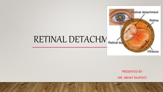
Retinal detachment is a disorder of the eye in which the retina separates from the layer underneath. Symptoms include an increase in the number of floaters, flashes of light, and worsening of the outer part of the visual field

Recomendados
Mais conteúdo relacionado
Mais procurados
Mais procurados (20)
Semelhante a Retinal Detachment
Semelhante a Retinal Detachment (20)
Mais de Abhay Rajpoot
Mais de Abhay Rajpoot (20)
Último
Último (20)
Retinal Detachment
- 1. RETINAL DETACHMENT PRESENTED BY: MR. ABHAY RAJPOOT
- 2. INTRODUCTION • Retinal detachment describes an emergency situation in which a thin layer of tissue (the retina) at the back of the eye pulls away from its normal position. • Retinal detachment separates the retinal cells from the layer of blood vessels that provides oxygen and nourishment.
- 3. DEFINITION Retinal detachment occurs when the retina separates from the back of the eye. This causes loss of vision that can be partial or total, depending on how much of the retina is detached.
- 4. RISK FACTORS • Aging — retinal detachment is more common in people over age 50 • Previous retinal detachment in one eye • Family history of retinal detachment • Extreme nearsightedness (myopia) • Previous eye surgery, such as cataract removal • Previous severe eye injury • Previous other eye disease or disorder, including retinoschisis, uveitis or thinning of the peripheral retina (lattice degeneration)
- 5. TYPES • Rhegmatogenous (reg-ma-TODGE-uh-nus). These types of retinal detachments are the most common. Rhegmatogenous detachments are caused by a hole or tear in the retina that allows fluid to pass through and collect underneath the retina, pulling the retina away from underlying tissues. The areas where the retina detaches lose their blood supply and stop working, causing loss of vision. • The most common cause of rhegmatogenous detachment is aging.
- 6. CONTI… • Tractional. This type of detachment can occur when scar tissue grows on the retina's surface, causing the retina to pull away from the back of the eye. Tractional detachment is typically seen in people who have poorly controlled diabetes or other conditions. • Exudative. In this type of detachment, fluid accumulates beneath the retina, but there are no holes or tears in the retina. Exudative detachment can be caused by age-related macular degeneration, injury to the eye, tumors or inflammatory disorders.
- 8. CLINICAL MANIFESTATION • The sudden appearance of many floaters — tiny specks that seem to drift through your field of vision • Flashes of light in one or both eyes (photopsia) • Blurred vision • Gradually reduced side (peripheral) vision • A curtain-like shadow over your visual field
- 9. DIAGNOSTIC EVALUATION • History • Physical examination • Retinal examination. The doctor may use an instrument with a bright light and special lenses to examine the back of eye, including the retina. This type of device provides a highly detailed view of whole eye, allowing the doctor to see any retinal holes, tears or detachments. • Ultrasound imaging. doctor may use this test if bleeding has occurred in the eye, making it difficult to see retina.
- 10. MANAGEMENT • Laser surgery (photocoagulation). The surgeon directs a laser beam into the eye through the pupil. The laser makes burns around the retinal tear, creating scarring that usually "welds" the retina to underlying tissue. • Freezing (cryopexy). After giving you a local anesthetic to numb your eye, the surgeon applies a freezing probe to the outer surface of the eye directly over the tear. The freezing causes a scar that helps secure the retina to the eye wall.
- 11. CONTI… • Pneumatic Retinopexy the surgeon injects a bubble of air or gas into the center part of the eye (the vitreous cavity). If positioned properly, the bubble pushes the area of the retina containing the hole or holes against the wall of the eye, stopping the flow of fluid into the space behind the retina. doctor also uses cryopexy during the procedure to repair the retinal break.
- 12. CONTI… • Scleral Buckling, involves the surgeon sewing (suturing) a piece of silicone material to the white of your eye (sclera) over the affected area. This procedure indents the wall of the eye and relieves some of the force caused by the vitreous tugging on the retina.
- 13. • Vitrectomy the surgeon removes the vitreous along with any tissue that is tugging on the retina. Air, gas or silicone oil is then injected into the vitreous space to help flatten the retina.