
Recomendados
Mais conteúdo relacionado
Mais procurados
Mais procurados (20)
Semelhante a chapter 3 CVS examination.pptx
Semelhante a chapter 3 CVS examination.pptx (20)
Mais de AbdiIsaq1
Mais de AbdiIsaq1 (9)
Último
Último (20)
chapter 3 CVS examination.pptx
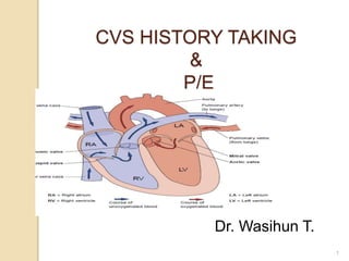
- 1. CVS HISTORY TAKING & P/E Dr. Wasihun T. 1
- 2. Introduction History Taking ◦ Features of common symptoms ◦ Presentation of common problems Examination Routine ◦ What do to ◦ Important physical signs 2
- 3. The History Symptoms of Heart Disease Dyspnea: This is a state of shortness of breath on exertion. But, it may occur at rest as the heart failure progresses. The degree dyspnea is graded based on the New York Heart Association Class (NHAC)
- 4. Cont… Paroxysmal Nocturnal Dyspnea: Is shortness of breath that occurs during sleep. The patient suddenly wakes up due the shortness of breath and then sits up or rush to open a window/door to get fresh air. Orthopnea: Shortness of breath that occurs during recumbent position. It is gauged by the number of pillows that are used to relieve the symptom
- 5. Cont… Body swelling: Usually which starts from the leg Palpitation: Is subjective unpleasant perception of one’s own heart beat. Cough: Which usually occurs at night (nocturnal) •Syncope: Sudden episode of fainting related to hemodynamic derangement. Pain: Angina pectoris is a cardiac pain. It arises in the precordial area usually on the retrosternal region and radiates to the left neck, shoulder and left upper arm. It has piercing, or squeezing character which is aggravated by exertion and relieved by rest
- 6. Cardiovascular History Record the date and time the history was taken. Name, Age, Occupation(s) Presenting Problem/ Complaint 6
- 7. Objectives To explain symptoms of cardiac disease To learn a step -wise approach in cardiovascular examination To appreciate the normal and abnormal cardiac findings To interpret cardiac findings
- 8. CARDINAL SYMPTOMS Dyspnea / shortness of breath Chest pain Palpitation Edema Cyanosis Syncope Cough Hemoptysis Fatigue Intermittent Claudication. 8
- 9. Symptoms: Chest Pain Important points to establish ◦ Site ◦ Radiation ◦ Character ◦ Exacerbating and Relieving factors ◦ Duration ◦ Associated symptoms 9
- 10. Symptoms: Chest Pain 10 CARDIAC Angina Myocardial Infarct Pericarditis Aortic dissection PULMONARY Pleurisy Pulmonary Embolus Pneumothorax GASTRO Ulcer or Reflux Gallstones Pancreatitis NON ORGANIC Anxiety
- 11. Symptoms: Breathlessness Breathlessness or dyspnoea can have a number of causes ◦ Heart Failure ◦ Valve disease ◦ Myocardial Ischaemia ◦ Pericardial disease 11 There are also non cardiac causes of dyspnoea - Pulmonary disease - Anaemia, Obesity or being unfit
- 12. PALPITATION: “Unpleasant Awareness of Forceful or Rapid Heart Beating.” Main Cause: Cardiac Arrhythmias. 12
- 13. EDEMA OF THE LOWER LIMBS. CAUSES: In general, the most common cause of limb edema is venous insufficiency. ◦ Cardiac: the most common cardiac cause is CHF ◦ Renal. Acute or chronic (nephrotic syndrome ). ◦ Hypoalbuminemia (Liver cirrhosis). ◦ In general, the most common cause of limb edema is venous insufficiency. 13
- 14. EDEMA OF THE LOWER LIMBS, cont’d Grades: 1+ Around ankle Joint. 2+ Below knee joint. 3+ Above knee joint. 4+ Scrotal edema: hydrocele, and edema of the ant. abdominal wall. ◦ Sacral edema can occur with 2nd,3rd, 4th degree if the patient is confined to bed. 14
- 15. History taking During the history consider (and ask about) the main risk factors for Ischemic Heart Disease: 1. Smoking 2. Hypertension 3. Diabetes mellitus 4. Hyper lipidaemia 5. Family history 15
- 16. History taking Past Medical History (may ask under presenting complaint) e.g. angina, myocardial infarction, bypass operation, rheumatic fever, stroke, intermittent claudication Social History Smoking (pack years), alcohol Family History At what age did the relative have illness? Drug History Allergies Systemic Review Summarize – does the patient have any questions? 16
- 18. Cont… Equipment Needed ◦ Stethoscope ◦ A Blood Pressure apparatus ◦ A Moveable Light Source or pen light
- 19. General Considerations The patient must be properly undressed above the waist. The examination room must be quiet to perform adequate auscultation. Observe the patient for general signs of cardiovascular disease ◦ Breathing pattern ◦ Cyanosis, ◦ Finger clubbing, ◦ Edema
- 20. Arterial Pulses Components of arterial examination include ◦ ƒ Rate ◦ ƒ Rhythm ◦ ƒ Character ◦ ƒ Volume (amplitude) ◦ ƒ Radio-femoral dela
- 21. Cont… Major Arteries: Major arteries Radial, Brachial, Carotid,Femoral, Popliteal, Posterior Tibial, Dorsalis pedis. All arteries should be palpated symmetrically at the same time except carotid arteries, as this could cut off the blood supply to the brain and cause syncope.
- 22. Cont… Rate and Rhythm: The radial artery is preferred Compress the radial artery with your index and middle fingers Note whether the pulse is regular or irregular. Count the pulse for one full minute. Record the rate and rhythm.
- 23. Cont… Pulse classification in adults 1. Based on the rate Normal: 60 - 100 beats / min Bradycardia: < 60 beats / min Tachycardia: > 100 beats / min 2. Based on rhythm Regular Regularly irregular; e.g. ectopic beats, 2nd degree heart block Irregularly irregular: e.g. atrial fibrillation
- 24. Blood Pressure 90 – 140, Diastolic: 60 – 85 millimeter of mercury 1. Position the patient's arm so that the anticubital fold is level with the heart. 2. Center the bladder of the cuff over the brachial artery approximately 2 cm above the anticubital fold. Proper cuff size is essential to obtain an accurate reading Physical Diagnosis 3. Palpate the radial pulse and inflate the cuff until the pulse disappears 4. Place the stethoscope over the brachial artery. 5. Inflate the cuff 20 to 30 mmHg above the estimated systolic pressure after the pulse disappears. 6. Release the pressure slowly, no greater than 5 mmHg per second. 7. The level at which you consistently hear beats is the systolic pressure. 8. Continue to lower the pressure until the sounds muffle and disappear. This is the diastolic pressure. 9. Record the blood pressure as systolic over diastolic (e.g. 120/70). 10. Blood pressure should be taken in both arms on the first encounter.
- 25. Cont… Character and Volume: best checked on carotid arteries Observe for carotid pulsations. Place fingers behind the patient's neck and compress the carotid on one side Assess the following: The amplitude of the pulse. The contour of the pulse wave. Variations in amplitude from beatto beat or with respiration. Repeat on the opposite side.
- 26. Venous system Jugular Venous Pressure (JVP):- is a reflection of the right atrial pressure and it is the most important part of venous system examination
- 27. Interpretation: Normal: - is less than or equal to 4 cm of water Elevated: - if greater than 4 cm of water above the sternal angle. This indicates raised right atrial pressure which is most often found in right ventricular failure (dysfunction)
- 28. The Precordium This is part of the anterior chest wall which overlies the heart. During examination of the precordium:- ◦ Position the patient supine with the head of the table slightly elevated ◦ Always examine from the patient's right side
- 29. Cont… Inspection ◦ Precordial bulge which may indicatelong standing cardiac diseases ◦ Precordial movement( activity ):- ◦ Multiple pulsations:- e.g. multivalvular lesions ◦ Quiet: - e.g. Pericardial effusion ◦ Apical beat: - which is the most laterally and downward positioned impulse
- 30. Palpation • Palpable heart sounds (at each valvular sites) • PMI: point of maximal impulse (which usually is located at the same area to the apical impulse,). It is normally located in the 4th or 5th intercostals space just medial to the mid clavicular line and properly characterizes the PMI.
- 31. Auscultation The stethoscope has two parts:- Diaphragm:preferred to auscultate high pitched sounds e.g. S1, S2, Holosystoic murmur etc Bell: preferred to auscultate low pitched sounds e.g. S3, S4, diastolic murmur of MS Position the patient supine with the head of the table slightly elevated. Always examine from the patient's right side
- 32. Areas of auscultation 1. The right 2nd inters space near the sternum (aortic area). 2. The left 2nd inter space near the sternum (pulmonic area). 3. The left 4th , and 5th inter spaces near the sternum (tricuspid area) 4. At the apex (mitral area).
- 33. Different maneuvers to accentuate cardiac auscultation ◦ Have the patient roll on their left side and auscultate at the apex. This position accentuates S3 and mitral murmurs. ◦ Have the patient sit up and lean forward. This position enhances diastolic murmur of aortic regurgitation, and pericardial friction rub
- 34. Cont… During auscultation focus on ◦ 1st Heart Sound, S1:- This signals the onset of systole and is caused by the closure of the mitral and tricuspid valves.
- 35. 1st Heart Sound , S1:- This signals the onset of systole and is caused by the closure of the mitral and tricuspid valves. S1 is loud in mitral stenosis tachycardia hyperdynamic circulation like e.g. anemia S1 is soft ( Muffled )in:- mitral regurgitation bradycardia and LVF
- 36. S2 Heart Sound Heard particularly during inspiration The sound is made by the closure of aortic and pulmonary valves 3rd Heart Sound, S3 This is produced by rapid ventricular filling and occurs in early-mid diastole i.e. soon after S2 occurs normally in young fit adults with bradycardia patients with heart failure left heart failure - S3 heard best in mitral area right heart failure - S3 heard best in tricuspid area patients with high ventricular filling pressure mitral regurgitation
- 37. 4th Heart Sound, S4 This is an atrial sound, occurring just before S1. It is always abnormal as it represents atrial contraction against a stiffened ventricle e.g. due to aortic stenosis or hypertensive heart disease. It may also occur in heart failure.
- 38. Cont… • 2nd Heart Sound, S2: The sound is made by the closure of aortic and pulmonary valves 3rd & 4th Heart Sounds ◦ These are low pitched sounds ◦ If either S3 or S4 is very loud it is often heard as gallop/triple rhythm
- 39. Murmur Murmur is abnormal sound due to turbulence of blood flow. It may be innocent (Physiologic) e.g. hyperdynamic states like anemia, pregnancy etc. or pathologic e.g. valvular lesions
- 40. Cont… Characterization of Murmur ◦ Timing: systole, diastole, continuous ◦ Point of maximum intensity (PMI) ◦ Direction of selective propagation (radiation) ◦ The character and quality of the murmur ◦ Intensity (grading)
- 41. Classification of Murmurs Systolic murmurs 1) Ejection systolic murmur ◦ This originates from the aortic outflow tract. It may be an innocent flow murmur which is ◦ common in childhood, pregnancy, anemia or ◦ pathological murmur as in Aortic stenosis and Aortic sclerosis Hypertrophic obstructive cardiomyopathy Pulmonary stenosis Atrial septal defect
- 42. Cont… 2. Pansystolic murmur: It is of uniform intensity and merges with S2. S1 is often muffled. It is found in:- Mitral or Tricuspid regurgitation Ventricular septal defect
- 43. Cont… Diastolic murmurs 1) Early diastolic murmur: This is high- pitched and blowing. It occurs due to:- ◦ Aortic or pulmonary regurgitation. 2) Mid-diastolic murmur: This is low- pitched and rumbling; it often starts after an opening snap. It is caused by:- mitral stenosis(common)
- 44. Thank you
Notas do Editor
- Description: – Fast or slow. – Regular or irregular. – Onset and offset: Sudden onset and offset in paroxysmal supraventricular tachycardia or paroxysmal atrial fibrillation. – Duration. – Associated symptoms: dizziness, presyncope and syncope, chest pain, dyspnea, polyuria, pallor.
- Face • Malar flush (thin face, purple cheeks) may be found in mitral stenosis. • Lips for (cyanosis). Eyes • Xanthelasma or corneal arcus indicates hyperlipidemia • Pallor of the conjunctiva indicates anemia • Icterus may be found in acutely congested liver • Exophthalmus may be seen in thyrotoxicosis. Hands • Clubbingof fingers may be seen : o Cyanotic congenital heart disease o Infective endocarditis • Peripheral cyanosisand Perfusion • Splinter hemorrhages:- vertical linear hemorrhages beneath the nails • Osler's nodes:- Tender lumps in pulp of fingertips which may be found in endocarditis • Janeway lesions:- are painless red macules on the wrist and palm which may be seen in patients with acute infective endocarditis • Nicotine stains:- indicate chronic smoking which is a major risk factor for atherosclerosis.
- Carotid pulse - for character • Normal • Hypokinetic (small volume):found in low output states like heart failure, shock, mitral stenosis etc. • Parvus et tardus ( Small Volume And Slow Rising pulse ): found in aortic stenosis • Bisferiens: a collapsing and slow rising pulse which occurs in mixed aortic disease (AS and AR ) • Pulsus alternans: alternating strong and week pulses • Jerky- hypertrophic cardiomyopathy • Pulsus Paradoxus: pulse weakens in inspiration, indicates tamponade or constrictive pericarditis • Collapsing(rapid up and rapid down): Water hammer pulse o strong radial pulse that taps hand up on lifting the arm o indicates wide pulse pressure of aortic regurgitation (also AV fistula or hyperdynamic circulation) • Bounding pulse o CO2retention o Liver failure o Sepsis • Radio-femoral delay- suggests coarctation or dissection of aorta
- Position the patient supine with the head of the table elevated 30 degrees Use tangential, side lighting to observefor venous pulsations in the neck. Look for a rapid, double (sometimestriple) wave with each heartbeat. Adjust the angle of table elevation to bring out the venous pulsation. 5. Identify the highest point ofpulsation. Using a horizontal line from this point, measure vertically from the sternal angle This measurement should be less than 4 cm in a normal healthy adult Interpretation: Normal: - is less than or equal to 4 cm of water Elevated: -if greater than 4 cm of water above the sternal angle. This indicates raised right atrial pressure which is most often found in right ventricular failure (dysfunction)
- Ask the patient to hold on breathing on inspiration which makes sounds arising from the right side of the hear louder , and if the patient holds on expiration sound originating from the left side of the heart are exaggerated.
- NB The 1st sound can be identified by palpating the carotid pulse while auscultating. The upstroke of the carotid pulse closely follows the 1st heart beat. S1is loud in o mitral stenosis o tachycardia o hyperdynamic circulation like e.g. anemia • S1is soft ( Muffled )in:-o mitral regurgitation bradycardia and LVF
- The aortic valve closes before the pulmonary valve and this splitting of the second sound is heard particularly during inspiration, as more blood is drawn into the right ventricle which is a normal phenomenon. S2 wide splittingoccurs in right bundle branch block pulmonary stenosis Wide and fixed splitting (i.e. not varying with respiration) occurs when there is an atrial septal defect A2is loud in systemic hypertension • A2 is soft in aortic stenosis • P2is loud in pulmonary hypertension • P2is soft in pulmonary stenosis
- The aortic regurgitation murmur is usually soft and is best heard with the patient leaning forward and in expiration. • mitral stenosis(common) exhibits pre-systolic accentuation in sinus rhythm o amplified by rolling the patient into the left lateral position Rheumatic fever thickens mitral valve leaflets Is called Carey Coombs' murmur aortic regurgitation regurgitant jet causes fluttering of anterior mitral valve cusp Is called Austin Flint murmur tricuspid stenosis(rare) large atrioseptal defect