Acute appendicitis
•Transferir como PPTX, PDF•
12 gostaram•1,057 visualizações

The document discusses acute appendicitis. It describes the anatomy of the appendix and explains that appendicitis is usually caused by obstruction of the appendix lumen by a fecalith. The clinical features of appendicitis include pain that initially starts around the umbilicus and later localizes to the right lower quadrant, along with nausea, vomiting, fever and tenderness at McBurney's point. Diagnosis is often made through ultrasound or CT scan. Treatment involves surgical removal of the appendix (appendicectomy) through an open or laparoscopic approach.

Recomendados
Mais conteúdo relacionado
Mais procurados
Mais procurados (20)
Semelhante a Acute appendicitis
Semelhante a Acute appendicitis (20)
Último
Último (20)
Acute appendicitis
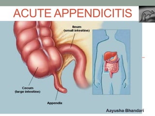
- 2. Anatomy Located at the terminal end of caecum, 2 cm below the ileocaecal junction. Length is about 5-10 cm .Diameter of appendix is 3-8 mm and diameter of lumen is 1-3 mm. Parts of appendix : base ,body and tip The mesentery attached to the appendix is known as mesoappendix which contains appendicular vessels. Mesoappendix doesn’t extend up to the tip of appendix so in obstructive type of appendicitis the commonest site of gangrene is the tip(the least vascular area).
- 3. • Appendix is supplied by appendicular artery which is a branch of ileocolic artery. The appendicular artery is an end artery. • The base of the apppendix is usually located at the MacBurney’s point. • Opening of appendix into the caecum is guarded by valve of Geralch. • Most common position of appendix is retrocaecal (78%) next is pelvic (21%).
- 4. Different positions of appendix
- 5. Acute appendicitis Etiology Common in young males and whites races Low fibre diet Viral infection can cause mucosal oedema and inflammation which later gets infected by bacteria 30% chances in first degree relatives Obstruction of lumen by faecoliths(most common), stricture, foreignbody and roundworm may cause obstructive appendicitis. Adhesion and kinking Distal colonic obstruction Abuse of purgatives
- 6. • Organisms are E.coli, enterococci, streptococci, anaerobic streptococci , Cl.welchii • Pseudoappendicitis is appendicitis due to acute ileitis due to yersinia infection. Pathogenesis Non obstructive appendicitis: acute inflammation of the mucus membrane with secondary infection. It may lead to resolution ,fibrosis ,recurrent appendicitis or eventual obstructive appendicitis.
- 7. Obstructive appendicitis : • luminal obstruction by faecoliths, FB, Carcinoma,lymphoid hyperplasia ,pinworm • Mucus and inflammatory fluid collects inside the lumen and increased intraluminal pressure • Blockage of lymphatic and venous drainage resulting in increased oedema of mucosa and wall • Mucosal ulceration and ischemia, bacterial translocation. If thrombosis of appendicular artery-ischaemic necrosis – gangrene of appendix and then perforation at the tip or base=peritonitis
- 8. • After perforation-localization at greater omentum and dilated ileum occurs-with suppuration and pus=appendicular abscess • In severe acute appendicitis, localization at G. omentum and dilated ileum occurs without pus formation = appendicular mass • Acute appendicitis with blockage at the opening of lumen- mucus collects inside the lumen resulting in enlargement of appendix = Mucocele of appendix
- 9. Types of appendicitis 1. Acute non-obstructive appendicitis 2. Acute obstructive appendicitis 3. Recurrent appendicitis: repeated attacks of non- obstructive 4. Subacute appendicitis 5. Stump appendicitis: due to retained stump of appendix after lap appendicitomy
- 11. • Pain:visceral pain around the umbilicus d/t distension of appendix later after few hours somatic pain in RIF d/t irritation of parietal peritoneum d/t inflamed appendix • Vomiting d/t reflex pylorospasm • Constipation/diarrhoea • Fever, tachycardia, fetor oris • Urinary frequency Tenderness and rebound tenderness at McBurney’s point in RIF P/R examination tenderness in the right side of rectum Hyperaesthesia in Sherren’s triangle( ASIS ,umbilicus and pubic symphisis )
- 12. Clinical signs in appendicitis Rovsing’s sign On pressing the LIF ,pain occurs in the RIF d/t shift of bowel loops which irritated the parietal peritoneum. Blumberg’s sign(release sign) pain upon removal of pressure rather than application of pressure to the abdomen. Cope psoas sign(hyperextension) and obturator sign(internal rotation) of the right hip causing pain in the RIF d/t irritation of the psoas muscle and obturator internus muscle respectively. Baldwing’s test: when legs are lifted off with knee extended, pain complains pain while pressing over the flanks.
- 13. Differential diagnosis 1) Perforated peptic ulcer 2) Ruptured or twisted ovarian cyst 3) Acute cholecystitis 4) Enterocolitis 5) Right ureteric colic 6) Right acute pyelonephritis 7) Crohn’s disease 8) Lobar pneumonia 9) Acute pancreatitis 10) Meckel’s diverticulum 11) Salphingitis 12) Ectopic pregnancy 13) Typhilitis
- 14. In children 1) Meckel’s diverticulum 2) Acute colitis 3) Intussusception 4) Roundworm colic 5) Lobar pneumonia 6) Acute iliac lymphadenitis In females 1. Ruptured ectopic gestation 2. Mittelschmerz rupture of ovarian follicle 3. Ovarian cyst torsion 4. Salpingo-oophoritis
- 15. Investigations • U/S to rule out stones, cyst, pancreatitis, ectopic pregnancy and confirm appendicular abscess or mass. • USG findings: size of appendix >6mm ,hyperechoic thickened appendix wall >2mm- target sign, appendicolith, interruption of submucosal continuity, periappendicular fluid. • Total leucocyte count is increased. • Contrast CT scan • C-reactive protein, MRI • Plain X-ray: to R/O duodenal ulcer perforation, intestinal obstruction and ureteric stone
- 18. Treatment • Surgery : Appendicectomy • Approaches • Grid iron incision at (incision perpendicular to the McBurney’s point) • Rutherford Morison incision • Lanz crease(centering at McBurney’s point) • Right lower paramedian incision or • Lower midline incision • Laparoscopic approach • Fowler-weir approach McBurney’s point is the lateral 1/3 and medial 2/3 of imaginary line joining ASIS and umbilicus.
- 20. Open appendectomy G.A is given. Mark McBurney’s point and grid-iron incision is given. Skin is incised. Subcutaneous tissue and superficial fascia (camper’s and scarpa’s) are cut using cautery. A nick is given to external oblique aponeurosis.it is opened in the line of incision and the incised free margins are lifted up using artery forceps. Internal oblique and tranversus muscle are split in the line of fibres.(retracted to reach the peritoneum) Peritoneum is held at 2 places by mosquito forceps.nick is given between two forceps. Peritoneal cavity entered.
- 21. Caecum is identified by the presence of taenia coli and ileocaecal junction. Appendix is held by Babcock’s forceps. Window is made in the mesoappendix with the help of curved artery forceps. Mesoappendix and appendicular artery is ligated using vicryl 2-0 Junction of caecum with appendicular base is identified. Now the appendix is crushed with straight clamp about 3- 5mm away from the caecum. Reapplied again Base of the appendix is double ligated using vicryl 2-0. appendix is cut distal to the suture ligature and removed. Stump is cleaned with antiseptics and exposed portion is cauterized. Internal oblique, T. abdominus and peritoneum closed with vicryl 1-0. gut preserved. E.O.A is closed vicryl 1-0 continuous
- 22. Complications after appendicectomy A. Reactionary haemorrhage d/t slipping of ligature of appendicular artery B. Paralytic ileus C. Residual abscess D. Pylephlebitis E. Adhesion, kinking and intestinal obstruction F. Right inguinal hernia G. wound sepsis H. Faecal fistula