
Recomendados
Mais conteúdo relacionado
Mais procurados
Mais procurados (20)
Semelhante a Perthes disease
Semelhante a Perthes disease (20)
Último
Último (20)
Perthes disease
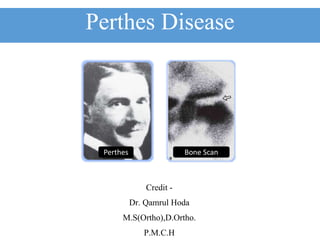
- 1. Perthes Disease Credit - Dr. Qamrul Hoda M.S(Ortho),D.Ortho. P.M.C.H Perthes Bone Scan
- 2. Perthes Disease • Also known as- o Legg-Calve-Perthes disease. o Calve Perthes disease. o Avascular necrosis. o Coxa plana o Pseudocoxalgia • It is an osteochondritis of the epiphysis of the femoral head. • In this disease the femoral head become partly or wholly avascular and deformed.
- 3. Epidemiology • Legg-Calve-Perthes disease is the most common hip disease that afflicts school-age children. • The disorder was described by three independent authors in 1910. • Arthur Legg • Jacques Calve • Georg Perthes • They might have surprised to see(if alive)how much and how little have changed in 100 years Legg Calve
- 4. What is Perthes Disease ? • Perthes disease is a childhood disorder which affects the head of the femur. • It is a vascular problem affect 1 in 10000 child (4 in 10000 in India) • In Perthes disease the blood supply to the growth plate of the bone at the end of the femur becomes inadequate. • As a result the bone softens and breaks down due to avascular necrosis.
- 5. Incidence • Legg-Calve-Perthes disease generally occurs in children between 4 to 8 years of age. • With males being affected about four to five times more frequently than females. • It is a unilateral in 90 percent of patients, while the other 10 percent of patients have bilateral hip. • Rates as low as 3.8 per 100,000 have been reported in Korea, and very similar rates of 4.4 per 100,000 have been identified in Southern India.
- 7. Vascular Phenomena • Up to the age of 4 months, the femoral head is supplied by - 1) Metaphyseal vessels which penetrate the growth disc 2) Lateral epiphyseal vessels running in the retinacula and 3) Scanty vessels in the ligamentum teres • The metaphyseal blood supply - 1) Gradually declines until, by the age of 4 years. 2) It has virtually disappeared; by the age of 7 years. 3) However, the vessels in the ligamentum teres have developed. • Between 4 and 7 years of age the femoral head may depend for its blood supply and venous drainage almost entirely on the Lateral Epiphyseal Vessels whose situation in the retinacula makes them susceptible to stretching and pressure from an effusion.
- 8. Pathophysiology • Another factor which needs to be taken into account Is that the vascular sinusoids which nourish the marrow and bone cells ,unlike arterial capillaries, have no adventitial layer and their patency is determined by the volume and pressure of the surrounding marrow tissue ,which itself is encased in unyielding bone. • The system functions essentially as a closed compartment within which one element can expand the presser(marrow presser)
- 9. Bones Vulnerable to Ischemia • Sites which are vulnerable to ischemic necrosis are- 1. Femoral Head, 2. Femoral Condyles 3. Head of the Humerus 4. Capitulum 5. Proximal Part of Scaphoid 6. Talus.
- 10. Pathology • Once a critical portion of blood flow to the femoral head has been compromised. • The femoral head will begin to take on a pathologic shape leading to - • Flatening • Fragmentation • Collapse Flatening Fragmentation Collapse Collapse
- 11. Etiology • It is not clear why this blood vessel problem occurs in the femoral head. • Factors which has been implicated are - 1) Heredity 2) Trauma 3) Endocrine 4) Inflammatory 5) Nutritional 6) Altered circulatory hemodynamic Bilateral -15% Cases Unilateral - 80% Cases
- 12. Clinical Features • It occur commonly in boys of age group from 5 to 10 years. • The signs and symptoms of -Perthes disease can be rather slow and may not be immediately recognized by parents or by treating doctor. • They may simply perceive a slightly abnormal gait • Since there is no complaint of pain, little attention on may be paid to it. • If hip/knee/groin pain is present, it may worsen towards the end of a strenuous day and is often relieved by rest. • Gait pattern disturbed-limp • Often present with a Trendelenburg gait, ( abductor lurch.) • ROM diminished-- loss of abduction and internal rotation.
- 13. Clinical Feature • Symptoms - 1) Hip, knee or groin pain, exacerbated by hip/leg movement, especially internal hip rotation . 2) Hip pain radiating to knee. 3) There may limping and stiffness. 4) Reduced range of motion – i. Abduction and ii. Internal rotation iii. Patient presents with a limp. 5) Pain is usually mild. • Signs - 1) Wasting of the muscles in the upper thigh. 2) Shortening of the leg 3) Stiffness of the Hip 4) Difficulty in Squatting PAIN IN KNEE- SHOULD BE CONSIDERED AS HIP IN CHILDREN UNLESS OTHERWISE PROVED
- 14. Stages of Perthes Disease • Necrosis: Initial period of ischemia/loss of blood supply to femoral head. • Fragmentation: Re-absorption of bone with femoral head collapse. • Re-ossification: New bone re-grows to reshape the femoral head. • Remodeling: Femoral head reshapes itself into normal spherical shape.
- 15. Diagnosis • X-rays of the hip • Finding 1) Femoral head broken or damaged. 2) Collapse and sclerosis of epiphysis of femoral head. 3) Hip joint space increase. 4) Flattened femoral head(Typical). • Bone Scan • Done for confirmation of Perthes Disease , when X-ray picture is normal. • MRI • Use to asses the extent of damage.
- 16. Differential Diagnosis • The irritable hip of early Perthes’ disease must be differentiated from other causes of irritability - 1) Morquio’s disease 2) Cretinism 3) Multiple epiphyseal dysplasia 4) Sickle-cell disease 5) Gaucher’s disease (may resemble Perthes’ disease radiologically, especially if they are bilateral; however, in bilateral Perthes’ disease the two sides are likely to be at different stages. ) • In Old Perthes deformities’ in adults, in the 10 per cent of cases with bilateral involvement, may resemble those of certain bone dysplasias, especially multiple epiphyseal dysplasia.
- 17. What is the Treatment for Perthes Disease ? • The overall goal of treatment is - 1) To reduce hip irritability and pain 2) Restore and maintain hip mobility 3) To prevent the femoral head from extruding or collapsing 4) To regain a spherical shape of the femoral head • It can be achieved by- • Conservative (or) • Operative Method
- 18. Conservative Treatment • Observation • Physical • Therapy/Traction to improve range of motion • Splints and abduction. Cast in internal rotation Ptrei Cast
- 19. Treatment Related to Age • Children under 6 years • No specific form of treatment has much influence on the outcome. Symptomatic treatment, including activity modification. • Bone age at or below 6 years • Lateral pillar group A and B (or Catterall stage I and II) – symptomatic treatment. • Lateral pillar group C (or Catterall stage III and IV)– abduction brace. • Bone age over 6 years • Lateral pillar group A and B (Catterall stage I and II) – abduction brace or osteotomy. • Lateral pillar group C (Catterall stage III and IV) – outcome probably unaffected by treatment, but some would operate. • Children 9 years and older • Operative containment is the treatment of choice.
- 20. Surgery • Femoral Varus Osteotomy • Acetabular Osteototomy( with internal fixation)
- 21. Operative Procedure • Schematic diagram of surgical options for containment of an extruded avascular femoral epiphysis – 1) Proximal femoral varus osteotomy 2) Innominate Osteotomy 3) Shelf Procedure
- 22. Total Hip Replacement • In older cases THR can be done.
- 23. Prognostic Feature • The outlook for children with Perthes’ disease, as a group, is well summarized by Herring (1994): • A small % of patients have a very difficult course, with recurrent loss of motion, pain, and an eventual poor outcome. • However, most children have moderate problems in the active phase of the disease and then improve steadily, leading to satisfactory outcome.’