Professor Yasser Metwally Edits Short Neuro Case
•
2 gostaram•392 visualizações

Short case...Lymphomatous leptomeningitis

Recomendados
Recomendados
Mais conteúdo relacionado
Mais procurados
Mais procurados (9)
Destaque
Destaque (20)
Semelhante a Professor Yasser Metwally Edits Short Neuro Case
Semelhante a Professor Yasser Metwally Edits Short Neuro Case (20)
Mais de Professor Yasser Metwally
Mais de Professor Yasser Metwally (20)
Último
Último (20)
Professor Yasser Metwally Edits Short Neuro Case
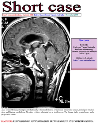
- 1. Short case publication... Version 2.11| Edited by professor Yasser Metwally | December 2008 Short case Edited by Professor Yasser Metwally Professor of neurology Ain Shams university school of medicine Cairo, Egypt Visit my web site at: http://yassermetwally.com A 22 years old male patient presented clinically with manifestations of increased intracranial tension, meningeal irritation signs and bilateral papilledema. No other evidence of cranial nerve involvement. The disease had a gradual onset and a progressive course. DIAGNOSIS: LYMPHOMATOUS MENINGITIS (BOTH LEPTOMENINGITIS AND PACHYMENINGITIS).
- 2. Figure 1. Precontrast MRI T1 images showing moderate hydrocephalic changes, more on the right side Figure 2.Postcontrast MRI T1 images showing enhancement of the basal cistern, both leptomeningeal and pachymeningeal enhancement and thick irregular enhancement of the ventricular walls. The leptomeningeal enhancement is thick and nodular and extends to the upper cervical spinal cord. Also notice the moderate hydrocephalic changes, more on the right side.
- 3. Figure 3. Postcontrast MRI T1 images showing ventricular dilatation, bilateral more or less symmetrical linear hypointensities in the presumed area of the cortico-spinal tract. Notice the bilateral fronto-partial hypointensities with probable subcortical cystic changes. The MRI T1 hypointense changes demonstrated in these MRI images correspond, pathologically, to edema, cystic changes, astrogliosis and demyelination involving mainly the cortical motor strip and the descending cortico-spinal tract in the internal capsule, thalamus and upper midbrain. Figure 4. Postcontrast MRI T1 images showing enhancement of the basal cistern, both leptomeningeal and pachymeningeal enhancement and thick irregular enhancement of the ventricular walls. The leptomeningeal enhancement is thick and nodular. Also notice the moderate hydrocephalic changes, more on the right side.
- 4. Figure 5. Postcontrast MRI T1 images showing enhancement of the basal cistern, both leptomeningeal and pachymeningeal enhancement and thick irregular enhancement of the ventricular walls. The leptomeningeal enhancement is thick and nodular. Notice enhancement of the tentorium cerebelli. Also notice the moderate hydrocephalic changes. Figure 6. Postcontrast MRI T1 images showing enhancement of the basal cistern, both leptomeningeal and pachymeningeal enhancement and thick irregular enhancement of the ventricular walls. The leptomeningeal enhancement is thick and nodular. Notice enhancement of the tentorium cerebelli. Also notice the moderate hydrocephalic changes, more on the right side.
- 5. Figure 7 . Postcontrast MRI T1 images showing enhancement of the basal cistern, both leptomeningeal and pachymeningeal enhancement and thick irregular enhancement of the ventricular walls. The leptomeningeal enhancement is thick and nodular and extends to the upper cervical spinal cord. Also notice the moderate hydrocephalic changes, more on the right side.
- 6. Figure 8. Postcontrast MRI T1 images showing enhancement of the basal cistern, both leptomeningeal and pachymeningeal enhancement and thick irregular enhancement of the ventricular walls. The leptomeningeal enhancement is thick and nodular and extends to the upper cervical spinal cord. Also notice the moderate hydrocephalic changes, more on the right side.
- 7. Figure 9. MRI FLAIR study showing periventricular nodular, irregular and thick hyperintensity almost completely ensheathing the ventricular system. Notice the parenchymal centrifugal fungation of the periventricular disease. References 1. Metwally, MYM: Textbook of neurimaging, A CD-ROM publication, (Metwally, MYM editor) WEB-CD agency for electronic publishing, version 9.4a October 2008 Addendum A new version of short case is uploaded in my web site every week (every Saturday and remains available till Friday.) To download the current version follow the link quot;http://pdf.yassermetwally.com/short.pdfquot;. You can download the long case version of this short case during the same week from: http://pdf.yassermetwally.com/case.pdf or visit web site: http://pdf.yassermetwally.com To download the software version of the publication (crow.exe) follow the link: http://neurology.yassermetwally.com/crow.zip At the end of each year, all the publications are compiled on a single CD-ROM, please contact the author to know more details. Screen resolution is better set at 1024*768 pixel screen area for optimum display For an archive of the previously reported cases go to www.yassermetwally.net, then under pages in the right panel, scroll down and click on the text entry quot;downloadable short cases in PDF formatquot; Also to view a list of the previously published case records follow the following link (http://wordpress.com/tag/ case-record/) or click on it if it appears as a link in your PDF reader