SlideShare uma empresa Scribd logo
1 de 94
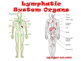
Lymphatic
System Organs


       DR KIRTI SOLANKE   1
INTRODUCTION
• Not a primary tissue type but a variety of
  connective tissue.
• Components-1.lymphatic vessels
                 2.lymphatic organs
                 3.cells
                 4.lymph


                                               2
Primary Lymphatic Organs and Tissues

• The primary (central) lymphatic organs and
  tissues include the thymus, bone marrow and gut
  associated lymphatic tissue (GALT).
• They are sites of
  – antigen-independent proliferation, and
  – differentiation into cells pre-programmed to
    recognise specific antigens.
• These immunocompetent cells then enter the
  blood and lymph:
  – get dispersed in the connective tissue and
  – penetrate into epithelia that line mucosal surfaces.

                                                           3
Secondary Lymphatic Organs and Tissues


• These are the effector lymphatic tissues.
• They include the lymphatic nodules, lymph
  nodes, tonsils and the spleen.
• Here lymphocytes undergo antigen-
  dependent proliferation and differentiation
  into effector lymphocytes and memory cells.


                                                4
5
Lymphatic capillaries are not ubiquitous-absent in
     cornea, cartilage, thymus, central and peripheral
               nervous sys & bone marrow.
Lymphatic circulation             Vascular circulation

•   Deep                          •   Superficial
•   Valves closely placed         •   Less closely placed
•   Blind ends                    •   Both ends open
•   No pericytes                  •   Pericyte
•   Irregular shape in cut sec.   •   Cylindrical form
•   Basal lamina incomplete       •   Continuous
•   Greater caliber               •   Less caliber


                                                            6
LYMPH
• Lighter specific gravity
  than blood
• Contains no
  RBCs,platelets,fibrinogen
  but it contains numerous
  Lymphocytes.
• Lymphocytes are added
  when it passes through
  LN.
• Coagulate at much
  slower rate than blood
• Does not carry O2 but
  may contain CO2.
• Chyle-fat globules,
  lacteal
                                      7
8
• Lymph vessels of- thyroid gland
                -oesophagus
                -coronary and triangular
                ligaments of liver
may drain directly into thoracic duct.


                                           9
Functions of lymphoid system

1) To maintain – pressure &
                 -volume of extracellular fluid by
  returning excess water to the circulation.
2) The site of clonal production of
  immunocompetent lymphocytes and
  macrophages in the specific immune
  response.


                                                 10
CELLS OF LYMPHATIC SYSTEM
Chief cells are lymphocytes-type of WBC’S.
• B lymphocytes
• T lymphocytes
• Natural killer cells
• epithelioreticular cells - MESENCHYMAL
• Supporting cells-
   – interact with lymphocyte
   – Present antigens to Lymphocytes
• OTHER TYPE OF WBC’S
  -monocyte
  -macrophages
  -neutruphils
  -eosinophils
  -basophils

                                             11
• ANTIGEN PRESENTING CELLS
  – dendriti cells-Bone marrow derived
  – follicular cells
  – langerhan’s cells


                                         12
Derivation and Distribution of Lymphocytes


                                             13
T Lymphocytes

• These evolve in the thymus
• part of the cell-mediated or thymus-
  dependent response to antigens.
• Upon interaction with an antigen, they will
  differentiate and proliferate into - 3 types
  of effector T lymphocytes:


                                                 14
1.Cytotoxic lymphocytes (killer T cells) (primary effector
   cells in cell-mediated immunity.)
   scan the surface of other cells for signs of viral
   infection or abnormality, killing them if necessary by
   causing them to lyse.
2.Helper T lymphocytes,
recognise foreign antigens presented by macrophages.
release interleukin hormones to stimulate ‘processed’ B
   cells to produce antibodies.
3.Suppresser T lymphocytes, suppress the activity of B
   cells.

                                                         15
HELPER T CELL SUBTYPES(CYTOKINES)
•      Th1 cell             •     Th2 cells
• interact with cytotoxic   • interact with B
  CD8,NK ,macrophages.        lymphocyte
• CMI                       • HIR
• For controling            • Exracellular pathogens
  intracellular pathogens
• Viruses &
  microorganisms.
• IL 2,Interferon gamma     • IL4, 5 ,10,13.

                                                       16
B Lymphocytes

• These evolve in bone marrow and GALT, and
  are part of the humoral response.
• They will only react with the antigen they
  have been genetically programmed for.
• Once activated by this antigen, they may
  differentiate and proliferate into either:
• Plasma cells, that produce antibodies.

                                               17
• The antibody binds, forming an antibody-antigen
  complex, that may be phagocytosed by
  macrophages.

• Memory cells, which, after exposure to the
  specific antigen, will be able to participate in a
  rapid, secondary response with the same
  antigen.
• They do not participate in an initial or primary
  response.
                                                       18
19
B – Lymphocytes
     T-Lymphocyte

–   Prog In Thymus           –   Prog In B M
–   Long life span           –   Variable lifespan
–   For CMI                  –   For H.I.
–   Graft Rejection          –   Plasma Cells – Ab
–   60% to 8o%               –   20 to 30%
–   CD 2,CD3,CD7             –   CD9,CD19,CD20 & CD24
–   T cell receptors(TCRs)   –   Bcell receptors
–   CD4,CD8                  –   MHC II


                                                    20
NK LYMPHOCYTE/NULL CELLS
• Neither T nor B cells
• Specialised to kill certain types of target cells
• 5 to 10%
• Do not mature in thymus
• Kill in the same way as that of CTLS
• After recognition of tranformed cell,secrete
  perforins & fragmentins
• CD 56, CD94.

                                                      21
Macrophages

• These are involved in both types of immune
  response.
• They can process and present the antigen to
  the B cells or helper T cells.
• Or they can destroy the antigen by digestion
  after it has been processed by other cells of
  the immune system.

                                                  22
CLASSIFICATION
I. FUNCTIONAL
       LYMPHOID ORGANS

        CENTRAL   PERIPHERAL
   THYMUS               LYMPH NODE

 BONE MARROW             SPLEEN

                        MALT, GALT


                                     23
II. MORPHOLOGICAL

           LYMPHOID ORGANS
         DISCRETE      DIFFUSE

    LN, SPLEEN
                    BM, PEYER’S PATCHES
  THYMUS, TONSIL


                                      24
LYMPH NODE
• Oval/Kidney Shape, 0.1 – 0.5cm Long
• Normal young body contains up to 450,of which 60
  to 70 in head and neck,100 in thorax,250 in abd&
  pelvis
• Greatest no. lie close to the viscera mainly
  mesenteries.


                                               25
Lymph Node: Gross Appearance


                               26
27
28
29
30
• Capsule – Trabeculae, hilus-
  collagenous framework
• Lymph flow
   –   Retculin Meshwork-sinuses
   –   Subcapsular Sinus
   –   Cortical Sinuses
   –   Medullary Sinuses
   –   Eff Lymph Ch


• Cortex medulla-diff in
  arrangement


                                   31
3

2

1


    32
• Role of germinal
  centre-affinity
  maturation
• Two zones in GC-dark
  zone-centroblast-
  undergoing rapid
  proliferation-
  hypermaturation of
  their antibody mol.
• light zone-centrocytes-
  ineract with the FDC-
  carrying unprocessed
  antigen on their
  surface.
                            33
Lymph Node


             34
35
Lymph Node

       A - Afferent lymphatic channels
       B - Subcapsular sinus
       C - Follicle
       D - Sinuses
       E - Paracortical region
       F - Medullary cords
       G - Efferent lymphatic channel


                                         36
Variations in nodes
•   In large nodes –trabeculae are prominent
•   In small nodes-thin and frequently interrupted
•   Hemal nodes
•   lymphadenitis


                                                 37
• Diffusely distributed lymphatic nodules
  represent local immune response to antigen
  that are present in tissue fluid.
• LN-lymph.
• Spleen-circulating blood-mainly HIR.


                                               38
The Spleen
• The spleen is the largest
  lymphatic organ, located
  in the upper left quadrant
  of the abdominal cavity.
• The spleen functions to
  filter the blood, and react
  immunologically to
  blood-borne antigens
  mainly HIR,to dispose of
  defective blood
  cells,store blood cells
  &platelets,hematopoiesis
                                  39
Structure of the Spleen
• There is an external capsule of dense connective
  tissue.
• trabeculae extend into the substance of the organ.
• myofibroblasts, and is thus contractile.
• medial surface of the spleen, the hilum allows
  passage of the splenic vessels, nerves and lymphatic
  vessels.
• The substance of the spleen is known as the splenic
  pulp.
• white pulp areas,
• surrounded by red pulp.

                                                         40
Spleen


         41
The White Pulp
• This mainly consists of lymphocytes.
• Branches of the splenic artery course through
  the trabeculae and then enter the white pulp,
  known as the central artery
• The lymphocytes aggregated around the
  central artery in a cylindrical fashion
  -periarterial lymphatic sheath (PALS) of the
  artery.
• Lymph nodules in the PALS may displace the
  central artery from its central position in the
  white pulp.
                                                42
The Red Pulp


• This has large numbers of red blood cells
  (RBCs).
• It consists of splenic sinuses, separated by
  splenic cords (of Billroth).


                                                 43
44
45
46
47
48
49
50
Spleen


         51
SPLENIC A
  SEGMENTAL A

 TRABECULAR A

   CENTRAL A

  FOLLICULAR A

  PENICILLAR A

   ELLIPSOIDS

PRE CAPILLARY A

   SINUSOIDS

TRABECULAR VEINS

  SPLENIC VEIN
53
Splenic Circulation
• The splenic artery branches into the
  trabecular, the central arteries.
• The central arteries of the white pulp then
  branch into penicillar arterioles in the red
  pulp.
• These are actually capillaries, and may be
  sheathed by aggregations of macrophages.
• Blood from these penicilli leaves the vascular
  system to populate the splenic pulp, before
  re-entering the red pulp.

                                                   54
Two theories
• Closed Circulation Model
• In this model, the splenic arterioles are a "continuous vascular
  channel".
• They only empty into the splenic sinuses of the red pulp.
• The blood then leaves the sinuses before re-entering them.
•
• Open Circulation Model
• In this model, the arterioles empty directly into the splenic cords.
• Thus, blood percolates through the reticular meshwork of the
  pulp.
• It then only enters the splenic sinuses from the extravascular side.
• This model has more supporting evidence than the former.
• FUNCTIONS


                                                                     55
THYMUS
• Introduction
• Gross anatomy
• development
• Histology
         -Circulation
          -Relation with
  immunology
• Functions
• Age changes
• Recent advances            56
The Thymus (neuroendocrine organ)
• The thymus -4-6 cm long
       2.5-5 cm wide
  1cm thick,20 gm             bi-lobed
  organ
• left lobe higher
       superior mediastinum,
                              anterior
  to the heart and great vessels
• Thyrothymic ligament
• b/d supply-inf thyroid A,Internal
  thorasic A.
• Lymphatics-
  parasternal,brachiocephalic,trache
  obronchial


                                         57
Structural components
• Stroma-capsule
        -trabeculae
• Parenchyma –epithelioreticular cells
             -lymphocytes
             -macrophages
             -mast cells


                                         58
Development
• 3rd &4th endodermal pouch-epithelial reticular
  cells
• Local cardiac neural crest mesenchyme
  control dev of gland.


                                                   59
Development
• .


                    60
61
Histology
• Important environment-acquiring immune
  tolerance
• Dev of T lymphocyte-interaction between
                      -thymocyte &
                      -epithelioreticular
  cells,APC,chemical factors
• Capsule-incomplete trabeculae-irregular
  lobule-0.5-2mm in dia.

                                            62
Thymus


         63
CIRCULATION


• Thymic cortex and medulla functionally independent.

     lymphoid stem cells from BM       outer
  cortex    progeny proliferate & differentiat
      inner cortex medullary venules at CM
  junction.


                                                    64
cortex                    medulla
• Dark staining-small L   • Paler staining-large L
• Less in no              • Macropgages,dendritic cells
                            are more
• Plasma cells absent     • Plasma cells present
• b/d supply mainly by    • Mainly thymic A,arteriole
  capillaries


                                                      65
Epithelioreticular cells
• Parenchyma: epithelioreticular cells.
  – are epithelial cells having stellate shape
  – form cytoplasmic reticulum: a framework for the
    thymic lymphocytes.
  – correspond to the other reticular cells in
    lymphatic tissues,
  – but no reticular fibres in the thymus.
  – Feature of both epithelial (intercellular junctions,
    intermediate filament)and reticular
    cells(framework)
      --thymic nurse cells
  – crosstalk                                          66
Epithelioreticular cells
• 6 types
• Type l-boundary of capsule & cortex
• Type ll-within cortex, compartmentalize the
  cortex
• Type lll-between cortex and medulla
• Type lV –b/w cortex & medulla,barrier at CM
  junction
• Type V-like type ll in cortex
• Type lV – corpuscles, keratohyaline granules,
  produce lL-4&7.
                                              67
OTHER CELLS
  •   Myeloid lineage-monocyte
  •   Macrophages-PAS cells
  •   Fibroblast
  •   Myoid cells
  •   Hassall’s corpuscles-distinguishing feature of
      thymic medulla.30-100μm in dia.
                     IL-4,IL-7;named after Arthur Hill
      Hassall British physician


• Plasma cells are rare in cortex.
• adipocytes                                             68
HASSALL’S CORPUSCLES
70
71
THYMIC HORMONES
• epithelial reticular cells -
                  1.thymopoietin
                  2.thymosin alpha
Induce expression of T lymphocyte surface
  markers


                                            72
THYMIC BARRIER
• No afferent lymphatics
• Physical barrier-Capillary endothelium
                  -Endothelial basal lamina
                   - pericyte
  -thin perivascular connective tissue sheath
  with macrophages
                   - Basal lamina of the
  epithelioreticular cells
                  - Epithelioreticular cell sheath

                                                     73
74
Changes of Thymic Structure with Age
            (involution)

• Largest at birth
• fully functional at 20 weeks of foetal life.
• progressive involution of adipose tissue.
   – Accelerated by adrenal corticosteroids and sex hormones
• In juveniles:
   – isolation of cortical compartments,
   – reduction of cortical and medullary volume, and
   – appearance of more, larger blood vessels,
• until the adult thymus is mainly dominated by fat.

                                                               75
76
Thymus


         77
78
Applied
• Myasthenia gravis
• Yellow fever vaccine
• DiGeorge syndrome-absent-thymus &
  parathyroid ,defect in cardiac outflow tract


                                                 79
QUANTIFICATION OF THYMIC
            FUNCTION
• RTE (Recent thymic emigrant)-
  – newly produced peripheral naive T cells
  – retain some phenotypic signature of recent thymic
    maturation
  – distinguishes them from long lived


                                                    80
81
THANK
 YOU


        82
APPLIED
-should ask about a history of thymus disorder or dysfunction,
  irrespective of age, including myasthenia gravis, thymoma,
  thymectomy, or DiGeorge syndrome, before administering
                      yellow fever vaccine.
(Barwick RS, Marfin AA, Cetron MS. Yellow fever vaccine-associated disease.
  In: . Washington: ASM Press, 2004: 25-34 vaccine was not one of the live
                      vaccines assessed in the study.)


                                                                          83
Involution of the thymus
 After puberty much of the parenchyma of the thymus, in particular cortical
  lymphoid tissue, is replaced by adipose tissue. The process, which is called
involution, initially proceeds rapidly but slows down in adulthood. Involution
 is under the control of steroid hormones (both sexual hormones and stress
     hormones). Although most pronounced in the thymus, involution is a
                     common feature of all lymphoid tissues.

 Another age-related phenomenon is the increase in size of the thymic (or
Hassall's) corpuscles. Thymic corpuscles are rounded eosinophilic structures,
 which consist of concentrically arranged, flattened cells. Thymic corpuscles
are likely to be formed by reticular cells. Similar structures occur also in the
tonsils. The size of these structures varies from 20 µm to more than 100 µm
 in diameter. Thymic corpuscles may calcify, and their core may "dissolve"
                       leading to the formation of a cyst.


                                                                               84
DIFFUSE LYMPHATIC ORGANS
• Lymphatic nodules-solitary lymphatic nodules
                   -temporary structers
                   -in the lamina propria
• Permanent aggregates


                                             85
PALATINE TONSIL
• Shape,location,bed of palatine
• Waldeyer’s ring


                                   86
87
LYMPHATIC NODULES/FOLLICLES
• In the lamina propria aggregations of small
  lymphocytes-a form of uncapulated lymphatic
  tissue
• Follicular associated epithelium-covering
  mucosa-associated L T.
• In small & large intestine these specialised
  cell have characterised short microvilli on
  their luminal surface called micrifold (M) cells
• In palatine tonsils-modified stratified
  squamous reticulated epithelial cells.
                                                 88
89
Gut Associated Lymphoid Tissue
• In the lamina propria and submucosal of the gastrointestinal
  tract from the tongue to the colon are collections of lymphoid
  tissue. In some areas, the lymphoid tissue is more prominent:
   – Lingual Tonsil:
       at the posterior tongue are larger
       collections of lymphoid tissue.

   – Pharyngeal Tonsil:
       these are the structures commonly
       called "tonsils" and comprise tissues
       functionally equivalent to lymph nodes.


                                                              90
91
92
Gut Associated Lymphoid
         Tissue


                          93
94

Mais conteúdo relacionado

Mais procurados

Lymphatic system- Thymus and MALT
Lymphatic system- Thymus and MALTLymphatic system- Thymus and MALT
Lymphatic system- Thymus and MALTKomal Parmar
 
Ch20-21lymphatic&immune system
Ch20-21lymphatic&immune systemCh20-21lymphatic&immune system
Ch20-21lymphatic&immune systemRonaldo Paulino
 
Haemopoisis and Histology of Bone Marrow
Haemopoisis and Histology of Bone MarrowHaemopoisis and Histology of Bone Marrow
Haemopoisis and Histology of Bone MarrowRadha Krishnan
 
Lymph and lymphatic system
Lymph and lymphatic systemLymph and lymphatic system
Lymph and lymphatic systemAswathi K S
 
Structure and functions of immune system
Structure and functions of immune systemStructure and functions of immune system
Structure and functions of immune systemGuddeti Prashanth Kumar
 
histology of cardiovascular system
histology of  cardiovascular systemhistology of  cardiovascular system
histology of cardiovascular systemMuhammad Amir Sohail
 
Histology and pathology of lymph nodes
Histology and pathology of lymph nodesHistology and pathology of lymph nodes
Histology and pathology of lymph nodesAnjum Baker
 
Histology of Muscle tissue
Histology of Muscle tissueHistology of Muscle tissue
Histology of Muscle tissueEneutron
 
Lymphoid organs
Lymphoid organs Lymphoid organs
Lymphoid organs KemUnited
 
Histology of endocrine glands
Histology of endocrine glands Histology of endocrine glands
Histology of endocrine glands Mehul Tandel
 
Muscle histology by dr. armaan singh
Muscle histology by  dr. armaan singhMuscle histology by  dr. armaan singh
Muscle histology by dr. armaan singhDr. Armaan Singh
 
Histology of the Respiratory System
Histology of the Respiratory SystemHistology of the Respiratory System
Histology of the Respiratory SystemGarry D. Lasaga
 
Histology of respiratory system
Histology of respiratory systemHistology of respiratory system
Histology of respiratory systemAddisu Alemu
 
Unit 11 Lymphatic System
Unit 11 Lymphatic SystemUnit 11 Lymphatic System
Unit 11 Lymphatic SystemNio Noveno
 
Histology of Connective Tissue.pptx
Histology of Connective Tissue.pptxHistology of Connective Tissue.pptx
Histology of Connective Tissue.pptxMathew Joseph
 

Mais procurados (20)

Lymphatic system- Thymus and MALT
Lymphatic system- Thymus and MALTLymphatic system- Thymus and MALT
Lymphatic system- Thymus and MALT
 
Ch20-21lymphatic&immune system
Ch20-21lymphatic&immune systemCh20-21lymphatic&immune system
Ch20-21lymphatic&immune system
 
Haemopoisis and Histology of Bone Marrow
Haemopoisis and Histology of Bone MarrowHaemopoisis and Histology of Bone Marrow
Haemopoisis and Histology of Bone Marrow
 
Histology of lymphatic system
Histology of lymphatic system Histology of lymphatic system
Histology of lymphatic system
 
Histology of lymphatic system
Histology of lymphatic systemHistology of lymphatic system
Histology of lymphatic system
 
Lymph and lymphatic system
Lymph and lymphatic systemLymph and lymphatic system
Lymph and lymphatic system
 
Structure and functions of immune system
Structure and functions of immune systemStructure and functions of immune system
Structure and functions of immune system
 
histology of cardiovascular system
histology of  cardiovascular systemhistology of  cardiovascular system
histology of cardiovascular system
 
Lymph node
Lymph nodeLymph node
Lymph node
 
Histology and pathology of lymph nodes
Histology and pathology of lymph nodesHistology and pathology of lymph nodes
Histology and pathology of lymph nodes
 
Histology of Muscle tissue
Histology of Muscle tissueHistology of Muscle tissue
Histology of Muscle tissue
 
Lymphoid organs
Lymphoid organs Lymphoid organs
Lymphoid organs
 
Histology of endocrine glands
Histology of endocrine glands Histology of endocrine glands
Histology of endocrine glands
 
Muscle histology by dr. armaan singh
Muscle histology by  dr. armaan singhMuscle histology by  dr. armaan singh
Muscle histology by dr. armaan singh
 
Histology of the Respiratory System
Histology of the Respiratory SystemHistology of the Respiratory System
Histology of the Respiratory System
 
Primary and secondary lymphoid organs
Primary and secondary lymphoid organsPrimary and secondary lymphoid organs
Primary and secondary lymphoid organs
 
The function of the thymus
The function of the thymusThe function of the thymus
The function of the thymus
 
Histology of respiratory system
Histology of respiratory systemHistology of respiratory system
Histology of respiratory system
 
Unit 11 Lymphatic System
Unit 11 Lymphatic SystemUnit 11 Lymphatic System
Unit 11 Lymphatic System
 
Histology of Connective Tissue.pptx
Histology of Connective Tissue.pptxHistology of Connective Tissue.pptx
Histology of Connective Tissue.pptx
 

Destaque

Lymphatic system
Lymphatic systemLymphatic system
Lymphatic systemakies22
 
The Lymphatic System & Lymphoid Organs And Tissues
The Lymphatic System & Lymphoid Organs And TissuesThe Lymphatic System & Lymphoid Organs And Tissues
The Lymphatic System & Lymphoid Organs And Tissuesmsu
 
Chapter 20: Lymphatic System
Chapter 20: Lymphatic SystemChapter 20: Lymphatic System
Chapter 20: Lymphatic SystemCaroline Tokarski
 
Chapter 21: The Immune System (#2)
Chapter 21: The Immune System (#2)Chapter 21: The Immune System (#2)
Chapter 21: The Immune System (#2)Caroline Tokarski
 
Lymphoid Organs
Lymphoid OrgansLymphoid Organs
Lymphoid Organsmsu
 
Primary and Secondary Lymphoid Organs
Primary and Secondary Lymphoid OrgansPrimary and Secondary Lymphoid Organs
Primary and Secondary Lymphoid OrgansAngelica Nhoj Gemora
 
2.disorders of prematurity; pediatric pathology
2.disorders of prematurity; pediatric  pathology2.disorders of prematurity; pediatric  pathology
2.disorders of prematurity; pediatric pathologyKrishna Tadepalli
 
General principles of endocrinal system
General principles of endocrinal systemGeneral principles of endocrinal system
General principles of endocrinal systemFarhan Ali
 
Circulatory system
Circulatory systemCirculatory system
Circulatory systemsekuhar
 
The lymphatic organs 2
The lymphatic organs 2The lymphatic organs 2
The lymphatic organs 2Eslam Massoud
 
The lymphatic organs
The lymphatic organsThe lymphatic organs
The lymphatic organsEslam Massoud
 
Immune Response From A Resource Allocation Prospective
Immune Response From A Resource Allocation ProspectiveImmune Response From A Resource Allocation Prospective
Immune Response From A Resource Allocation ProspectiveOssama Motawae
 
Immuniy, Antigen and Antibody, Hypersensitivity reactions and ocular corelati...
Immuniy, Antigen and Antibody, Hypersensitivity reactions and ocular corelati...Immuniy, Antigen and Antibody, Hypersensitivity reactions and ocular corelati...
Immuniy, Antigen and Antibody, Hypersensitivity reactions and ocular corelati...Raju Kaiti
 
The Spleen : Trauma & Portal Hypertension
The Spleen : Trauma & Portal HypertensionThe Spleen : Trauma & Portal Hypertension
The Spleen : Trauma & Portal HypertensionMuhammad Eimaduddin
 
Organs Immune System 1
Organs Immune System 1Organs Immune System 1
Organs Immune System 11395872
 
Epitope prediction and its algorithms
Epitope prediction and its algorithmsEpitope prediction and its algorithms
Epitope prediction and its algorithmsPrasanthperceptron
 

Destaque (20)

Lymphatic system
Lymphatic systemLymphatic system
Lymphatic system
 
The Lymphatic System & Lymphoid Organs And Tissues
The Lymphatic System & Lymphoid Organs And TissuesThe Lymphatic System & Lymphoid Organs And Tissues
The Lymphatic System & Lymphoid Organs And Tissues
 
Lymphoid organs
Lymphoid organsLymphoid organs
Lymphoid organs
 
Chapter 20: Lymphatic System
Chapter 20: Lymphatic SystemChapter 20: Lymphatic System
Chapter 20: Lymphatic System
 
Chapter 21: The Immune System (#2)
Chapter 21: The Immune System (#2)Chapter 21: The Immune System (#2)
Chapter 21: The Immune System (#2)
 
Lymphoid Organs
Lymphoid OrgansLymphoid Organs
Lymphoid Organs
 
Lymphoid organs ppt
Lymphoid organs  pptLymphoid organs  ppt
Lymphoid organs ppt
 
Primary and Secondary Lymphoid Organs
Primary and Secondary Lymphoid OrgansPrimary and Secondary Lymphoid Organs
Primary and Secondary Lymphoid Organs
 
Lymphatic System Notes
Lymphatic System NotesLymphatic System Notes
Lymphatic System Notes
 
2.disorders of prematurity; pediatric pathology
2.disorders of prematurity; pediatric  pathology2.disorders of prematurity; pediatric  pathology
2.disorders of prematurity; pediatric pathology
 
General principles of endocrinal system
General principles of endocrinal systemGeneral principles of endocrinal system
General principles of endocrinal system
 
Circulatory system
Circulatory systemCirculatory system
Circulatory system
 
The lymphatic organs 2
The lymphatic organs 2The lymphatic organs 2
The lymphatic organs 2
 
The lymphatic organs
The lymphatic organsThe lymphatic organs
The lymphatic organs
 
Blood (RBCs)
Blood (RBCs)Blood (RBCs)
Blood (RBCs)
 
Immune Response From A Resource Allocation Prospective
Immune Response From A Resource Allocation ProspectiveImmune Response From A Resource Allocation Prospective
Immune Response From A Resource Allocation Prospective
 
Immuniy, Antigen and Antibody, Hypersensitivity reactions and ocular corelati...
Immuniy, Antigen and Antibody, Hypersensitivity reactions and ocular corelati...Immuniy, Antigen and Antibody, Hypersensitivity reactions and ocular corelati...
Immuniy, Antigen and Antibody, Hypersensitivity reactions and ocular corelati...
 
The Spleen : Trauma & Portal Hypertension
The Spleen : Trauma & Portal HypertensionThe Spleen : Trauma & Portal Hypertension
The Spleen : Trauma & Portal Hypertension
 
Organs Immune System 1
Organs Immune System 1Organs Immune System 1
Organs Immune System 1
 
Epitope prediction and its algorithms
Epitope prediction and its algorithmsEpitope prediction and its algorithms
Epitope prediction and its algorithms
 

Semelhante a 13 lymphoid organs dr kirti 21 dec (2)

EMH and Bubbleology.pptx
EMH and Bubbleology.pptxEMH and Bubbleology.pptx
EMH and Bubbleology.pptxOsmanHassan35
 
structureandfunctionsofimmunesystem-lecture 2.pptx
structureandfunctionsofimmunesystem-lecture 2.pptxstructureandfunctionsofimmunesystem-lecture 2.pptx
structureandfunctionsofimmunesystem-lecture 2.pptxOsmanHassan35
 
structureandfunctionsofimmunesystem- lecture 2.pptx
structureandfunctionsofimmunesystem- lecture 2.pptxstructureandfunctionsofimmunesystem- lecture 2.pptx
structureandfunctionsofimmunesystem- lecture 2.pptxOsmanHassan35
 
Cell & tissue of Immune System
Cell & tissue of Immune SystemCell & tissue of Immune System
Cell & tissue of Immune SystemDr Alok Tripathi
 
L1B. Anatomy and biology of the Immune system-2023 ppt - Copy.pptx
L1B. Anatomy and biology of the Immune system-2023 ppt - Copy.pptxL1B. Anatomy and biology of the Immune system-2023 ppt - Copy.pptx
L1B. Anatomy and biology of the Immune system-2023 ppt - Copy.pptxdanielmwandu
 
Organs of immune system
Organs of immune systemOrgans of immune system
Organs of immune systemLIFE SCIENCES
 
Secondary lymphatic system in humans system
Secondary lymphatic system in humans systemSecondary lymphatic system in humans system
Secondary lymphatic system in humans systemAbhay jha
 
components of systemic defense - tissue and blood
components of systemic defense - tissue and bloodcomponents of systemic defense - tissue and blood
components of systemic defense - tissue and bloodvinothiyagarajan1
 
Immunology [autosaved]
Immunology [autosaved]Immunology [autosaved]
Immunology [autosaved]DrsmritiGupta1
 
Basic ap chapter 21 powerpoint 2017
Basic ap chapter 21 powerpoint  2017Basic ap chapter 21 powerpoint  2017
Basic ap chapter 21 powerpoint 2017Kathy Richards
 
Lymphatic system and immunity
Lymphatic system and immunityLymphatic system and immunity
Lymphatic system and immunityAbhijeet Lokras
 

Semelhante a 13 lymphoid organs dr kirti 21 dec (2) (20)

Lymphopoiesis
LymphopoiesisLymphopoiesis
Lymphopoiesis
 
EMH and Bubbleology.pptx
EMH and Bubbleology.pptxEMH and Bubbleology.pptx
EMH and Bubbleology.pptx
 
Organs of the immune system
Organs of the immune systemOrgans of the immune system
Organs of the immune system
 
structureandfunctionsofimmunesystem-lecture 2.pptx
structureandfunctionsofimmunesystem-lecture 2.pptxstructureandfunctionsofimmunesystem-lecture 2.pptx
structureandfunctionsofimmunesystem-lecture 2.pptx
 
Organs of immune system
Organs of immune systemOrgans of immune system
Organs of immune system
 
structureandfunctionsofimmunesystem- lecture 2.pptx
structureandfunctionsofimmunesystem- lecture 2.pptxstructureandfunctionsofimmunesystem- lecture 2.pptx
structureandfunctionsofimmunesystem- lecture 2.pptx
 
Immunology
ImmunologyImmunology
Immunology
 
Cell & tissue of Immune System
Cell & tissue of Immune SystemCell & tissue of Immune System
Cell & tissue of Immune System
 
L1B. Anatomy and biology of the Immune system-2023 ppt - Copy.pptx
L1B. Anatomy and biology of the Immune system-2023 ppt - Copy.pptxL1B. Anatomy and biology of the Immune system-2023 ppt - Copy.pptx
L1B. Anatomy and biology of the Immune system-2023 ppt - Copy.pptx
 
Organs of immune system
Organs of immune systemOrgans of immune system
Organs of immune system
 
Secondary lymphatic system in humans system
Secondary lymphatic system in humans systemSecondary lymphatic system in humans system
Secondary lymphatic system in humans system
 
components of systemic defense - tissue and blood
components of systemic defense - tissue and bloodcomponents of systemic defense - tissue and blood
components of systemic defense - tissue and blood
 
Cells of immune
Cells of immuneCells of immune
Cells of immune
 
Immunology [autosaved]
Immunology [autosaved]Immunology [autosaved]
Immunology [autosaved]
 
LYMPH NODE
LYMPH NODELYMPH NODE
LYMPH NODE
 
Lymphatic system.pptx
Lymphatic system.pptxLymphatic system.pptx
Lymphatic system.pptx
 
cells of immune system.pdf
cells of immune system.pdfcells of immune system.pdf
cells of immune system.pdf
 
Lymphatics
LymphaticsLymphatics
Lymphatics
 
Basic ap chapter 21 powerpoint 2017
Basic ap chapter 21 powerpoint  2017Basic ap chapter 21 powerpoint  2017
Basic ap chapter 21 powerpoint 2017
 
Lymphatic system and immunity
Lymphatic system and immunityLymphatic system and immunity
Lymphatic system and immunity
 

Mais de Nepalese army institute of health sciences

Mais de Nepalese army institute of health sciences (20)

Introduction upper limb
Introduction upper limbIntroduction upper limb
Introduction upper limb
 
Lymphoid tissue
Lymphoid tissueLymphoid tissue
Lymphoid tissue
 
Anatomy of Rectum
Anatomy of RectumAnatomy of Rectum
Anatomy of Rectum
 
Muscles
MusclesMuscles
Muscles
 
Joints and movements
Joints  and movementsJoints  and movements
Joints and movements
 
Cell & tissue nursing
Cell & tissue nursingCell & tissue nursing
Cell & tissue nursing
 
General concepts of anatomy for nursing
General concepts of anatomy for nursingGeneral concepts of anatomy for nursing
General concepts of anatomy for nursing
 
Blood supply of brain
Blood supply of brainBlood supply of brain
Blood supply of brain
 
Female external genitalia & penis
Female external genitalia & penisFemale external genitalia & penis
Female external genitalia & penis
 
Cerebrum
CerebrumCerebrum
Cerebrum
 
Hepatic Portal vein and portocaval anatomosis
Hepatic Portal vein and portocaval anatomosisHepatic Portal vein and portocaval anatomosis
Hepatic Portal vein and portocaval anatomosis
 
Vessels of head, neck & face
Vessels of head, neck & faceVessels of head, neck & face
Vessels of head, neck & face
 
Axilla
AxillaAxilla
Axilla
 
Brachial plexus
Brachial plexusBrachial plexus
Brachial plexus
 
Skin
SkinSkin
Skin
 
Muscle
MuscleMuscle
Muscle
 
Thoracic cavity & mediastinum
Thoracic cavity & mediastinumThoracic cavity & mediastinum
Thoracic cavity & mediastinum
 
Development & anomalies respiratory system embryology
Development & anomalies respiratory system embryologyDevelopment & anomalies respiratory system embryology
Development & anomalies respiratory system embryology
 
Connective tissue
Connective tissueConnective tissue
Connective tissue
 
Epithelium
EpitheliumEpithelium
Epithelium
 

13 lymphoid organs dr kirti 21 dec (2)

  • 1. Lymphatic System Organs DR KIRTI SOLANKE 1
  • 2. INTRODUCTION • Not a primary tissue type but a variety of connective tissue. • Components-1.lymphatic vessels 2.lymphatic organs 3.cells 4.lymph 2
  • 3. Primary Lymphatic Organs and Tissues • The primary (central) lymphatic organs and tissues include the thymus, bone marrow and gut associated lymphatic tissue (GALT). • They are sites of – antigen-independent proliferation, and – differentiation into cells pre-programmed to recognise specific antigens. • These immunocompetent cells then enter the blood and lymph: – get dispersed in the connective tissue and – penetrate into epithelia that line mucosal surfaces. 3
  • 4. Secondary Lymphatic Organs and Tissues • These are the effector lymphatic tissues. • They include the lymphatic nodules, lymph nodes, tonsils and the spleen. • Here lymphocytes undergo antigen- dependent proliferation and differentiation into effector lymphocytes and memory cells. 4
  • 5. 5
  • 6. Lymphatic capillaries are not ubiquitous-absent in cornea, cartilage, thymus, central and peripheral nervous sys & bone marrow. Lymphatic circulation Vascular circulation • Deep • Superficial • Valves closely placed • Less closely placed • Blind ends • Both ends open • No pericytes • Pericyte • Irregular shape in cut sec. • Cylindrical form • Basal lamina incomplete • Continuous • Greater caliber • Less caliber 6
  • 7. LYMPH • Lighter specific gravity than blood • Contains no RBCs,platelets,fibrinogen but it contains numerous Lymphocytes. • Lymphocytes are added when it passes through LN. • Coagulate at much slower rate than blood • Does not carry O2 but may contain CO2. • Chyle-fat globules, lacteal 7
  • 8. 8
  • 9. • Lymph vessels of- thyroid gland -oesophagus -coronary and triangular ligaments of liver may drain directly into thoracic duct. 9
  • 10. Functions of lymphoid system 1) To maintain – pressure & -volume of extracellular fluid by returning excess water to the circulation. 2) The site of clonal production of immunocompetent lymphocytes and macrophages in the specific immune response. 10
  • 11. CELLS OF LYMPHATIC SYSTEM Chief cells are lymphocytes-type of WBC’S. • B lymphocytes • T lymphocytes • Natural killer cells • epithelioreticular cells - MESENCHYMAL • Supporting cells- – interact with lymphocyte – Present antigens to Lymphocytes • OTHER TYPE OF WBC’S -monocyte -macrophages -neutruphils -eosinophils -basophils 11
  • 12. • ANTIGEN PRESENTING CELLS – dendriti cells-Bone marrow derived – follicular cells – langerhan’s cells 12
  • 13. Derivation and Distribution of Lymphocytes 13
  • 14. T Lymphocytes • These evolve in the thymus • part of the cell-mediated or thymus- dependent response to antigens. • Upon interaction with an antigen, they will differentiate and proliferate into - 3 types of effector T lymphocytes: 14
  • 15. 1.Cytotoxic lymphocytes (killer T cells) (primary effector cells in cell-mediated immunity.) scan the surface of other cells for signs of viral infection or abnormality, killing them if necessary by causing them to lyse. 2.Helper T lymphocytes, recognise foreign antigens presented by macrophages. release interleukin hormones to stimulate ‘processed’ B cells to produce antibodies. 3.Suppresser T lymphocytes, suppress the activity of B cells. 15
  • 16. HELPER T CELL SUBTYPES(CYTOKINES) • Th1 cell • Th2 cells • interact with cytotoxic • interact with B CD8,NK ,macrophages. lymphocyte • CMI • HIR • For controling • Exracellular pathogens intracellular pathogens • Viruses & microorganisms. • IL 2,Interferon gamma • IL4, 5 ,10,13. 16
  • 17. B Lymphocytes • These evolve in bone marrow and GALT, and are part of the humoral response. • They will only react with the antigen they have been genetically programmed for. • Once activated by this antigen, they may differentiate and proliferate into either: • Plasma cells, that produce antibodies. 17
  • 18. • The antibody binds, forming an antibody-antigen complex, that may be phagocytosed by macrophages. • Memory cells, which, after exposure to the specific antigen, will be able to participate in a rapid, secondary response with the same antigen. • They do not participate in an initial or primary response. 18
  • 19. 19
  • 20. B – Lymphocytes T-Lymphocyte – Prog In Thymus – Prog In B M – Long life span – Variable lifespan – For CMI – For H.I. – Graft Rejection – Plasma Cells – Ab – 60% to 8o% – 20 to 30% – CD 2,CD3,CD7 – CD9,CD19,CD20 & CD24 – T cell receptors(TCRs) – Bcell receptors – CD4,CD8 – MHC II 20
  • 21. NK LYMPHOCYTE/NULL CELLS • Neither T nor B cells • Specialised to kill certain types of target cells • 5 to 10% • Do not mature in thymus • Kill in the same way as that of CTLS • After recognition of tranformed cell,secrete perforins & fragmentins • CD 56, CD94. 21
  • 22. Macrophages • These are involved in both types of immune response. • They can process and present the antigen to the B cells or helper T cells. • Or they can destroy the antigen by digestion after it has been processed by other cells of the immune system. 22
  • 23. CLASSIFICATION I. FUNCTIONAL LYMPHOID ORGANS CENTRAL PERIPHERAL THYMUS LYMPH NODE BONE MARROW SPLEEN MALT, GALT 23
  • 24. II. MORPHOLOGICAL LYMPHOID ORGANS DISCRETE DIFFUSE LN, SPLEEN BM, PEYER’S PATCHES THYMUS, TONSIL 24
  • 25. LYMPH NODE • Oval/Kidney Shape, 0.1 – 0.5cm Long • Normal young body contains up to 450,of which 60 to 70 in head and neck,100 in thorax,250 in abd& pelvis • Greatest no. lie close to the viscera mainly mesenteries. 25
  • 26. Lymph Node: Gross Appearance 26
  • 27. 27
  • 28. 28
  • 29. 29
  • 30. 30
  • 31. • Capsule – Trabeculae, hilus- collagenous framework • Lymph flow – Retculin Meshwork-sinuses – Subcapsular Sinus – Cortical Sinuses – Medullary Sinuses – Eff Lymph Ch • Cortex medulla-diff in arrangement 31
  • 32. 3 2 1 32
  • 33. • Role of germinal centre-affinity maturation • Two zones in GC-dark zone-centroblast- undergoing rapid proliferation- hypermaturation of their antibody mol. • light zone-centrocytes- ineract with the FDC- carrying unprocessed antigen on their surface. 33
  • 35. 35
  • 36. Lymph Node A - Afferent lymphatic channels B - Subcapsular sinus C - Follicle D - Sinuses E - Paracortical region F - Medullary cords G - Efferent lymphatic channel 36
  • 37. Variations in nodes • In large nodes –trabeculae are prominent • In small nodes-thin and frequently interrupted • Hemal nodes • lymphadenitis 37
  • 38. • Diffusely distributed lymphatic nodules represent local immune response to antigen that are present in tissue fluid. • LN-lymph. • Spleen-circulating blood-mainly HIR. 38
  • 39. The Spleen • The spleen is the largest lymphatic organ, located in the upper left quadrant of the abdominal cavity. • The spleen functions to filter the blood, and react immunologically to blood-borne antigens mainly HIR,to dispose of defective blood cells,store blood cells &platelets,hematopoiesis 39
  • 40. Structure of the Spleen • There is an external capsule of dense connective tissue. • trabeculae extend into the substance of the organ. • myofibroblasts, and is thus contractile. • medial surface of the spleen, the hilum allows passage of the splenic vessels, nerves and lymphatic vessels. • The substance of the spleen is known as the splenic pulp. • white pulp areas, • surrounded by red pulp. 40
  • 41. Spleen 41
  • 42. The White Pulp • This mainly consists of lymphocytes. • Branches of the splenic artery course through the trabeculae and then enter the white pulp, known as the central artery • The lymphocytes aggregated around the central artery in a cylindrical fashion -periarterial lymphatic sheath (PALS) of the artery. • Lymph nodules in the PALS may displace the central artery from its central position in the white pulp. 42
  • 43. The Red Pulp • This has large numbers of red blood cells (RBCs). • It consists of splenic sinuses, separated by splenic cords (of Billroth). 43
  • 44. 44
  • 45. 45
  • 46. 46
  • 47. 47
  • 48. 48
  • 49. 49
  • 50. 50
  • 51. Spleen 51
  • 52. SPLENIC A SEGMENTAL A TRABECULAR A CENTRAL A FOLLICULAR A PENICILLAR A ELLIPSOIDS PRE CAPILLARY A SINUSOIDS TRABECULAR VEINS SPLENIC VEIN
  • 53. 53
  • 54. Splenic Circulation • The splenic artery branches into the trabecular, the central arteries. • The central arteries of the white pulp then branch into penicillar arterioles in the red pulp. • These are actually capillaries, and may be sheathed by aggregations of macrophages. • Blood from these penicilli leaves the vascular system to populate the splenic pulp, before re-entering the red pulp. 54
  • 55. Two theories • Closed Circulation Model • In this model, the splenic arterioles are a "continuous vascular channel". • They only empty into the splenic sinuses of the red pulp. • The blood then leaves the sinuses before re-entering them. • • Open Circulation Model • In this model, the arterioles empty directly into the splenic cords. • Thus, blood percolates through the reticular meshwork of the pulp. • It then only enters the splenic sinuses from the extravascular side. • This model has more supporting evidence than the former. • FUNCTIONS 55
  • 56. THYMUS • Introduction • Gross anatomy • development • Histology -Circulation -Relation with immunology • Functions • Age changes • Recent advances 56
  • 57. The Thymus (neuroendocrine organ) • The thymus -4-6 cm long 2.5-5 cm wide 1cm thick,20 gm bi-lobed organ • left lobe higher superior mediastinum, anterior to the heart and great vessels • Thyrothymic ligament • b/d supply-inf thyroid A,Internal thorasic A. • Lymphatics- parasternal,brachiocephalic,trache obronchial 57
  • 58. Structural components • Stroma-capsule -trabeculae • Parenchyma –epithelioreticular cells -lymphocytes -macrophages -mast cells 58
  • 59. Development • 3rd &4th endodermal pouch-epithelial reticular cells • Local cardiac neural crest mesenchyme control dev of gland. 59
  • 61. 61
  • 62. Histology • Important environment-acquiring immune tolerance • Dev of T lymphocyte-interaction between -thymocyte & -epithelioreticular cells,APC,chemical factors • Capsule-incomplete trabeculae-irregular lobule-0.5-2mm in dia. 62
  • 63. Thymus 63
  • 64. CIRCULATION • Thymic cortex and medulla functionally independent. lymphoid stem cells from BM outer cortex progeny proliferate & differentiat inner cortex medullary venules at CM junction. 64
  • 65. cortex medulla • Dark staining-small L • Paler staining-large L • Less in no • Macropgages,dendritic cells are more • Plasma cells absent • Plasma cells present • b/d supply mainly by • Mainly thymic A,arteriole capillaries 65
  • 66. Epithelioreticular cells • Parenchyma: epithelioreticular cells. – are epithelial cells having stellate shape – form cytoplasmic reticulum: a framework for the thymic lymphocytes. – correspond to the other reticular cells in lymphatic tissues, – but no reticular fibres in the thymus. – Feature of both epithelial (intercellular junctions, intermediate filament)and reticular cells(framework) --thymic nurse cells – crosstalk 66
  • 67. Epithelioreticular cells • 6 types • Type l-boundary of capsule & cortex • Type ll-within cortex, compartmentalize the cortex • Type lll-between cortex and medulla • Type lV –b/w cortex & medulla,barrier at CM junction • Type V-like type ll in cortex • Type lV – corpuscles, keratohyaline granules, produce lL-4&7. 67
  • 68. OTHER CELLS • Myeloid lineage-monocyte • Macrophages-PAS cells • Fibroblast • Myoid cells • Hassall’s corpuscles-distinguishing feature of thymic medulla.30-100μm in dia. IL-4,IL-7;named after Arthur Hill Hassall British physician • Plasma cells are rare in cortex. • adipocytes 68
  • 70. 70
  • 71. 71
  • 72. THYMIC HORMONES • epithelial reticular cells - 1.thymopoietin 2.thymosin alpha Induce expression of T lymphocyte surface markers 72
  • 73. THYMIC BARRIER • No afferent lymphatics • Physical barrier-Capillary endothelium -Endothelial basal lamina - pericyte -thin perivascular connective tissue sheath with macrophages - Basal lamina of the epithelioreticular cells - Epithelioreticular cell sheath 73
  • 74. 74
  • 75. Changes of Thymic Structure with Age (involution) • Largest at birth • fully functional at 20 weeks of foetal life. • progressive involution of adipose tissue. – Accelerated by adrenal corticosteroids and sex hormones • In juveniles: – isolation of cortical compartments, – reduction of cortical and medullary volume, and – appearance of more, larger blood vessels, • until the adult thymus is mainly dominated by fat. 75
  • 76. 76
  • 77. Thymus 77
  • 78. 78
  • 79. Applied • Myasthenia gravis • Yellow fever vaccine • DiGeorge syndrome-absent-thymus & parathyroid ,defect in cardiac outflow tract 79
  • 80. QUANTIFICATION OF THYMIC FUNCTION • RTE (Recent thymic emigrant)- – newly produced peripheral naive T cells – retain some phenotypic signature of recent thymic maturation – distinguishes them from long lived 80
  • 81. 81
  • 82. THANK YOU 82
  • 83. APPLIED -should ask about a history of thymus disorder or dysfunction, irrespective of age, including myasthenia gravis, thymoma, thymectomy, or DiGeorge syndrome, before administering yellow fever vaccine. (Barwick RS, Marfin AA, Cetron MS. Yellow fever vaccine-associated disease. In: . Washington: ASM Press, 2004: 25-34 vaccine was not one of the live vaccines assessed in the study.) 83
  • 84. Involution of the thymus After puberty much of the parenchyma of the thymus, in particular cortical lymphoid tissue, is replaced by adipose tissue. The process, which is called involution, initially proceeds rapidly but slows down in adulthood. Involution is under the control of steroid hormones (both sexual hormones and stress hormones). Although most pronounced in the thymus, involution is a common feature of all lymphoid tissues. Another age-related phenomenon is the increase in size of the thymic (or Hassall's) corpuscles. Thymic corpuscles are rounded eosinophilic structures, which consist of concentrically arranged, flattened cells. Thymic corpuscles are likely to be formed by reticular cells. Similar structures occur also in the tonsils. The size of these structures varies from 20 µm to more than 100 µm in diameter. Thymic corpuscles may calcify, and their core may "dissolve" leading to the formation of a cyst. 84
  • 85. DIFFUSE LYMPHATIC ORGANS • Lymphatic nodules-solitary lymphatic nodules -temporary structers -in the lamina propria • Permanent aggregates 85
  • 86. PALATINE TONSIL • Shape,location,bed of palatine • Waldeyer’s ring 86
  • 87. 87
  • 88. LYMPHATIC NODULES/FOLLICLES • In the lamina propria aggregations of small lymphocytes-a form of uncapulated lymphatic tissue • Follicular associated epithelium-covering mucosa-associated L T. • In small & large intestine these specialised cell have characterised short microvilli on their luminal surface called micrifold (M) cells • In palatine tonsils-modified stratified squamous reticulated epithelial cells. 88
  • 89. 89
  • 90. Gut Associated Lymphoid Tissue • In the lamina propria and submucosal of the gastrointestinal tract from the tongue to the colon are collections of lymphoid tissue. In some areas, the lymphoid tissue is more prominent: – Lingual Tonsil: at the posterior tongue are larger collections of lymphoid tissue. – Pharyngeal Tonsil: these are the structures commonly called "tonsils" and comprise tissues functionally equivalent to lymph nodes. 90
  • 91. 91
  • 92. 92
  • 94. 94

Notas do Editor

  1. FG23_07.JPG Title: Derivation and Distribution of Lymphocytes Notes: Hemocytoblast divisions produce lymphocytic stem cells with two different fates. One group remains in the bone marrow, producing B cells and NK cells. The second group migrates to the thymus, producing T cells. All three types circulate in the bloodstream Keywords: lymphocytes, derivation, distribution, hemocytoblast, lymphoid stem cells, B cells, NK cells, migratory lymphoid stem, thymus, T cells, red bone marrow, peripheral tissue
  2. Primary l org.
  3. Middle by CT,capsule attached to fibrous pericardium,ant to aorta inf to lt brachioc v,inf reaches at 4 th costal c sup may extend into neck dev,strap muscles in front-ST,SH,S,F,ITV,3 COSTAL carti,pleura lateraly,phrenic anterolateraly,inf.
  4. Hollow diverticula from v endo,solid ,grow caudally nc mesenchyme,ventral 2 aortic sac,2meet but do not fuse,ant mediastinum-neck is fully formed& heart has descended,connection with 3 rd pouch soon lost,defective development of cardiac neural crest affect heart& thymic def digeoge s.angiogenic mesenchyme s L stem cells invade by 10 w.by 17 w fully differenciated
  5. Thymus processed t lym,T cell precursors(thymocyte),immune body’s own comp,to describe histo dev-pharyngeal,mesenchyme,haemo,vascular(angiogenic m)
  6. Subcapsular cortex,b/d supply
  7. Innervation -sympathetic chain via cervicothoracic(stellate)ganglion & from vagus, capsule- phrenic & descending cervical n
  8. Epithelial –framework 4 dev T cells;reticular-correspond 2 RC &their assoc Rfiber in other lymph organ,large EC associated with 50 more thymocyte called nurse.
  9. Same origine but diff function,morphology.intermediate filament bundles of cytokeratin in cyto.
  10. KERATANISATION-dev from oropharyngeal epithelium
  11. Promoted by direct cellular interaction ERC &close proximity 2 stromal macrophages
  12. After puberty much of the parenchyma of the thymus, in particular cortical lymphoid tissue, is replaced by adipose tissue. The process, which is called involution, initially proceeds rapidly but slows down in adulthood. Involution is under the control of steroid hormones (both sexual hormones and stress hormones). Although most pronounced in the thymus, involution is a common feature of all lymphoid tissues. Another age-related phenomenon is the increase in size of the thymic (or Hassall's) corpuscles. Thymic corpuscles are rounded eosinophilic structures, which consist of concentrically arranged, flattened cells. Thymic corpuscles are likely to be formed by reticular cells. Similar structures occur also in the tonsils. The size of these structures varies from 20 µm to more than 100 µm in diameter. Thymic corpuscles may calcify, and their core may "dissolve" leading to the formation of a cyst.
  13. provides essential protection to people travelling to or living in areas where yellow fever is endemic or epidemic.