3. Synopsis of MRI
1) Put subject in big magnetic field
2) Transmit radio waves into subject [2~10 ms]
3) Turn off radio wave transmitter
4) Receive radio waves re-transmitted by subject0
5) Convert measured RF data to image
4. History of fMRI
The First “Brain Imaging Experiment”
E = mc2
???
Angelo Mosso
Italian physiologist
(1846-1910)
“[In Mosso’s experiments] the subject to be observed lay on a delicately balanced table which
could tip downward either at the head or at the foot if the weight of either end were increased.
The moment emotional or intellectual activity began in the subject, down went the balance at
the head-end, in consequence of the redistribution of blood in his system.”
-- William James, Principles of Psychology (1890)
5. History of fMRI
NMR -nuclear magnetic resonance
Felix Block and Edward Purcell
1946: atomic nuclei absorb and re-emit radio frequency energy
1952: Nobel prize in physics
MRI
-1973: Lauterbur suggests NMR could be used to form images
-1977: clinical MRI scanner patented
-1977: Mansfield proposes echo-planar imaging (EPI) to acquire images faster
fMRI
-1990: Ogawa observes BOLD effect with T2*
blood vessels became more visible as blood oxygen decreased
-1991: Belliveau observes first functional images using a contrast agent
-1992: Ogawa & Kwong publish first functional images using BOLD signal
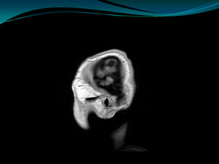
7. The Brain Before fMRI (1957)
Polyak, in Savoy, 2001, Acta Psychologica
8. The Brain After fMRI (Incomplete)
reaching and
pointing
motor
control
touch
eye
retinotopic visual maps
movements grasping
executive
control motion
near head
memory orientation selectivity
motion perception
moving bodies static faces objects
scenes social cognition bodies
9. MRI vs. fMRI
high resolution
MRI fMRI low resolution
(1 mm) (~3 mm but can be better)
one image
…
many images
(e.g., every 2 sec for 5 mins)
12. How to measure neural activity?
The physiology of neural activity involves many complex processes.
MR has the capability to measure parameters related to several
neural physiological functions, including:
changes in phosphorus metabolism and metabolic byproducts
blood flow
blood volume
blood oxygenation
Capillary beds within the cortex
13. BOLD
Blood Oxygenation Level Dependent (BOLD) signal
indirect measure of neural activity
Commonly used method
neural activity blood flow
oxyhemoglobin T2* MR signal
16. What is fMRI?
Functional MRI creates a series of images that
capture blood oxygen levels in parts of the brain
that are responsible for
movement, perception, and cognition.
fMRI is becoming the diagnostic method of
choice for learning how a normal, diseased or
injured brain is working, as well as for assessing
the potential risks of surgery or other invasive
treatments of the brain.
17. What is fMRI?
Functional magnetic resonance imaging is a relatively
new research tool that provides important insight
into the physiology of cognition, memory, emotion
and creativity.
Functional MRI is proving its worth in clinical
practice and is a safe and noninvasive imaging
modality that does not expose the patient to x-
rays, contrast media or invasive diagnostic
procedures.
fMRI, a technological variant of magnetic resonance
imaging, allows researchers and clinicians literally to
watch the brain in action.
20. Why is fMRI Used?
Examine the anatomy of the brain
Determine precisely which part of the brain is handling
critical functions such as thought, speech, movement and
sensation, which is called brain mapping
Help assess the effects of stroke, trauma or degenerative
disease (such as Alzheimer's) on brain functions
Monitor the growth and function of brain tumors
Guide the planning of surgery, radiation therapy, or other
surgical treatments for the brain
21. Potential Clinical Applications
fMRI, while used primarily as a research tool, does
have clinical applications that span a broad range of
disease processes and medical fields including:
Epilepsy
Schizophrenia
Diabetes
Overactive Bladder
Parkinson’s Disease
Depression
22. Potential Clinical Applications
Epilepsy
Epilepsy is a neurological disorder of the brain
that affects nearly 3 million people living in the
United States.
Seizures are the outcome of abnormal electrical
activity in the brain. There are many types of
seizures, and their signs and symptoms depend
on the part of the brain affected.
The benefits of fMRI in epilepsy surgery include
the reduced need for the services of a complex
medical and surgical team, reduced patient
exposure to invasive diagnostic testing, reduced
medical and surgical complications and reduced
medical costs.
23. Potential Clinical Applications
Schizophrenia
Schizophrenia, considered the most
chronic, disabling and expensive mental
illness, affects approximately 2.2 million people in the
United States.
Functional MRI studies are helping researchers
connect the clinical signs and symptoms associated
with schizophrenia, such as deficits in working
memory and language function, thought disorder and
impaired social cognition, with decreased brain
activity.
fMRI is also a source of physical clinical evidence that
can help the clinician make a diagnosis, develop and
monitor treatment and offer a prognosis.
Functional MRI studies eventually may help clinicians
evaluate the efficacy of medical interventions.
24. Potential Clinical Applications
Diabetes
Diabetes, a disease characterized by the inability
to maintain normal blood glucose levels, affects
more than 20 million children and adults in the
United States.
Recent research using fMRI shows that, under
certain circumstances, having type 1 diabetes can
affect cognition.
Furthermore, there is an association between
early onset (childhood) diabetes and the
development of mild brain atrophy, lower non-
verbal intellectual ability, and slower
psychomotor speed in adulthood.
25. Potential Clinical Applications
Overactive Bladder
Urinary urgency, or overactive bladder, is a
common condition that affects more than 17
million people in the United States.
Physicians currently treat this condition using
behavior modification to re-establish normal
brain-bladder communication and medication to
suppress detrusor muscle contractions.
fMRI studies have revealed the potential for drug
treatment strategies.
Functional MRI research findings eventually
could lead to therapeutic approaches directed at
specific areas of the brain rather than the bladder.
26. Potential Clinical Applications
Parkinson’s Disease
Parkinson disease (PD), a progressively degenerative
neurologic illness, affects more than an estimated 1
million people in the United States.
Functional MRI studies are revealing new information
about the effects of PD on the brain and behavior.
Functional MRI helps us better understand how PD
disrupts spontaneous movements by showing how
people learn and perform automated or quot;built-inquot;
motions such as accurate typing.
More importantly, fMRI studies show that specialized
training regimens help patients with PD learn how to
use other parts of their brain in efforts to re-establish
fluid movement.
27. Potential Clinical Applications
Depression
Depression, one of the most common psychiatric
illnesses in the world, affects nearly 19 million
people in the United States.
Functional MRI studies are helping researchers
decipher the relationship between depression
and brain activity and develop evidence-based
medical care strategies.
fMRI studies also could help clinicians predict
which patients will benefit from cognitive
behavioral therapy (CBT) that helps them stop
ruminating over negative information.