Biology Form 5 Chapter 6 Variation 6.2
•
73 gostaram•17,120 visualizações

Biology Form 5 Chapter 6 Variation

Recomendados
Mais conteúdo relacionado
Mais procurados
Mais procurados (20)
Semelhante a Biology Form 5 Chapter 6 Variation 6.2
Semelhante a Biology Form 5 Chapter 6 Variation 6.2 (20)
Mais de Nirmala Josephine
Mais de Nirmala Josephine (20)
Último
Último (20)
Biology Form 5 Chapter 6 Variation 6.2
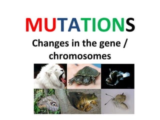
- 1. MUTATIONS Changes in the gene / chromosomes
- 2. Genetic Factor that cause variation GENETIC MUTATION SEXUAL REPRODUCTION CHROMOSOMAL MUTATION GENE MUTATION INDEPENDENT ASSORTMENT CROSSING OVER RANDOM FERTILISATION duplication inversion translocation deletion deletion insertion substitution
- 3. MUTATION • Mutation: the spontaneous change to the structure of genes or chromosomes • Resulting in irreversible & permanent change in the organism
- 4. a. Mutation can involve the ENTIRE chromosome or a SINGLE gene b. May HELP, HARM, or have LITTLE to no effect on future generations o Changes may be SILENT or may cause phenotypic changes
- 7. The causes of mutations 1. DNA fails to copy accurately:- When a cell divides, it makes a copy of its DNA — and sometimes the copy is not quite perfect. That small difference from the original DNA sequence is a mutation.
- 8. High energy ionising radiation - X-rays, gamma rays, ultraviolet rays. Chemical mutagens - benzene, asbestos, formaldehyde, pesticide, mustard gas, tar in tobacco smoke 2. Chemicals / Radiation These agents cause the DNA to break down. Mutagens = agents that cause mutation
- 10. GENE MUTATION
- 11. Eg of Diseases Caused by Gene Mutation Sickle cell Anemia Polydactylism Albinism
- 12. Colour blindness Mutant gene (recessive gene) on the X chromosome. •Not able to differentiate colours especially green and red •More common in males because male has only one X chromosome
- 13. Albinism The gene for skin colour is mutated (recessive gene) •Unable to produce melanin (black pigment) •White hair •Pink eyes and pinkish skin •Skin is sensitive to sunlight
- 14. Albinism is caused by an alteration of the gene that makes the pigment melanin. ALBINISM not limited to humans
- 15. Sickle-cell anemia Mutation in gene that produce haemoglobin •Sickle-cell shaped red blood cells •Not efficient in transporting oxygen •Yellowing of eyes
- 16. Haemophilia Mutation in chromosome X (for blood clotting) • Have problem in blood clotting • May bleed continuously or for a longer period • A woman carrier may pass the disease to a son
- 17. DNA 4 nucleotide bases: A, T, G, C. (adenine, thymine, guanine, and cytosine. ) The sequence of these bases encodes instructions to make amino acids.
- 18. Sets of three bases that specify an amino acid - codon The amino acid that corresponds to "GCA" - Alanine 20 different amino acids are synthesized in humans.
- 19. Gene Mutations 3 types: 1. Base Substitution 2. Base Deletion 3. Base Insertion Changes in the structure of DNA
- 20. 1. Base substitution one base is replaced by another. ATT/CGC/TTA ATG/CGC/TTA Only affects a single amino acid. Little effect unless active site of an enzyme is affected. Eg. sickle-cell anemia (caused by a single amino acid substitution)
- 21. Analogy 3 letter words because codons are 3 letters The cat ate the rat. SUBSTITUTION Thc cat ate the rat. May have little effect. You still have the idea like a typo on a test. The hat ate the rat. Changes the thought of the sentence. The effect depends on where the substitution happens
- 22. Sickle Cell Hemoglobin GUG CAC CUG ACU CCU GAG GAG AAG val his leu thr pro glu glu lys 1 2 3 4 5 6 7 8 GUG CAC CUG ACU CCU GUG GAG AAG val his leu thr pro val glu lys 1 2 3 4 5 6 7 8 Mutation (in DNA) Normal mRNA Normal protein Mutant mRNA Mutant protein Glutamate (glu), a negatively charged amino acid, is replaced by valine (val), which has no charge.
- 23. Sickle Cell Hemoglobin Significant change in structure caused by the single mutation
- 24. 2. Base Insertion Base insertion means a single base is added. This will affect all subsequent amino acids in that protein and the effects are therefore likely to be severe ATT/CCT/GTC ATG/TCC/TGT/C G
- 25. Normal DNA: CGA – TGC – ATC Base Insertion Mutated DNA: CGA – TAG – CAT – C Alanine – Threonine – stop Alanine – Isoleucine – Valine An adenine was inserted thereby pushing all the other bases over a frame.
- 26. Analogy Insertion The cat ate the rat. The cca tat eth era t. Inserting the c causes a FRAMESHIFT THE SENTENCE NO LONGER MAKES SENSE!! Insertions may have huge effects.
- 27. Gene Mutations Huntington’s Disease is caused by an insertion mutation. People with this disorder have involuntary movement and loss of motor control. They eventually have memory loss and dementia. The disease is terminal. Huntington Disease Located on chromosome 4 First Gene Disease Mapped
- 28. 3. Base Deletion Base deletion means a single base is omitted. This will affect all subsequent amino acids in that protein and the effects are therefore likely to be severe ATT/CCT/GTC ATT/CTG/TC
- 29. Mutated DNA: CGA – TCA- TC A guanine was deleted, thereby pushing all the bases down a frame. Alanine – Threonine – stop Alanine – Serine Base Deletion Normal DNA: CGA – TGC – ATC
- 30. Analogy DELETION The cat ate the rat. Thc ata tet her at FRAMESHIFT The sentence no longer makes sense!! Deletions can have huge effects.
- 32. CHROMOSOMAL MUTATION Caused by •An increase/ decrease in the number of the chromosomes. •Changes in the structure of the chromosome.
- 33. 4 types of chromosomal mutations : 1. deletion 2. inversion 3. duplication 4. translocation . CHROMOSOMAL MUTATION
- 34. 1. Deletion Deletions are mutations in which a section of DNA is lost, or deleted.
- 35. 2. Inversion Chromosome segment breaks off Segment flips around backwards Segment reattaches
- 36. 3. Duplication Occurs when a gene sequence is repeated.
- 37. 4. Translocation • Involves two chromosomes that aren’t homologous. • Part of one chromosome is transferred to another chromosomes
- 38. Chromosomal Mutation – 4 types
- 39. A Boy with Cri-du-Chat Syndrome – a Debilitating Disorder Caused by Chromosome 5 Deletion
- 40. Cri-du-Chat - Caused by the Loss of the Short Arm of One Copy of Chromosome 5
- 41. Deletions - loss of a chromosomal segment Eg. Prader-Willi Syndrome Cri du Chat Syndrome
- 42. Translocations Lead to a Number of Human Cancers Burkitt’s lymphoma - Caused by chromosome translocation
- 43. Video on chromosome abnormalities
- 44. 1. Alterations in chromosome number. Euploid - normal set (2n) Polyploidy-extra set of the entire genome(3n, 4n etc) (duplication of the entire set of chromosomes) Aneuploidy – the number of chromosomes is not a multiple of the normal haploid number. (a) Monosomy - one member of a chromosome pair is missing (2n-1) (b) Trisomy - one chromosome set consists of 3 copies of a chromosome (2n+1) Chromosomal abnormalities
- 45. Occasionally, there is an accident during Meiosis 1 or Meiosis 2 called nondisjunction: How Accidents During Meiosis Can Alter Chromosome Number – The members of a chromosome pair fail to separate during anaphase I or II – Gametes with an incorrect number of chromosomes are produced
- 46. Meiosis I Nondisjunction n + 1 n + 1 n - 1 n - 1 n + 1 n - 1 n n Number of chromosomes Gametes Nondisjunction Meiosis II (a) Nondisjunction in meiosis I (b) Nondisjunction in meiosis II NONDISJUNCTION IN MEIOSIS I / MEIOSIS II
- 47. The result of nondisjunction Figure 8.22 Egg cell n (normal) Zygote 2n + 1 n + 1 Sperm cell Upon Fertilisation
- 49. Down Syndrome: An Extra Chromosome 21 –Is also called trisomy 21 Figure 8.19
- 51. Down Syndrome Additional chromosome at autosome 21. •Physical retardation •Thick neck •Slanted eyes •Protuding tongue •Reduced resistance to diseases
- 52. Round face flattened nose bridge small, irregular teeth short stature heart defects A shorter life span mental retardation Underdeveloped sexual organs sterility
- 53. The incidence of Down Syndrome increases with the age of the mother Figure 8.20
- 54. KLINEFELTER SYNDROME (44 + XXY)
- 56. Turner Syndrome (44 + XO)
- 57. Turner Syndrome (44 + XO) Constriction of aorta (b) A woman with Turner syndrome (XO) Poor breast development Web of skin Under-developed ovaries •Infertile , Short, Mentally retarded •Has no ovaries, Small breasts •Folded skin on neck •No menstrual cycle
- 58. Genetic causes
- 59. Environmental Factors That cause Variation Climate Diet Physical accidents Temperature pH Humidity Light intensity Soil fertility Culture Lifestyle PHENOTYPE = GENOTYPE + EFFECTS OF ENVIRONMENT
- 66. The Importance of Variation in the Survival of A Spesies Provide necessary adaptation to help a species survive in an ever changing environment Can withstand severe weather Become more resistant to pests and diseases.
- 67. Interactions between environmental factors and genetic factors Dark & grey moths – Biston betularia in England Unpolluted environment, lichen covered tree trunks - Dark moths not camouflaged. More easily detected & eaten by predators. Dark moth population decreased. - Grey moth population
- 68. Industrial Revolution - pollution, soot on trees, lichen killed - Dark moths camouflaged. Dark moth population increased - Grey moth population decreased (more obvious to predator) 1960, Clean Air Act- air quality improved. - Dark moth population decreased because again more obvious to predator. - Grey moth population increased.
- 69. ~~ENDEND~~
- 70. Polyploidy – The number of chromosomes is increased by one or more sets. – More common in plants.
- 71. Searching for Chromosomal Defects - Amniocentesis and Chorionic Villus Sampling Many new techniques for learning about individual genes rather than whole chromosomes are available or under development.
- 72. Searching for Chromosome and Gene Defects – Pre-Implantation Genetic Diagnosis (PGD) Removing a cell for diagnosis from a human embryo. The diagnosis: trisomy 21 (Down syndrome).
- 73. loss of a chromosomal segment extra copy of a chromosomal segment order of a chromosome segment has been reversed chromosomal segment transferred from one chromosome to another
Notas do Editor
- High energy ionising radiation.- (Enter body cells and change structures of genes & chromosomes) Chemical mutagens - (Can damage DNA, causing genetic disorders)
- Ask students if they can figure out what is happening in this mutation. Answer is on the next slide.
- A genetic marker linked to Huntington disease was found on chromosome 4 in 1983, making Huntington disease, or HD, the first genetic disease mapped using DNA polymorphisms. HD is inherited as an autosomal dominant disease.
- Ask students if they can figure out what is happening in this mutation. Answer is on the next slide.
- When chromosome 7 loses an end piece, the result is Williams syndrome. These children, although unrelated, have the same appearance, health, and behavioral problems. Children with this syndrome look like pixies because they have turned-up noses, wide mouths, a small chin, and large ears. Although their academic skills are poor, they exhibit excellent verbal and musical abilities.
- This karyotype shows an individual (male) with Down syndrome having an extra chromosome 21.