SlideShare uma empresa Scribd logo
1 de 58
 
LECTURE  №8 THEME :  Demyelination diseases  of nervous system
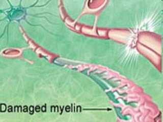
Myelinated axons Most axons are surrounded by a  myelin sheath . The myelin   sheaths of the central nervous system are composed   of the cell membrane of  oligodendroglia , which is   wrapped around the axon to form a multilaminar structure.
The myelin sheath enables the axon to conduct impulses   more rapidly; thus, loss of myelin lowers conduction velocity, producing clinical manifestations of neuronal dysfunction. Each layer of a myelin sheath is 7.5 microns thick and is composed of  two lipoid  and  two proteinaceous monomolecular layers .  The common feature of all diseases affecting myelin is a  pathological abnormality   or  total destruction of myelin sheaths , primarily in the central nervous system. In the genetic  myelinopathies , the  primary development of myelin is deficient .
B ut, in  many cases , myelin is lost later in life,  in the metabolic and autoimmune demyelinating diseases . They call this group of  demyelinating  diseases  myelinoclasthies .   Myelinopathies : adrenoleikodystrophies, phenylke-tonuria, mitochondrial leukoencephalopathies,  leukodys - trophies  Krabbe and disease of Kanavan, disease of Alexander. The most common  myelinoclasthies  are  multiple sclerosis,  acute disseminated encephalomyelitis concentric sclerosis (Balo sclerosis), Neuromyelitis optica (Devic disease) diffuse encephalitis of Schilder and subacute leukoencephalitis of Van Bogart .
Multiple sclerosis (MS)  is a  chronic   disease of the  CNS that begins most commonly in young adults and is characterized pathologically by multiple areas of CNS white matter inflammation, demyelination, and glial scarring (sclerosis) . It usually presents with   episodic neurological deficits , which, later on in   the course of the disease, tend not to reverse fully,   leaving increasingly severe residual deficits whose   summation causes  progressively severe disability .   The clinical   manifestations are very diverse because   widely   separated areas of the CNS are affected   and  the   temporal course of the disease is   variable .   “Disseminated encephalomyelitis”  is a synonym   for multiple sclerosis.
An important characteristic of MS is that the number of lesions, or plaques, that are discovered scattered throughout the white matte (with occasional extension into gray matter) at autopsy is invariably larger than the number of lesions detected clinically or by means of laboratory tests. These silent lesions may be responsible for aspects of the disease that are poorly understood, especially mental and cognitive disturbances.
Epidemiology.  MS is particularly common in northern Europe ,  the northern U.S.A., southern Canada, New Zealand, and southwest Australia.  Although environmental factors may be important in etiology, the observation of increased risk of MS in families of an MS patient cannot be lightly dismissed; both environmental and genetic factors are probably important.  There are the main of peculiriaties: 1. MS affects women more than men in a ratio of about 1.5 to 1. 2. There are also some clinical differences in different localities. 3. MS is predominantly a disease of young adults.
Pathological anatomy.  There are  disseminated foci of   demyelination in the  CNS   (brain and   spinal cord), sometimes with destruction of axons as   well. Local, reactive gliosis is found at the sites of older   foci. Thus, “sclerosis” develops at “multiple” locations,   giving the disease its English name.
MRI of the brain and spinal cord in patient with MS
MRI of the brain in multiple sclerosis.   a   Asymmetrically   scattered foci of abnormal signal, affecting only the white matter,   are seen in the periventricular regions and at the anterior and posterior   ends of the lateral ventricles. There is mild internal hydrocephalus.   b   There are typical signal abnormalities in the corpus callosum,   extending into the white   matter of the hemispheres.
[object Object],[object Object],[object Object],[object Object],[object Object],[object Object],[object Object]
Pathogenesis.  Two major theories about the pathogenesis of MS are being pursued. The first  rests on the concept of an infection (presumably by a viral agent) with a long incubation period.   The other theory  postulates altered immune regulation.   The major  histocompatibility complex (MHC)  on  chromosome 6  has been identified as one genetic determinant for MS. The MHS encodes the genes for the  histocompatibility antigens  (the human leucocyte antigen [HLA] system) involved in antigen presentation to T-cell. The class II haplotype DR15, DQ6, Dw2 is associated with increased risk of MS.
Therefore, different infectious agents or other environmental factors may cause or predispose to autoimmune attack upon central myelin. The result of this process is myelin destruction. At the beginning of the process auto-allergic processes prevail over the other ones. Then immunodeficiency is developed. Restoration of a balance between immunosuppressor and cytotoxic cells might induce clinical remission.
Active demyelination Inflammation
New data on the ontogeny of human immunity indicate that adult levels of humoral and cellular immunity develop gradually during childhood and may not be mature until the second half of the first decade. In childhood, when there is a natural imbalance of immune components, individuals may be particularly susceptible to a demyelinating type of host response.
[object Object],[object Object],[object Object],[object Object],[object Object],[object Object]
Clinical forms I. Cerebral forms: - cortical (epileptic attacks, psychiatric disorders) - visual - brain stem - cerebellar II. Spinal forms: - cervical - thoracic - lumbar-sacral - pseudotabes III. Cerebrospinal
The course of the disease: - remittent (exacerbation and remission without progression) - remitting-progressive - secondary progressive (by episodic worsening at first with 2-3 remission, followed by steady progression) - primary progressive ( by steady progression from the beginning ). The periods of the disease: - exacerbation - remission (complete, incomplete) - stable periods
Clinical manifestations and neurological findings. The neurological deficits present in each individual   patient depend on the number and location of   the demyelinating foci.  Exacerbations and remissions occur frequently.  The main characteristics of the symptoms of MS are : - multiplicity - the tendency to vary in nature - severity with the passage of time - complete remission of the first symptoms occurs frequently but, with subsequent attacks, remissions do not occur or are incomplete. - the clinical course extends for one or many decades in most cases, but a few may terminate in death within a few months of onset.
The following are among the   more characteristic disease   manifestations   and   physical   findings: - retrobulbar neuritis - disturbances of ocular motility -  Lhermitte sign - p yramidal tract signs - cerebellar signs - gait impairement - sensory deficits - bladder dysfunction - ictal phenomena - mental disturbances
Retrobulbar neuritis  is usually unilateral. Over the course of a few days, the patient develops an  impairment of color vision , followed by a  marked impairment of visual acuity   (finger counting is barely possible).  Orbital pain   is often present and the patient may see  flashes of light on movement of the globe .  These problems begin to improve in one or two weeks and usually resolve completely. The temporal side of the optic disc becomes pale three or four weeks after the onset of symptoms. Retrobulbar neuritis rarely affects both eyes, either at the same time or in rapid succession. Recurrences are rare.
If retrobulbar neuritis is an isolated event in a patient otherwise free of neurological disease, the probability that other clinical signs of multiple sclerosis will appear in the future is roughly 50%. This probability is greater if pathological changes are seen in the CSF or on an MRI scan.  Temporal papillary atrophy (after optic neuritis)
Disturbances of ocular motility.   Diplopia , particularly   due to  abducens palsy , is a common early symptom but   nearly always resolves spontaneously. Later, typical   findings are  nystagmus   (often dissociated) and  internuclear   ophthalmoplegia , often without any subjective   correlate.  Internuclear ophthalmoplegia  in a   young patient is relatively specific for multiple sclerosis. This condition is caused by a  lesion of the medial longitudinal   fasciculus (MLF).  When the patient attempts to   look away from the side of the lesion, the ipsilateral (adducting)   eye cannot fully adduct, and the contralateral   (abducting) eye exhibits end-gaze nystagmus.
The inability   of the ipsilateral eye to adduct is not due to a lesion   of the oculomotor n. nucleus, as is demonstrated by   a preserved ability to adduct   (converge) in the near reflex.   Internuclear ophthalmoplegia (INO) can also be bilateral if the MLF is damaged on both sides. Right internuclear   ophthalmoplegia in a   patient with   multiple   sclerosis.  In the initial   phase of leftward gaze (upper photograph),   only   the left eye is abducted.   The right eye follows,   after a   delay (lower   photograph).
Test for visual field defects (confrontation test) Central scotoma
Lhermitte sign (positive neck-flexion sign).  Active or passive forward flexion of the neck induces an “electric”   paresthesia running down the spine and/or into the   limbs. Retrobulbar neuritis, disturbances of ocular motility, sensory deficits, and Lhermitte sign are common early   findings in multiple sclerosis. Pyramidal tract signs  and exaggerated intrinsic muscle   reflexes may be present early in the course of the disease.   The abdominal cutaneous reflexes are absent .  Later   on, in almost all patients,  spastic paraparesis  or  quadriparesis   develops.
Motor disturbances   (central paresis,   spasticity,   abnormal fatigability)
Cerebellar signs  are practically always present in advanced   MS, incl u ding   impaired   coordination ,   ataxia , and,   frequently, a very characteristic  intention tremor Gait impairment  often becomes severe early in the   course of the disease. Typically, the   combination of   spastic   paraparesis  and  ataxia  results in a spastic−ataxic,   uneven,   uncoordinated, and stiff gait .
Sensory deficits  are found early in the course of the disease in about half of all patients.  Vibration sense in the lower limbs is nearly always impaired. Pain   is not uncommon; sometimes there is even a  dissociated sensory deficit.   Bladder dysfunction  is present in about three-quarters of all patients (generally in association with spasticity); disturbances of defecation are much rarer.  Bladder dysfunction is sometimes an early manifestation  of the disease.
Urge incontinence   is highly characteristic, i. e., a sudden, almost uncontrollable need to urinate, perhaps leading to “accidents” and bedwetting. Patients often do not mention bladder dysfunction until they are directly asked about it.
Ictal phenomena  of various types are not uncommon. About 1.5% of persons with MS suffer from  trigeminal   neuralgia ,  which may alternate from one side to the other.  Acute dizzy spells   can occur, as can  paroxysmal dystonia ,  dysarthria , or  ataxia.   The characteristic so - called  tonic brainstem seizures   consist of paroxysmal, often painful, tonic stiffness of the muscles on one side of the body. The lower limb is hyperextended, the upper limb flexed (Wernicke−Mann posture).
Mental disturbances  are not severe early in the course of the disease. Later on, however, ma - ny patients develop  psychoor - ganic changes   and  psycho - re - active and depressive   distur - bances.   Psychosis is very rare.
Adiadochokinesia Gait impairment
Intention tremor (fingeer-nose test and heel-to-knee test)
Nystagmus   Internuclear ophthalmoplegia
 
 
MS degree: I – patient has difficulty to walk only after physical training II – patient has difficulty to walk and weakness on 2-3 km III – patient has spastic-paretic gait, difficulty to walk and weakness on 200-300m IV – patient can not to walk without help V – patient can not to walk or has blindness
[object Object],[object Object],[object Object],[object Object],[object Object],[object Object]
MRI in patient with MS
Evoked potentials. Visual evoked potential (VEP) studies reliably detect optic nerve lesions, VEP reveals   prolongation of the P100 latency in one eye and/or an abnormally large discrepancy between the   latencies in the two eyes in roughly 40% of MS   patients   without known optic neuritis, and in almost   half of those with early optic neuritis.  Somatosensory   evoked potential (SEP)   studies of   the median or tibial nerve typically reveal prolonged   latencies in MS. Low amplitude of   evoked potentials, on the other hand, often indicates   a pathological process of another type, e. g.   tumor. SEP abnormalities are found in up to 60%   of MS patients with predominantly sensory manifestations.
Auditory evoked potential (AEP)   studies are less sensitive in MS than VEP or SEP.   The most common AEP change is prolongation   of latency. AEP studies are   helpful for the further   classification of vertigo, tinnitus, and hearing   loss.  Motor evoked potential (MEP)   studies reveal   prolonged central conduction times when   CNS lesions involve the pyramidal pathway. The   sensitivity of MEP in MS is approximately the   same as that of SEP. MEP studies can provide   supporting evidence for MS in patients with   latent paresis, gait disturbances, abnormal reflexes,   or movement disorders that are difficult   to classify.
Tests of bladder function.   The  residual urine volume   can be measured by ultrasound. It should not exceed 100 ml in patients with a normal bladder capacity of 400–450 ml; in general, it should normally be 15–20% of the cystomanometrically   determined bladder volume. Urodynamic electromyography  (EMG) provides more specific data concerning bladder dysfunction.
CSF examination.   CSF abnormalities are found in   more than 95% of MS patients. The cell count   rarely exceeds 20 cells/mm3. The total protein   concentration is elevated in ca. 40% of patients,   and intrathecal IgG synthesis (IgG index) in ca.   90%. Oligoclonal IgG is found in 95% of MS   pati - ents, and antibodies to mumps, measles and   herpes zoster in 80%. Oligoclonal bands in the serum  (a)   and CSF  (b)   of a   patient with multiple sclerosis ;  compare with the serum  ( c )  and   CSF  ( d )  of a normal control subject.
Differential Diagnosis There is no single clinical test, imaging study, or   laboratory finding that alone establishes the diagnosis   of MS. A meticulous   differential diagnostic evaluation is   needed in every case. (Cerebral) Vasculitis.   Systemic lupus   erythematosus, Sjögren syndrome, Behçet syndrome,   granulomatous angiitis, polyarteritis   nodosa, antiphospholipid syndrome, chronic inflammatory   demyelinating   polyradiculoneuro - pathy.
Inflammatory diseases.   Neurosarcoidosis, neuroborreliosis,   neurosyphilis, Whipple disease, postinfectious acute disseminated encephalomyelitis (ADEM), progressive multifocal   leukoencephalopathy (PML), subacute sclerosing   panencephalitis (SSPE), HIV infection,   HTLV-1 infection. Neurovascular disorders.   Arteriovenous fistula of spinal dura mater, cavernoma. Hereditary/metabolic disorders.   Adrenoleukodystro - phy, endocrine   diseases, mitochondrial encephalo - myelopathy,   vitamin B12 deficiency (funicular myelosis).
Tumors   of the brain or spinal cord  ( lymphoma,   glioma,meningioma).     Skull base anomalies .  Arnold–Chiari malformation, platybasia. Myelopathy .  Cervical myelopathy (spinal stenosis). Somatoform disturbances   in the context of   mental illness. Prognosis Favorable prognostic indicators in MS include   onset b e- fore age 40, monosymptomatic onset,   absence of cere - bellar involvement at onset,   rapid resolution of the initial symptom(s), a relapsing-remitting course, short duration of relapses,   and long-term preservation of the ability   to walk.
A relatively favorable course is also predicted if, after the first 5 years of illness, the MRI reveals no more than a few, small lesions without rapid radiological progression and the clinical manifestations of cerebellar disease and central paresis are no more than mild. A  benign course , defined as a low frequency of recurrences and only mild disability in the first 15 years of illness, is seen in 20–30% of patients. The disease takes a  malignant course ,   with major disability within 5 years, in fewer than 5% of patients. Half of all MS patients have a second relapse within 2 years of disease onset.
[object Object],[object Object],[object Object],[object Object],[object Object],[object Object],[object Object],[object Object]
[object Object],[object Object],[object Object],[object Object],[object Object],[object Object],[object Object]
[object Object],[object Object],[object Object],[object Object],Neuroprotections
Symptomatic therapy/rehabilitation.   Medications,   physical, occupational, and speech therapy,   social, psychological, and dietary counseling,   and mechanical aids (e. g., walking aids,   wheelchair) are provided as needed. The   possible benefits of oligodendrocyte precursor   cell transplantation for remyelination, and of   growth factors and immunoglobulins for the   promotion of endogenous remyelination, are   currently under investigation in both experimental   and clinical studies.
Gait problems   are the most common disability of MS; the problems respond to treatment if spasticity is the major cause. In these cases, careful use of baclofen is helpful, but if too much is administered, spasticity gives way to excessive flaccidity and makes the gait disorder worse.  Intention tremor   occasionally responds to  propranolol 320 mg  per day in divided dosage. Cryothalamotomy  has been used, but the complication of pseudobulbar palsy makes this too hazardous.
Diffuse encephalitis of Schilder.   The diagnostic criteria established by Poser in 1985 require 6 elements:  1. One or 2 roughly symmetrical large plaques are manifest, and if more than 1 is present, 1 should be in each brain hemisphere, chiefly in the centrum semiovale.  2. No other lesions are demonstrable by clinical, paraclinical, or imaging data. 3. No abnormalities of the peripheral nervous system are demonstrable. 4. Results of adrenal function studies are normal. 5. Serum very long chain fatty acids are normal. 6. Pathological analysis by autopsy or biopsy demonstrates histologic changes consistent with subacute or chronic myelinoclastic diffuse sclerosis,   changes which in essence cannot be distinguished from those of multiple sclerosis.
No features of the general examination are characteristic features of the presentation of Schilder disease.  Aphasia ,  memory disturbances ,  mental dullness ,  irritability ,  changes in personality ,  confusion, disorientation , and  behavioral disturbances  are not infrequently encountered. Patients may appear to be  psychotic .  Deafness  is common. Other brainstem or cerebellar deficits that are encountered include  vertigo ;  paresis of eye movements , including  internuclear ophthalmoplegia  and  nystagmus ;  facial palsy ;  dysarthria ; or  dysphagia .
Peripheral cranial nerve abnormalities  that are sometimes encountered include optic neuritis and optic atrophy. Cortical blindness is common, and various field cuts may be found, particularly hemianopsia.  Hemiparesis  or  cortical sensory deficits  may be found.  Seizures  may occur but are not common. Some patients manifest psychosis. Extrapyramidal manifestations  are rare but have been described. Findings of variable or uncertain localization include generalized spasticity and incontinence of bowel and bladder function.  Malnutrition  and  cachexia  are commonly reported in the middle or late chronic stages of illness, especially in instances where patients fall to a low level of neurologic function such as a chronic vegetative state.
Thank you for your attention !

Mais conteúdo relacionado

Mais procurados

Charcot-Marie-Tooth disease
Charcot-Marie-Tooth diseaseCharcot-Marie-Tooth disease
Charcot-Marie-Tooth diseaseVihari Rajaguru
 
Approach to dystonia
Approach to dystoniaApproach to dystonia
Approach to dystoniaNeurologyKota
 
Multiple sclerosis
Multiple sclerosisMultiple sclerosis
Multiple sclerosisazad4v
 
Neuromyelitis Optica
Neuromyelitis OpticaNeuromyelitis Optica
Neuromyelitis Opticasm171181
 
multiple sclerosis
multiple sclerosismultiple sclerosis
multiple sclerosissunil bobade
 
Multiple sclerosis: Introduction, Risk Factors, Diagnosis and Treatment
Multiple sclerosis: Introduction, Risk Factors, Diagnosis and TreatmentMultiple sclerosis: Introduction, Risk Factors, Diagnosis and Treatment
Multiple sclerosis: Introduction, Risk Factors, Diagnosis and TreatmentEnriqueAlvarez93
 
Movement disorders
Movement disordersMovement disorders
Movement disordersHelao Silas
 
Motor Neuron Disease and PT Mx
Motor Neuron Disease and PT MxMotor Neuron Disease and PT Mx
Motor Neuron Disease and PT Mxrichamistry3
 
Myotonic muscle disorders
Myotonic muscle disordersMyotonic muscle disorders
Myotonic muscle disordersNeurologyKota
 
Temporal lobe epilepsy
Temporal lobe epilepsyTemporal lobe epilepsy
Temporal lobe epilepsySiva Pesala
 
Mononeritis multiplex
Mononeritis multiplex Mononeritis multiplex
Mononeritis multiplex NeurologyKota
 
Neurological manifestations of HIV
Neurological manifestations of HIVNeurological manifestations of HIV
Neurological manifestations of HIVGarima Aggarwal
 

Mais procurados (20)

Charcot-Marie-Tooth disease
Charcot-Marie-Tooth diseaseCharcot-Marie-Tooth disease
Charcot-Marie-Tooth disease
 
Approach to dystonia
Approach to dystoniaApproach to dystonia
Approach to dystonia
 
Multiple sclerosis
Multiple sclerosisMultiple sclerosis
Multiple sclerosis
 
Peripheral neuropathy
Peripheral neuropathyPeripheral neuropathy
Peripheral neuropathy
 
MYOTONIC DYSTROPHY
MYOTONIC DYSTROPHYMYOTONIC DYSTROPHY
MYOTONIC DYSTROPHY
 
Polyneuropathy
PolyneuropathyPolyneuropathy
Polyneuropathy
 
Neuromyelitis Optica
Neuromyelitis OpticaNeuromyelitis Optica
Neuromyelitis Optica
 
multiple sclerosis
multiple sclerosismultiple sclerosis
multiple sclerosis
 
Multiple sclerosis: Introduction, Risk Factors, Diagnosis and Treatment
Multiple sclerosis: Introduction, Risk Factors, Diagnosis and TreatmentMultiple sclerosis: Introduction, Risk Factors, Diagnosis and Treatment
Multiple sclerosis: Introduction, Risk Factors, Diagnosis and Treatment
 
approach to polyneuropathy
approach to polyneuropathyapproach to polyneuropathy
approach to polyneuropathy
 
Movement disorders
Movement disordersMovement disorders
Movement disorders
 
Myoclonus
MyoclonusMyoclonus
Myoclonus
 
Multiple sclerosis
Multiple sclerosisMultiple sclerosis
Multiple sclerosis
 
Diabetic POLYNEUROPATHY
Diabetic POLYNEUROPATHYDiabetic POLYNEUROPATHY
Diabetic POLYNEUROPATHY
 
Motor Neuron Disease and PT Mx
Motor Neuron Disease and PT MxMotor Neuron Disease and PT Mx
Motor Neuron Disease and PT Mx
 
Myotonic muscle disorders
Myotonic muscle disordersMyotonic muscle disorders
Myotonic muscle disorders
 
PLEDS
PLEDSPLEDS
PLEDS
 
Temporal lobe epilepsy
Temporal lobe epilepsyTemporal lobe epilepsy
Temporal lobe epilepsy
 
Mononeritis multiplex
Mononeritis multiplex Mononeritis multiplex
Mononeritis multiplex
 
Neurological manifestations of HIV
Neurological manifestations of HIVNeurological manifestations of HIV
Neurological manifestations of HIV
 

Destaque

Multiple Sclerosis.ppt
Multiple Sclerosis.pptMultiple Sclerosis.ppt
Multiple Sclerosis.pptShama
 
Multiple sclerosis adjusted to publish
Multiple sclerosis adjusted to publishMultiple sclerosis adjusted to publish
Multiple sclerosis adjusted to publishAhmad Amirdash
 
Multiple sclerosis
Multiple sclerosisMultiple sclerosis
Multiple sclerosisIrfan Ziad
 
Multiple Sclerosis ppt
Multiple Sclerosis pptMultiple Sclerosis ppt
Multiple Sclerosis pptStacey Turner
 

Destaque (6)

Multiple Sclerosis.ppt
Multiple Sclerosis.pptMultiple Sclerosis.ppt
Multiple Sclerosis.ppt
 
Multiple sclerosis 2015
Multiple sclerosis 2015 Multiple sclerosis 2015
Multiple sclerosis 2015
 
Multiple sclerosis adjusted to publish
Multiple sclerosis adjusted to publishMultiple sclerosis adjusted to publish
Multiple sclerosis adjusted to publish
 
Multiple sclerosis
Multiple sclerosisMultiple sclerosis
Multiple sclerosis
 
Multiple Sclerosis ppt
Multiple Sclerosis pptMultiple Sclerosis ppt
Multiple Sclerosis ppt
 
Multiple Sclerosis
Multiple SclerosisMultiple Sclerosis
Multiple Sclerosis
 

Semelhante a Multiple sclerosis

Multiple sclerosis
Multiple sclerosisMultiple sclerosis
Multiple sclerosisAHLAM MAJALI
 
UMNLand AHC.pptx
UMNLand AHC.pptxUMNLand AHC.pptx
UMNLand AHC.pptxNonaElarabi
 
multiplesclerosis-171219042211.pdf
multiplesclerosis-171219042211.pdfmultiplesclerosis-171219042211.pdf
multiplesclerosis-171219042211.pdffirdauseah2
 
Neurology 8th multiple sclerosis
Neurology 8th multiple sclerosisNeurology 8th multiple sclerosis
Neurology 8th multiple sclerosisRamiAboali
 
1.multiple sclerosis
1.multiple sclerosis1.multiple sclerosis
1.multiple sclerosisanzilmaharjan
 
04 lecture Neuro-ophthalmology
04 lecture Neuro-ophthalmology04 lecture Neuro-ophthalmology
04 lecture Neuro-ophthalmologyAnisur Rahman
 
Multiple Sclerosis MS(lecture)abcd.pptx
Multiple Sclerosis  MS(lecture)abcd.pptxMultiple Sclerosis  MS(lecture)abcd.pptx
Multiple Sclerosis MS(lecture)abcd.pptxwosade3943
 
MULTIPLE SCLEROSIS (1).pdf
MULTIPLE SCLEROSIS  (1).pdfMULTIPLE SCLEROSIS  (1).pdf
MULTIPLE SCLEROSIS (1).pdfAfiqAsraaf
 
Demyelination by Dr Sabu Augustine
Demyelination by Dr Sabu AugustineDemyelination by Dr Sabu Augustine
Demyelination by Dr Sabu Augustinedrsabuaugustine
 
Multiple Sclerosis.pptx
Multiple Sclerosis.pptxMultiple Sclerosis.pptx
Multiple Sclerosis.pptxKetakiPatani1
 
Multiple sclerosis
Multiple sclerosisMultiple sclerosis
Multiple sclerosisJincy Ashish
 
Multiple sclerosis
Multiple  sclerosisMultiple  sclerosis
Multiple sclerosisDR.
 
Multiple Sclerosis And The Central Nervous System
Multiple Sclerosis And The Central Nervous SystemMultiple Sclerosis And The Central Nervous System
Multiple Sclerosis And The Central Nervous SystemAmanda Brady
 
Demyelinating diseases & Multiple Sclerosis
Demyelinating diseases  & Multiple SclerosisDemyelinating diseases  & Multiple Sclerosis
Demyelinating diseases & Multiple SclerosisDR MUKESH SAH
 
1-141102051233-conversion-gate01.pdf
1-141102051233-conversion-gate01.pdf1-141102051233-conversion-gate01.pdf
1-141102051233-conversion-gate01.pdffirdauseah2
 
CNS Lecture Data.pdf
CNS Lecture Data.pdfCNS Lecture Data.pdf
CNS Lecture Data.pdfSohailaGoda
 
Multiple Sclerosis (MS)
Multiple Sclerosis (MS)Multiple Sclerosis (MS)
Multiple Sclerosis (MS)rawazabdullah1
 
Multiple sclerosis
Multiple sclerosisMultiple sclerosis
Multiple sclerosissgpharshida
 

Semelhante a Multiple sclerosis (20)

Multiple sclerosis
Multiple sclerosisMultiple sclerosis
Multiple sclerosis
 
UMNLand AHC.pptx
UMNLand AHC.pptxUMNLand AHC.pptx
UMNLand AHC.pptx
 
multiplesclerosis-171219042211.pdf
multiplesclerosis-171219042211.pdfmultiplesclerosis-171219042211.pdf
multiplesclerosis-171219042211.pdf
 
Neurology 8th multiple sclerosis
Neurology 8th multiple sclerosisNeurology 8th multiple sclerosis
Neurology 8th multiple sclerosis
 
Multiple sclerosis
Multiple sclerosisMultiple sclerosis
Multiple sclerosis
 
1.multiple sclerosis
1.multiple sclerosis1.multiple sclerosis
1.multiple sclerosis
 
04 lecture Neuro-ophthalmology
04 lecture Neuro-ophthalmology04 lecture Neuro-ophthalmology
04 lecture Neuro-ophthalmology
 
Multiple Sclerosis MS(lecture)abcd.pptx
Multiple Sclerosis  MS(lecture)abcd.pptxMultiple Sclerosis  MS(lecture)abcd.pptx
Multiple Sclerosis MS(lecture)abcd.pptx
 
Motor neuron diseases
Motor neuron diseases Motor neuron diseases
Motor neuron diseases
 
MULTIPLE SCLEROSIS (1).pdf
MULTIPLE SCLEROSIS  (1).pdfMULTIPLE SCLEROSIS  (1).pdf
MULTIPLE SCLEROSIS (1).pdf
 
Demyelination by Dr Sabu Augustine
Demyelination by Dr Sabu AugustineDemyelination by Dr Sabu Augustine
Demyelination by Dr Sabu Augustine
 
Multiple Sclerosis.pptx
Multiple Sclerosis.pptxMultiple Sclerosis.pptx
Multiple Sclerosis.pptx
 
Multiple sclerosis
Multiple sclerosisMultiple sclerosis
Multiple sclerosis
 
Multiple sclerosis
Multiple  sclerosisMultiple  sclerosis
Multiple sclerosis
 
Multiple Sclerosis And The Central Nervous System
Multiple Sclerosis And The Central Nervous SystemMultiple Sclerosis And The Central Nervous System
Multiple Sclerosis And The Central Nervous System
 
Demyelinating diseases & Multiple Sclerosis
Demyelinating diseases  & Multiple SclerosisDemyelinating diseases  & Multiple Sclerosis
Demyelinating diseases & Multiple Sclerosis
 
1-141102051233-conversion-gate01.pdf
1-141102051233-conversion-gate01.pdf1-141102051233-conversion-gate01.pdf
1-141102051233-conversion-gate01.pdf
 
CNS Lecture Data.pdf
CNS Lecture Data.pdfCNS Lecture Data.pdf
CNS Lecture Data.pdf
 
Multiple Sclerosis (MS)
Multiple Sclerosis (MS)Multiple Sclerosis (MS)
Multiple Sclerosis (MS)
 
Multiple sclerosis
Multiple sclerosisMultiple sclerosis
Multiple sclerosis
 

Mais de Kapil Dhital

Personality disorder and mental retardation.
Personality disorder and mental retardation.Personality disorder and mental retardation.
Personality disorder and mental retardation.Kapil Dhital
 
Chronic tonsillitis
Chronic tonsillitisChronic tonsillitis
Chronic tonsillitisKapil Dhital
 
Bipolar affective disorder
Bipolar affective disorderBipolar affective disorder
Bipolar affective disorderKapil Dhital
 
Diseases of the paranasal sinuses.
Diseases of the paranasal sinuses.Diseases of the paranasal sinuses.
Diseases of the paranasal sinuses.Kapil Dhital
 
L7post traumatic stress disorder
L7post traumatic stress disorderL7post traumatic stress disorder
L7post traumatic stress disorderKapil Dhital
 
Peptic ulcer disease
Peptic ulcer diseasePeptic ulcer disease
Peptic ulcer diseaseKapil Dhital
 
Otitis externa and media
Otitis externa and mediaOtitis externa and media
Otitis externa and mediaKapil Dhital
 
lecture psychiatry
lecture psychiatrylecture psychiatry
lecture psychiatryKapil Dhital
 
Urinary+system+anatomy+and+physiology
Urinary+system+anatomy+and+physiologyUrinary+system+anatomy+and+physiology
Urinary+system+anatomy+and+physiologyKapil Dhital
 
Anatomy and physiology of ENT organs
Anatomy and physiology of ENT organsAnatomy and physiology of ENT organs
Anatomy and physiology of ENT organsKapil Dhital
 
Anatomy and physiology of ENT organs
Anatomy and physiology of ENT organsAnatomy and physiology of ENT organs
Anatomy and physiology of ENT organsKapil Dhital
 

Mais de Kapil Dhital (17)

Personality disorder and mental retardation.
Personality disorder and mental retardation.Personality disorder and mental retardation.
Personality disorder and mental retardation.
 
Chronic tonsillitis
Chronic tonsillitisChronic tonsillitis
Chronic tonsillitis
 
Bipolar affective disorder
Bipolar affective disorderBipolar affective disorder
Bipolar affective disorder
 
schizophrenia
schizophreniaschizophrenia
schizophrenia
 
Diseases of the paranasal sinuses.
Diseases of the paranasal sinuses.Diseases of the paranasal sinuses.
Diseases of the paranasal sinuses.
 
L7post traumatic stress disorder
L7post traumatic stress disorderL7post traumatic stress disorder
L7post traumatic stress disorder
 
Peptic ulcer disease
Peptic ulcer diseasePeptic ulcer disease
Peptic ulcer disease
 
Otitis part 3
Otitis part 3Otitis part 3
Otitis part 3
 
Otitis part 2
Otitis part 2Otitis part 2
Otitis part 2
 
Otitis externa and media
Otitis externa and mediaOtitis externa and media
Otitis externa and media
 
lecture psychiatry
lecture psychiatrylecture psychiatry
lecture psychiatry
 
Urinary+system+anatomy+and+physiology
Urinary+system+anatomy+and+physiologyUrinary+system+anatomy+and+physiology
Urinary+system+anatomy+and+physiology
 
rheumatic fever
rheumatic feverrheumatic fever
rheumatic fever
 
Anatomy and physiology of ENT organs
Anatomy and physiology of ENT organsAnatomy and physiology of ENT organs
Anatomy and physiology of ENT organs
 
Acoustic analyzer
Acoustic analyzerAcoustic analyzer
Acoustic analyzer
 
Anatomy and physiology of ENT organs
Anatomy and physiology of ENT organsAnatomy and physiology of ENT organs
Anatomy and physiology of ENT organs
 
Mal presentation
Mal presentationMal presentation
Mal presentation
 

Multiple sclerosis

  • 1.  
  • 2. LECTURE №8 THEME : Demyelination diseases of nervous system
  • 3. Myelinated axons Most axons are surrounded by a myelin sheath . The myelin sheaths of the central nervous system are composed of the cell membrane of oligodendroglia , which is wrapped around the axon to form a multilaminar structure.
  • 4. The myelin sheath enables the axon to conduct impulses more rapidly; thus, loss of myelin lowers conduction velocity, producing clinical manifestations of neuronal dysfunction. Each layer of a myelin sheath is 7.5 microns thick and is composed of two lipoid and two proteinaceous monomolecular layers . The common feature of all diseases affecting myelin is a pathological abnormality or total destruction of myelin sheaths , primarily in the central nervous system. In the genetic myelinopathies , the primary development of myelin is deficient .
  • 5. B ut, in many cases , myelin is lost later in life, in the metabolic and autoimmune demyelinating diseases . They call this group of demyelinating diseases myelinoclasthies . Myelinopathies : adrenoleikodystrophies, phenylke-tonuria, mitochondrial leukoencephalopathies, leukodys - trophies Krabbe and disease of Kanavan, disease of Alexander. The most common myelinoclasthies are multiple sclerosis, acute disseminated encephalomyelitis concentric sclerosis (Balo sclerosis), Neuromyelitis optica (Devic disease) diffuse encephalitis of Schilder and subacute leukoencephalitis of Van Bogart .
  • 6. Multiple sclerosis (MS) is a chronic disease of the CNS that begins most commonly in young adults and is characterized pathologically by multiple areas of CNS white matter inflammation, demyelination, and glial scarring (sclerosis) . It usually presents with episodic neurological deficits , which, later on in the course of the disease, tend not to reverse fully, leaving increasingly severe residual deficits whose summation causes progressively severe disability . The clinical manifestations are very diverse because widely separated areas of the CNS are affected and the temporal course of the disease is variable . “Disseminated encephalomyelitis” is a synonym for multiple sclerosis.
  • 7. An important characteristic of MS is that the number of lesions, or plaques, that are discovered scattered throughout the white matte (with occasional extension into gray matter) at autopsy is invariably larger than the number of lesions detected clinically or by means of laboratory tests. These silent lesions may be responsible for aspects of the disease that are poorly understood, especially mental and cognitive disturbances.
  • 8. Epidemiology. MS is particularly common in northern Europe , the northern U.S.A., southern Canada, New Zealand, and southwest Australia. Although environmental factors may be important in etiology, the observation of increased risk of MS in families of an MS patient cannot be lightly dismissed; both environmental and genetic factors are probably important. There are the main of peculiriaties: 1. MS affects women more than men in a ratio of about 1.5 to 1. 2. There are also some clinical differences in different localities. 3. MS is predominantly a disease of young adults.
  • 9. Pathological anatomy. There are disseminated foci of demyelination in the CNS (brain and spinal cord), sometimes with destruction of axons as well. Local, reactive gliosis is found at the sites of older foci. Thus, “sclerosis” develops at “multiple” locations, giving the disease its English name.
  • 10. MRI of the brain and spinal cord in patient with MS
  • 11. MRI of the brain in multiple sclerosis. a Asymmetrically scattered foci of abnormal signal, affecting only the white matter, are seen in the periventricular regions and at the anterior and posterior ends of the lateral ventricles. There is mild internal hydrocephalus. b There are typical signal abnormalities in the corpus callosum, extending into the white matter of the hemispheres.
  • 12.
  • 13. Pathogenesis. Two major theories about the pathogenesis of MS are being pursued. The first rests on the concept of an infection (presumably by a viral agent) with a long incubation period. The other theory postulates altered immune regulation. The major histocompatibility complex (MHC) on chromosome 6 has been identified as one genetic determinant for MS. The MHS encodes the genes for the histocompatibility antigens (the human leucocyte antigen [HLA] system) involved in antigen presentation to T-cell. The class II haplotype DR15, DQ6, Dw2 is associated with increased risk of MS.
  • 14. Therefore, different infectious agents or other environmental factors may cause or predispose to autoimmune attack upon central myelin. The result of this process is myelin destruction. At the beginning of the process auto-allergic processes prevail over the other ones. Then immunodeficiency is developed. Restoration of a balance between immunosuppressor and cytotoxic cells might induce clinical remission.
  • 16. New data on the ontogeny of human immunity indicate that adult levels of humoral and cellular immunity develop gradually during childhood and may not be mature until the second half of the first decade. In childhood, when there is a natural imbalance of immune components, individuals may be particularly susceptible to a demyelinating type of host response.
  • 17.
  • 18. Clinical forms I. Cerebral forms: - cortical (epileptic attacks, psychiatric disorders) - visual - brain stem - cerebellar II. Spinal forms: - cervical - thoracic - lumbar-sacral - pseudotabes III. Cerebrospinal
  • 19. The course of the disease: - remittent (exacerbation and remission without progression) - remitting-progressive - secondary progressive (by episodic worsening at first with 2-3 remission, followed by steady progression) - primary progressive ( by steady progression from the beginning ). The periods of the disease: - exacerbation - remission (complete, incomplete) - stable periods
  • 20. Clinical manifestations and neurological findings. The neurological deficits present in each individual patient depend on the number and location of the demyelinating foci. Exacerbations and remissions occur frequently. The main characteristics of the symptoms of MS are : - multiplicity - the tendency to vary in nature - severity with the passage of time - complete remission of the first symptoms occurs frequently but, with subsequent attacks, remissions do not occur or are incomplete. - the clinical course extends for one or many decades in most cases, but a few may terminate in death within a few months of onset.
  • 21. The following are among the more characteristic disease manifestations and physical findings: - retrobulbar neuritis - disturbances of ocular motility - Lhermitte sign - p yramidal tract signs - cerebellar signs - gait impairement - sensory deficits - bladder dysfunction - ictal phenomena - mental disturbances
  • 22. Retrobulbar neuritis is usually unilateral. Over the course of a few days, the patient develops an impairment of color vision , followed by a marked impairment of visual acuity (finger counting is barely possible). Orbital pain is often present and the patient may see flashes of light on movement of the globe . These problems begin to improve in one or two weeks and usually resolve completely. The temporal side of the optic disc becomes pale three or four weeks after the onset of symptoms. Retrobulbar neuritis rarely affects both eyes, either at the same time or in rapid succession. Recurrences are rare.
  • 23. If retrobulbar neuritis is an isolated event in a patient otherwise free of neurological disease, the probability that other clinical signs of multiple sclerosis will appear in the future is roughly 50%. This probability is greater if pathological changes are seen in the CSF or on an MRI scan. Temporal papillary atrophy (after optic neuritis)
  • 24. Disturbances of ocular motility. Diplopia , particularly due to abducens palsy , is a common early symptom but nearly always resolves spontaneously. Later, typical findings are nystagmus (often dissociated) and internuclear ophthalmoplegia , often without any subjective correlate. Internuclear ophthalmoplegia in a young patient is relatively specific for multiple sclerosis. This condition is caused by a lesion of the medial longitudinal fasciculus (MLF). When the patient attempts to look away from the side of the lesion, the ipsilateral (adducting) eye cannot fully adduct, and the contralateral (abducting) eye exhibits end-gaze nystagmus.
  • 25. The inability of the ipsilateral eye to adduct is not due to a lesion of the oculomotor n. nucleus, as is demonstrated by a preserved ability to adduct (converge) in the near reflex. Internuclear ophthalmoplegia (INO) can also be bilateral if the MLF is damaged on both sides. Right internuclear ophthalmoplegia in a patient with multiple sclerosis. In the initial phase of leftward gaze (upper photograph), only the left eye is abducted. The right eye follows, after a delay (lower photograph).
  • 26. Test for visual field defects (confrontation test) Central scotoma
  • 27. Lhermitte sign (positive neck-flexion sign). Active or passive forward flexion of the neck induces an “electric” paresthesia running down the spine and/or into the limbs. Retrobulbar neuritis, disturbances of ocular motility, sensory deficits, and Lhermitte sign are common early findings in multiple sclerosis. Pyramidal tract signs and exaggerated intrinsic muscle reflexes may be present early in the course of the disease. The abdominal cutaneous reflexes are absent . Later on, in almost all patients, spastic paraparesis or quadriparesis develops.
  • 28. Motor disturbances (central paresis, spasticity, abnormal fatigability)
  • 29. Cerebellar signs are practically always present in advanced MS, incl u ding impaired coordination , ataxia , and, frequently, a very characteristic intention tremor Gait impairment often becomes severe early in the course of the disease. Typically, the combination of spastic paraparesis and ataxia results in a spastic−ataxic, uneven, uncoordinated, and stiff gait .
  • 30. Sensory deficits are found early in the course of the disease in about half of all patients. Vibration sense in the lower limbs is nearly always impaired. Pain is not uncommon; sometimes there is even a dissociated sensory deficit. Bladder dysfunction is present in about three-quarters of all patients (generally in association with spasticity); disturbances of defecation are much rarer. Bladder dysfunction is sometimes an early manifestation of the disease.
  • 31. Urge incontinence is highly characteristic, i. e., a sudden, almost uncontrollable need to urinate, perhaps leading to “accidents” and bedwetting. Patients often do not mention bladder dysfunction until they are directly asked about it.
  • 32. Ictal phenomena of various types are not uncommon. About 1.5% of persons with MS suffer from trigeminal neuralgia , which may alternate from one side to the other. Acute dizzy spells can occur, as can paroxysmal dystonia , dysarthria , or ataxia. The characteristic so - called tonic brainstem seizures consist of paroxysmal, often painful, tonic stiffness of the muscles on one side of the body. The lower limb is hyperextended, the upper limb flexed (Wernicke−Mann posture).
  • 33. Mental disturbances are not severe early in the course of the disease. Later on, however, ma - ny patients develop psychoor - ganic changes and psycho - re - active and depressive distur - bances. Psychosis is very rare.
  • 35. Intention tremor (fingeer-nose test and heel-to-knee test)
  • 36. Nystagmus Internuclear ophthalmoplegia
  • 37.  
  • 38.  
  • 39. MS degree: I – patient has difficulty to walk only after physical training II – patient has difficulty to walk and weakness on 2-3 km III – patient has spastic-paretic gait, difficulty to walk and weakness on 200-300m IV – patient can not to walk without help V – patient can not to walk or has blindness
  • 40.
  • 41. MRI in patient with MS
  • 42. Evoked potentials. Visual evoked potential (VEP) studies reliably detect optic nerve lesions, VEP reveals prolongation of the P100 latency in one eye and/or an abnormally large discrepancy between the latencies in the two eyes in roughly 40% of MS patients without known optic neuritis, and in almost half of those with early optic neuritis. Somatosensory evoked potential (SEP) studies of the median or tibial nerve typically reveal prolonged latencies in MS. Low amplitude of evoked potentials, on the other hand, often indicates a pathological process of another type, e. g. tumor. SEP abnormalities are found in up to 60% of MS patients with predominantly sensory manifestations.
  • 43. Auditory evoked potential (AEP) studies are less sensitive in MS than VEP or SEP. The most common AEP change is prolongation of latency. AEP studies are helpful for the further classification of vertigo, tinnitus, and hearing loss. Motor evoked potential (MEP) studies reveal prolonged central conduction times when CNS lesions involve the pyramidal pathway. The sensitivity of MEP in MS is approximately the same as that of SEP. MEP studies can provide supporting evidence for MS in patients with latent paresis, gait disturbances, abnormal reflexes, or movement disorders that are difficult to classify.
  • 44. Tests of bladder function. The residual urine volume can be measured by ultrasound. It should not exceed 100 ml in patients with a normal bladder capacity of 400–450 ml; in general, it should normally be 15–20% of the cystomanometrically determined bladder volume. Urodynamic electromyography (EMG) provides more specific data concerning bladder dysfunction.
  • 45. CSF examination. CSF abnormalities are found in more than 95% of MS patients. The cell count rarely exceeds 20 cells/mm3. The total protein concentration is elevated in ca. 40% of patients, and intrathecal IgG synthesis (IgG index) in ca. 90%. Oligoclonal IgG is found in 95% of MS pati - ents, and antibodies to mumps, measles and herpes zoster in 80%. Oligoclonal bands in the serum (a) and CSF (b) of a patient with multiple sclerosis ; compare with the serum ( c ) and CSF ( d ) of a normal control subject.
  • 46. Differential Diagnosis There is no single clinical test, imaging study, or laboratory finding that alone establishes the diagnosis of MS. A meticulous differential diagnostic evaluation is needed in every case. (Cerebral) Vasculitis. Systemic lupus erythematosus, Sjögren syndrome, Behçet syndrome, granulomatous angiitis, polyarteritis nodosa, antiphospholipid syndrome, chronic inflammatory demyelinating polyradiculoneuro - pathy.
  • 47. Inflammatory diseases. Neurosarcoidosis, neuroborreliosis, neurosyphilis, Whipple disease, postinfectious acute disseminated encephalomyelitis (ADEM), progressive multifocal leukoencephalopathy (PML), subacute sclerosing panencephalitis (SSPE), HIV infection, HTLV-1 infection. Neurovascular disorders. Arteriovenous fistula of spinal dura mater, cavernoma. Hereditary/metabolic disorders. Adrenoleukodystro - phy, endocrine diseases, mitochondrial encephalo - myelopathy, vitamin B12 deficiency (funicular myelosis).
  • 48. Tumors of the brain or spinal cord ( lymphoma, glioma,meningioma). Skull base anomalies . Arnold–Chiari malformation, platybasia. Myelopathy . Cervical myelopathy (spinal stenosis). Somatoform disturbances in the context of mental illness. Prognosis Favorable prognostic indicators in MS include onset b e- fore age 40, monosymptomatic onset, absence of cere - bellar involvement at onset, rapid resolution of the initial symptom(s), a relapsing-remitting course, short duration of relapses, and long-term preservation of the ability to walk.
  • 49. A relatively favorable course is also predicted if, after the first 5 years of illness, the MRI reveals no more than a few, small lesions without rapid radiological progression and the clinical manifestations of cerebellar disease and central paresis are no more than mild. A benign course , defined as a low frequency of recurrences and only mild disability in the first 15 years of illness, is seen in 20–30% of patients. The disease takes a malignant course , with major disability within 5 years, in fewer than 5% of patients. Half of all MS patients have a second relapse within 2 years of disease onset.
  • 50.
  • 51.
  • 52.
  • 53. Symptomatic therapy/rehabilitation. Medications, physical, occupational, and speech therapy, social, psychological, and dietary counseling, and mechanical aids (e. g., walking aids, wheelchair) are provided as needed. The possible benefits of oligodendrocyte precursor cell transplantation for remyelination, and of growth factors and immunoglobulins for the promotion of endogenous remyelination, are currently under investigation in both experimental and clinical studies.
  • 54. Gait problems are the most common disability of MS; the problems respond to treatment if spasticity is the major cause. In these cases, careful use of baclofen is helpful, but if too much is administered, spasticity gives way to excessive flaccidity and makes the gait disorder worse. Intention tremor occasionally responds to propranolol 320 mg per day in divided dosage. Cryothalamotomy has been used, but the complication of pseudobulbar palsy makes this too hazardous.
  • 55. Diffuse encephalitis of Schilder. The diagnostic criteria established by Poser in 1985 require 6 elements: 1. One or 2 roughly symmetrical large plaques are manifest, and if more than 1 is present, 1 should be in each brain hemisphere, chiefly in the centrum semiovale. 2. No other lesions are demonstrable by clinical, paraclinical, or imaging data. 3. No abnormalities of the peripheral nervous system are demonstrable. 4. Results of adrenal function studies are normal. 5. Serum very long chain fatty acids are normal. 6. Pathological analysis by autopsy or biopsy demonstrates histologic changes consistent with subacute or chronic myelinoclastic diffuse sclerosis, changes which in essence cannot be distinguished from those of multiple sclerosis.
  • 56. No features of the general examination are characteristic features of the presentation of Schilder disease. Aphasia , memory disturbances , mental dullness , irritability , changes in personality , confusion, disorientation , and behavioral disturbances are not infrequently encountered. Patients may appear to be psychotic . Deafness is common. Other brainstem or cerebellar deficits that are encountered include vertigo ; paresis of eye movements , including internuclear ophthalmoplegia and nystagmus ; facial palsy ; dysarthria ; or dysphagia .
  • 57. Peripheral cranial nerve abnormalities that are sometimes encountered include optic neuritis and optic atrophy. Cortical blindness is common, and various field cuts may be found, particularly hemianopsia. Hemiparesis or cortical sensory deficits may be found. Seizures may occur but are not common. Some patients manifest psychosis. Extrapyramidal manifestations are rare but have been described. Findings of variable or uncertain localization include generalized spasticity and incontinence of bowel and bladder function. Malnutrition and cachexia are commonly reported in the middle or late chronic stages of illness, especially in instances where patients fall to a low level of neurologic function such as a chronic vegetative state.
  • 58. Thank you for your attention !