SlideShare uma empresa Scribd logo
1 de 24
,

RATIFICATION PAGE
Complete report of basic biology practicum with title ’’ How To Use
Microscope’’ that arranged by :
Name

: Jeny ayu hardiah ningrum

Registrasion Number

: 1114040162

Group

: III (Three)

Class

: ICP A

After checked by assistant and assistant coordinator so this report was
accepted
Makassar, November 4th 2011
Assistant Coordinator

Assistant

( Djumarirmanto,S.Pd)

(Fitri)
ID.091404168

CHAPTER I
,

INTRODUCTION
A.Background
Microscope ,this word not new for us,I have use when I in junior high school
and senior high school,we had now practicum of microscope is one of practicum in
biology department ,biology is one of branch of science wich explained about
matter and the energy which has connection with human life and the process of
their life,human life concist of seme cell very small,for the first time we must to
know how to use the microscope because we just can see cell with use the
microscope which can help us to see cell and structure of cell
Do this practicum is the challenge for us because we must know components
or part of microscope before we do this practicum about microscope, we can not
get shadow, but my teacher help me,and my observation have succesed, and this
practicum very accostumed but I always try although the tenses in report very
hurd, not only present tense but past tens also. light very important for this
practicum if don’t have light,I can not see cell and I can not find something
This practicum is very interest and it is very challenge, so we have to try and
try use it better than past time when we still studied ,I always studied in junior high
school and senior high scool because I hope in future I can become a biology
teacher.the reasons to do this practicum very many because it is one of the subject
which we must know it.seeing happening problem, I interest to do praktikum this
and to know more about microscope, therefore of that i hopes more know a lot of
about microscope and part. not only sectioned but logistic and way utilizes also
need at knows that deep utilize tool not false again.

B.Purpose
,

The purpose of practicum is the students of University can use the biology
microscope and safe to see a simple preparate.
C.Benefit
The benefit this practicum is the student of university more to know,part of
microscope and function.the students can use microscope well more than senior
high school,we can know the purpose of this practicum ,the manner to keep the
microscope and the right procedure wor k to use microscope.
,

CHAPTER II
PREVIEW OF LITERATURE
Light microscopes is an indispensable technique for cell

and molecular

biologist to study cellular structures and biological processes in both living and fixed
cells.this chapter provides and overview of light microscopy .describes the important
parts of the microscope and goes on to explain how to set up a standard research
microscope for bright field and phase contrast microscopy.there is also a short section
on concfocalmicroscopy,more comprehensive descriptions of the different forms of
light microscopy are found elsewhere.Microsopes are instruments that produce an
enlarged image of a specimen. The eyepieces and the objectives are the main
components

of the magnification system of the microscope,the product of the

magnification of the objective lens and the ocular give the total magnification of the
microscope.the visibility of the magnifed specimen depends on contrast and
resolution. Contrast is the difference in light bintensity between an object and its
background. some biological samples

contain coloured compouns. The key

components of the compound microscope consist of : the eyepieces ,body tubes,nose
pieces and objectives are part of the magnification system of the microscope.To use a
microscope properly,and to get the most out of it ,it is important to understand the
purpose and function of each of the microscope’s components (Harris,2006)
The simple example of a microscope is a double convex lens of the type that
is used as a magnifiying glass .in the late 1500s to dutch spectacle makers developed
the compound microscope .their device had two convex lenses placed at either and of
a tube and was capable of magnifiying an object to 10 times.its actual size
today,developments in microscopy provide scientist with a wide selection of
instruments with wich to view the smallest organism and even the components of
individual cells.These microscopes range in complexity from the relatively simple
models you will use in the laboratory today to highty sophisticated scanning and
transmission electron microscopes (Helms R.Dorris ,2006)
,

A compound microscope contains two lens system .one is located in the
ocular and the other in the objective ,each of the lenses magnifies the object
independently .the ocular eyepiece magnification is disagnated in a similar manner,in
order to determine the total magnification of acombination a two lenses multiply
themanification of your microscope for both low and high power (windell,1975)
There are two objectives mounted on a movable stage instead of a movable
body tube,on each on side of the arm near the base you will see a large knob and
represents both the coarse and fine adjustment,turning the adjustment one quarter turn
in either direction represents fine adjustment ,turning it a greater amount represents
coarse adjustment (windell,1957).
There are two objecteves the nosepiece called the resolving nosepiece.a spring
catch enganges the nose piece to hold each objective in position,the shorter objective
is the low power objective and is marked 10x.the high power objective is longer and
is ,arked 43x or 45x.The microscope is parfocal which means that anobject is in focus
when the noce piece is rotated from one position to another .when switching from
low to high power watch from the side to see whether or not the objective will touch
the slide .if it does touch,lower the stage slightly before placing the objective into
position (windell,1957).
A microscope is an instrument used to see objects that are too small for the naked
eye. The science of investigating small objects using such an instrument is called microscopy.
Microscopic means invisible to the eye unless aided by a microscope. There are many types
of microscopes, the most common and first to be invented is the optical microscope which
uses light to image the sample. Other major types of microscopes are the electron microscope
(both the transmission electron microscope and the scanning electron microscope) and the
various types of scanning probe microscope(anonymous a,2011)

The first microscope to be developed was the optical microscope, although the
original inventor is not easy to identify. An early microscope was made in 1590 in
Middelburg, Netherlands. Two eyeglass makers are variously given credit: Hans
,

Lippershey (who developed an early telescope) and Zacharias Janssen. Giovanni
Faber coined the name microscope for Galileo Galilei's compound microscope in
1625 (Galileo had called it the "occhiolino" or "little eye"). The first detailed account
of the interior construction of living tissue based on the use of a microscope did not
appear until 1644, in Giambattista Odierna's L'occhio della mosca, or The Fly's Eye.
It was not until the 1660s and 1670s that the microscope was used extensively for
research in Italy, Holland and England. Marcelo Malpighi in Italy began the analysis
of biological structures beginning with the lungs. Robert Hooke's Micrographia had a
huge impact, largely because of its impressive illustrations. The greatest contribution
came from Antoni van Leeuwenhoek who discovered red blood cells and
spermatozoa and helped popularise microscopy as a technique. On 9 October 1676,
Leeuwenhoek reported the discovery of micro-organisms. In 1893 August Köhler
developed a key technique for sample illumination, Köhler illumination, which is
central to modern light microscopy. This method of sample illumination gives rise to
extremely even lighting and overcomes many limitations of older techniques of
sample illumination. Further developments in sample illumination came from Fritz
Zernike in 1953 and George Nomarski 1955 for their development of phase contrast
and differential interference contrast illumination which allow imaging of transparent
samples. Specialized techniques may exceed this magnification but the resolution is
diffraction limited. The use of shorter wavelengths of light, such as the ultraviolet, is
one way to improve the spatial resolution of the optical microscope, as are devices
such as the near-field scanning optical microscope.Sarfus, a recent optical technique
increases the sensitivity of standard optical microscope to a point it becomes possible
to directly visualize nanometric films (down to 0.3 nanometre) and isolated nanoobjects (down to 2 nm-diameter). The technique is based on the use of non-reflecting
substrates for cross-polarized reflected light microscopy (anonymous b,2011).

CHAPTER III
PRACTICUM METHOD
,

A.Time and Place
Day / Date :Monday/October 25th 2011
Time

:10.00 A.M until 12.30 P.M

Place

:Biology laboratory 3th flour at FMIPA UNM

B.Tool and Material
1. Tool
a. Biology microscope
b. Tool box have contents:
1)

Object glass

2)

Cover glass

3)

Petri dish

4)

Tweezers

5)

Hand pipette

c. The equipments are served by the student of university:
1)

New razor blade

2)

New flannel cloth

3)

Cotton cloth

4)

Drawing Book

5)

Tooth picks

2. Material
,

a. The materials are served by laboratory:
1)

Flute water

2)

Filter paper

3)

Cotton or barn

b. The materials are served by the student of university:
1)

Hibiscus rosa-sinensis

2)

Hibiscus tillaceus

3)

Cucurbita moschata

4)

Allium cepa

C.Work produce
1. Prepared the microscope
a.

Put the microscope on the work table in front of you

b. Cleaned up the body of microscope by using flannel eloth. Didn’t shine the
lens of microscope with cloth.
c. Opened toolbox than took petri dish which have object glass and cover glass.
Cleaned up the object glass with cotton cloth or filter paper.
d. Only there were microscope, toolbox and is contents, guide book and note
book, the materials of practicum on the table, besides it put on the other place
which had been served.
2. Straightened up the light for enter to the tube
a. Paid attention to your practicum room condition, from where the bright light
came (from front, left or right ). Directed the mirror of microscope to the light
source. Opened diaphragm or revolved the slab to the medium hole position.
,

The microscope which have condenser straightened the position near the stage
and used thin mirror. The microscope without condenser used concave mirror.
b. Settled the revolver position until the short objective lens looked out to the
stage until click sound.
c. Brought the tube to down until the objective tip with the stage 5-10 mm or the
tube maximize down.
d. With the left eye closed the right eye looked the ocular will appeared white
round area. If the light didn’t flat, moved mirror position until the light flat. If
more dazzled, constricted the diaphragm or the hole of slab. If white round
area still hazy, it was need the light, opened diaphragm put on the biggest lab.
e. The microscope ready to used for observed the preparate.
3. The method to set the lens distance with preparate
a. Revolved the macro meter to the thumb, dropped the tube, objective distance
with stage become small.
b. Put the object glass which have preserved preparate on the stage so that the
material which observed been in the middle in the hole stage. Hold the
preparate with stage clips so that it didn’t move.
c. Paid attention the objective distance with the object glass not more 10 mm. If
the distance loose, the hand revolved macro meter to down the tube while
looked from beside objective tip approach the object glass until 5-10 mm.
d. Looked from ocular while the hand revolved macro meter to up the tube
slowly. Observed the while round area until the shadow appear. If that had
done and the shadow had not appeared, it is had past. Repeat again from 3.3.
,

if had there was a shadow but still hazy so saw it while turned the micro meter
up and down until the shadow clearly visible.
e. Looked into the ocular (what increases is used?) and the objective (what
increase is used?) counted the shadow increase which we see.
f. Put out side the preparat when we had observed.
4. Made simple preparate
a. Took the object glass which was cleaned, held everywhere.
b. Sprinkled water on the middle.
c. Pulped the cotton fibers with tweezers and put it on the middle drops of water.
d. One of the hand held cover glass between the thumb and index finger on the
contrary side.
e. The side of cover glass touched on the object glass near the drops of water
with 45 declivity than released it so it cover the drops of water. Reserved the
surplus of water which ooze out on the glass side by using filter paper.
f. Put on the preparate make on the stage and inspected it like the step 3.2,
3.3,3.4, and 3.5.
5. Changed the magnification
a. The 4.6 surveillance had success, 3.4 and 3.5, increased the shadow which
measles. Didn’t touch preparate position.
b. Revolved until the longest of objective lens (strong) vertical on the stage and
click sound (looked the increase).
,

c. Looked while turned the micro meter until maesles the shadow whish more
big. Inspected the shadow.
d. Failed to found the biggest shadow, upward the tube with turned the macro
meter contrary direction with the thumb. Revolved again revolved to put the
small objective lens on the position from the beginning. Without change
preparate position, did the step 3.3, 3.4, 3.5, and continued to 5.1, 5.2, 5.3
until success.
e. If would observe another object, so upward the tube. Put out preparate which
was observed and cleaned object glass and cover glass.
f. Made a new preparate conform with step 4.1 -4.6.
g. The end of activity which used the microscope, take note of this metter.
1) Could not store preparate on the atage
2) Cleaned the wet preparate with filter paper.
3) (object glass + cover glass). Stored it in petri dish and put into the tool
box.
4) Cleaned the microscope body with flannel cloth.
5) Put the microscope into the microscope box.
6) Cleaned the all epuipments were used with cotton cloth and put into its
box.
7) Put each our equipments into box for used at the next practicum.
,

CHAPTER IV
OBSERVATION RESULT
A. Observation result
We had done this practicum and got bthe result of it,The microscope,its
components and its functions ,the observation result are the result of the
practicum.The picture of microscope and its components:
,

The information of the picture
1. Ocular lens
2. Macro meter
3. Micro meter
4. Microscope arm
5. Mechanic activator
6. Inclination point
7. Supervisor condenser
8. Mast
9. Base
10. Mirror
11. Diapraghm
12. Condenser
13. Object table
14. Clippers
15. Objective lens
16. Revolver
17. Tube

:
,

The observation result :
,

OBSERVATION PAGE
Allium cepa

Magnification 10 x 10

Notes:
Cell wall
Cytoplasm
Neucleus
Cell membran
From internet
,

OBSERVATION PAGE
Hibiscus rosa-sinensis
Magnification 10 x 10

Notes:
Trichome star
Cell wall
Cell membrane
Cytoplasm
Neucleus
stomata

From internet
,
OBSERVATION PAGE
Cucurbitha moschata
,
Magnification 10 x 10

Notes :
Neucleus
Cytoplasm
Cell wall

From internet

OBSERVATION PAGE
Hibiscus tilachius
Magnification 10x10
Notes:
Trichome star
Cell wall
Neucleus
,

B.Discussion

Microscope is one of important tool for activity in biology laboratory,for see
small things structure.it has some components .like accept a shadow,move the tube
up and down,and many function other.like a 1st practicum i have many information
from assistant about microscope and I have try use biology microscope.I have see cell
of Hibiscus rosa-sinensis ,Hibiscus tillaceus ,Cucurbita moschata and Allium cepa
and when I see in internet not difference for my research.
The component and function of microscope are:The function of ocular lens is
to accept a shadow from objective and magnify the shadow.mechanic activator is as
the regulator of the place of the object glass on the stage .supervisor condenser is up
and down the condenser. base is the place microscope standing.mirror is the captor
and the reflection of the light.diaphragm is control the light that will enter to the
condenser.condenser is provides a bright from the mirror which will eneter to the hole
of stage and many other part.The microscope have objective amd ocular lense which
have enlargement of objective lens and ocular lens

CHAPTER V
,

CONCLUSION AND SUGGESTION
A.Conclusion
Student at university can use the biology microscope and safe to see a simple
preparate,and the practicum have succes
B. Suggestion
1. Suggestion for laboratory
I hope for next practicum student can use microscope for each self .and the
laboratory can become good place or suitable for practicum.
2. Suggestion for Assistant
I hope assistant can give information and directive about practicum ,may be
can give time to make the result observation
3. Suggestion for the all friends
I hope all friend can hear and can see if assistant and coordinator assistant
give information,so we can do practicum

BIBLIOGRAPHY
,

Anonymous a.2011.Microscope,www.wikipedia.com,accesed at October 30 th 2011
Anonymous b.2011.Microscope,www.wikipedia.com,accesed at October 30 th 2011
Doris,R.doris.2006.Biology in the laboratory.city:publisher
Robin,Harris.2006.cell biology protocol.Usa:john willey and soon,ltd
Tim pengajar,2011.penuntun praktikum biologi dasar.Makassar UNM
Windell,john.1975.Intestigations for practicing biology

THE ANSWER OF QUESTION
1. The name of mechanic part of the microscop are
,

a. Ocular lens
b. Objective lens
c. Condenser
d. Mirror
2. The name of mechanic parts of the microscop are
a. Tube
b. Revolver
c. Stage
d. Clippers
e. Diaphragm
f. Arm
g. Mast
h. Micro meter
i. Macro meter
j. Condenser setting
k. Mechanic activator
3. The function of mechanic are
a) Tube function as the place of ocular lens
b) Revolver as place of objective lens
c) Stage as the place of object glass
,

d) Clippers function to clip the object glass
e) Diaphragm as the regulator of the light which enter to condenser
f) Arm function to held when we want to move microscope
g) Mast function is the place of microscope standing
h) Micro meter as the tool for move up and down the tube smoothly
i) Macro meter as the tool for move up and down the tube roughly
j) Condenser setting if we turn can up and down the condenser
k) Mechanic activator function manage the place of object glass on the table
4.

When the shadow in the white round area will be shifted to the front left, we
must shift the object glass to the opposite direction, because the quality of ocular
lens will receive with the objective lens shadow

5. Because it can make the lens of microscope will be broken. And if the lens
broken, we can not use it again.
,

Mais conteúdo relacionado

Mais procurados

Microscopic techniques
Microscopic techniquesMicroscopic techniques
Microscopic techniquesRammeharSingh4
 
Microscope and its types
Microscope and its typesMicroscope and its types
Microscope and its typesKetan Patil
 
Microscopes and microscopy
Microscopes and microscopyMicroscopes and microscopy
Microscopes and microscopyDakota Boswell
 
microscopes- a brief introduction
 microscopes- a brief introduction microscopes- a brief introduction
microscopes- a brief introductionMalathi Murugesan
 
Phase contrast microscopy Likhith K
Phase contrast microscopy Likhith KPhase contrast microscopy Likhith K
Phase contrast microscopy Likhith KLIKHITHK1
 
Types and functions of Microscope
Types and functions of MicroscopeTypes and functions of Microscope
Types and functions of MicroscopeAbu Reza
 
Optical Coherence Tomography: Technology and applications for neuroimaging
Optical Coherence Tomography: Technology and applications for neuroimagingOptical Coherence Tomography: Technology and applications for neuroimaging
Optical Coherence Tomography: Technology and applications for neuroimagingManish Kumar
 
Compendium.imaging.physics
Compendium.imaging.physicsCompendium.imaging.physics
Compendium.imaging.physicspooja joshi
 
Optical/ Metallurgical Microscopy
Optical/ Metallurgical MicroscopyOptical/ Metallurgical Microscopy
Optical/ Metallurgical MicroscopyGulfam Hussain
 

Mais procurados (20)

Microscopy
MicroscopyMicroscopy
Microscopy
 
Microscopy
MicroscopyMicroscopy
Microscopy
 
Microscopic techniques
Microscopic techniquesMicroscopic techniques
Microscopic techniques
 
Microscope and its types
Microscope and its typesMicroscope and its types
Microscope and its types
 
Microscopes and microscopy
Microscopes and microscopyMicroscopes and microscopy
Microscopes and microscopy
 
microscopes- a brief introduction
 microscopes- a brief introduction microscopes- a brief introduction
microscopes- a brief introduction
 
Types of microscopy
Types of microscopyTypes of microscopy
Types of microscopy
 
Electron microscope
Electron microscopeElectron microscope
Electron microscope
 
Phase contrast microscopy Likhith K
Phase contrast microscopy Likhith KPhase contrast microscopy Likhith K
Phase contrast microscopy Likhith K
 
Light microscope
Light microscopeLight microscope
Light microscope
 
Nano tech
Nano techNano tech
Nano tech
 
Light microscope
Light microscopeLight microscope
Light microscope
 
Medical imaging
Medical imagingMedical imaging
Medical imaging
 
Types and functions of Microscope
Types and functions of MicroscopeTypes and functions of Microscope
Types and functions of Microscope
 
Optical Coherence Tomography: Technology and applications for neuroimaging
Optical Coherence Tomography: Technology and applications for neuroimagingOptical Coherence Tomography: Technology and applications for neuroimaging
Optical Coherence Tomography: Technology and applications for neuroimaging
 
Magnetic resonance
Magnetic resonanceMagnetic resonance
Magnetic resonance
 
Compendium.imaging.physics
Compendium.imaging.physicsCompendium.imaging.physics
Compendium.imaging.physics
 
Microscope ppt
Microscope pptMicroscope ppt
Microscope ppt
 
Optical/ Metallurgical Microscopy
Optical/ Metallurgical MicroscopyOptical/ Metallurgical Microscopy
Optical/ Metallurgical Microscopy
 
Compound microscope
Compound microscopeCompound microscope
Compound microscope
 

Semelhante a How to use microscope (basic biology) unm

Nt1310 Unit 3 Lab Report Essay
Nt1310 Unit 3 Lab Report EssayNt1310 Unit 3 Lab Report Essay
Nt1310 Unit 3 Lab Report EssayAshley Jean
 
Microscope & Microscopy.pptxvjvgbngn bjjfhbxjb
Microscope & Microscopy.pptxvjvgbngn bjjfhbxjbMicroscope & Microscopy.pptxvjvgbngn bjjfhbxjb
Microscope & Microscopy.pptxvjvgbngn bjjfhbxjbAmirAmeerAli
 
PP3_History_of_microscopy_1453876435.ppt
PP3_History_of_microscopy_1453876435.pptPP3_History_of_microscopy_1453876435.ppt
PP3_History_of_microscopy_1453876435.pptMuhammad Rawat
 
اجهزة مختبرية -م2.pptx
اجهزة مختبرية -م2.pptxاجهزة مختبرية -م2.pptx
اجهزة مختبرية -م2.pptxsaraso888
 
Bright field microscopy, Principle and applications
Bright field microscopy, Principle and applicationsBright field microscopy, Principle and applications
Bright field microscopy, Principle and applicationsKAUSHAL SAHU
 
Biology book layout, Layout Design, Book Layout, book inner page design
Biology book layout, Layout Design, Book Layout, book inner page designBiology book layout, Layout Design, Book Layout, book inner page design
Biology book layout, Layout Design, Book Layout, book inner page designKS Designers
 
Microscopy_and_its_Application_in_Microbiology_and.pdf
Microscopy_and_its_Application_in_Microbiology_and.pdfMicroscopy_and_its_Application_in_Microbiology_and.pdf
Microscopy_and_its_Application_in_Microbiology_and.pdfImeneFl
 
Types of Light Microscopes used in Histological Studies.pptx
Types of Light Microscopes used in Histological Studies.pptxTypes of Light Microscopes used in Histological Studies.pptx
Types of Light Microscopes used in Histological Studies.pptxssuserab552f
 
maitri218principlesandapplicationoflightphaseconstrastfluorescencemicroscope-...
maitri218principlesandapplicationoflightphaseconstrastfluorescencemicroscope-...maitri218principlesandapplicationoflightphaseconstrastfluorescencemicroscope-...
maitri218principlesandapplicationoflightphaseconstrastfluorescencemicroscope-...AbhaySharma686520
 
Principles and application of light, phase constrast and fluorescence microscope
Principles and application of light, phase constrast and fluorescence microscopePrinciples and application of light, phase constrast and fluorescence microscope
Principles and application of light, phase constrast and fluorescence microscopeMaitriThakor
 
1. Grade-7-Q2-Module-1-Parts-and-Functions-of-the-Compound-Microscope-and-its...
1. Grade-7-Q2-Module-1-Parts-and-Functions-of-the-Compound-Microscope-and-its...1. Grade-7-Q2-Module-1-Parts-and-Functions-of-the-Compound-Microscope-and-its...
1. Grade-7-Q2-Module-1-Parts-and-Functions-of-the-Compound-Microscope-and-its...rosepalmajalandoni
 

Semelhante a How to use microscope (basic biology) unm (20)

Nt1310 Unit 3 Lab Report Essay
Nt1310 Unit 3 Lab Report EssayNt1310 Unit 3 Lab Report Essay
Nt1310 Unit 3 Lab Report Essay
 
Microscope & Microscopy.pptxvjvgbngn bjjfhbxjb
Microscope & Microscopy.pptxvjvgbngn bjjfhbxjbMicroscope & Microscopy.pptxvjvgbngn bjjfhbxjb
Microscope & Microscopy.pptxvjvgbngn bjjfhbxjb
 
PP3_History_of_microscopy_1453876435.ppt
PP3_History_of_microscopy_1453876435.pptPP3_History_of_microscopy_1453876435.ppt
PP3_History_of_microscopy_1453876435.ppt
 
اجهزة مختبرية -م2.pptx
اجهزة مختبرية -م2.pptxاجهزة مختبرية -م2.pptx
اجهزة مختبرية -م2.pptx
 
Microscopy - Study
Microscopy - StudyMicroscopy - Study
Microscopy - Study
 
Microscopy - Study
Microscopy - StudyMicroscopy - Study
Microscopy - Study
 
Microscope.pptx
Microscope.pptxMicroscope.pptx
Microscope.pptx
 
Chapter 1 Microscopy
Chapter 1 MicroscopyChapter 1 Microscopy
Chapter 1 Microscopy
 
The microscope
The microscopeThe microscope
The microscope
 
Bright field microscopy, Principle and applications
Bright field microscopy, Principle and applicationsBright field microscopy, Principle and applications
Bright field microscopy, Principle and applications
 
Biology book layout, Layout Design, Book Layout, book inner page design
Biology book layout, Layout Design, Book Layout, book inner page designBiology book layout, Layout Design, Book Layout, book inner page design
Biology book layout, Layout Design, Book Layout, book inner page design
 
Microscope.pptx
Microscope.pptxMicroscope.pptx
Microscope.pptx
 
Microscope.pptx
Microscope.pptxMicroscope.pptx
Microscope.pptx
 
Microscopy_and_its_Application_in_Microbiology_and.pdf
Microscopy_and_its_Application_in_Microbiology_and.pdfMicroscopy_and_its_Application_in_Microbiology_and.pdf
Microscopy_and_its_Application_in_Microbiology_and.pdf
 
Microscope.ppt
Microscope.pptMicroscope.ppt
Microscope.ppt
 
Types of Light Microscopes used in Histological Studies.pptx
Types of Light Microscopes used in Histological Studies.pptxTypes of Light Microscopes used in Histological Studies.pptx
Types of Light Microscopes used in Histological Studies.pptx
 
maitri218principlesandapplicationoflightphaseconstrastfluorescencemicroscope-...
maitri218principlesandapplicationoflightphaseconstrastfluorescencemicroscope-...maitri218principlesandapplicationoflightphaseconstrastfluorescencemicroscope-...
maitri218principlesandapplicationoflightphaseconstrastfluorescencemicroscope-...
 
Principles and application of light, phase constrast and fluorescence microscope
Principles and application of light, phase constrast and fluorescence microscopePrinciples and application of light, phase constrast and fluorescence microscope
Principles and application of light, phase constrast and fluorescence microscope
 
Microscopy
MicroscopyMicroscopy
Microscopy
 
1. Grade-7-Q2-Module-1-Parts-and-Functions-of-the-Compound-Microscope-and-its...
1. Grade-7-Q2-Module-1-Parts-and-Functions-of-the-Compound-Microscope-and-its...1. Grade-7-Q2-Module-1-Parts-and-Functions-of-the-Compound-Microscope-and-its...
1. Grade-7-Q2-Module-1-Parts-and-Functions-of-the-Compound-Microscope-and-its...
 

Mais de Jeny Hardiah

Rpp4 peredaran darah
Rpp4  peredaran darahRpp4  peredaran darah
Rpp4 peredaran darahJeny Hardiah
 
Lp4 peredaran darah
Lp4  peredaran darahLp4  peredaran darah
Lp4 peredaran darahJeny Hardiah
 
Rpp3 peredaran darah
Rpp3 peredaran darahRpp3 peredaran darah
Rpp3 peredaran darahJeny Hardiah
 
Lp3 peredaran darah
Lp3 peredaran darahLp3 peredaran darah
Lp3 peredaran darahJeny Hardiah
 
Rpp2 peredaran darah
Rpp2 peredaran darahRpp2 peredaran darah
Rpp2 peredaran darahJeny Hardiah
 
Lp2 peredaran darah
Lp2  peredaran darahLp2  peredaran darah
Lp2 peredaran darahJeny Hardiah
 
Rpp1 peredaran darah
Rpp1 peredaran darahRpp1 peredaran darah
Rpp1 peredaran darahJeny Hardiah
 
Soal ulangan harian peredaran darah
Soal ulangan harian peredaran darahSoal ulangan harian peredaran darah
Soal ulangan harian peredaran darahJeny Hardiah
 
Lks 6 sistem peredaran darah
Lks 6 sistem peredaran darahLks 6 sistem peredaran darah
Lks 6 sistem peredaran darahJeny Hardiah
 
Lks 5 sistem peredaran darah
Lks 5 sistem peredaran darahLks 5 sistem peredaran darah
Lks 5 sistem peredaran darahJeny Hardiah
 
Lks 4 sistem peredaran darah
Lks 4 sistem peredaran darahLks 4 sistem peredaran darah
Lks 4 sistem peredaran darahJeny Hardiah
 
Lks 2 sistem peredaran darah
Lks 2 sistem peredaran darahLks 2 sistem peredaran darah
Lks 2 sistem peredaran darahJeny Hardiah
 
Lks 1 sistem peredaran darah
Lks 1 sistem peredaran darahLks 1 sistem peredaran darah
Lks 1 sistem peredaran darahJeny Hardiah
 
Lks 3 sistem peredaran darah
Lks 3 sistem peredaran darahLks 3 sistem peredaran darah
Lks 3 sistem peredaran darahJeny Hardiah
 
Program tahunan kelas x kurikulum 2013
Program tahunan kelas x kurikulum 2013Program tahunan kelas x kurikulum 2013
Program tahunan kelas x kurikulum 2013Jeny Hardiah
 
Program semester genap kelas x kurikulum 2013
Program semester genap  kelas x kurikulum 2013Program semester genap  kelas x kurikulum 2013
Program semester genap kelas x kurikulum 2013Jeny Hardiah
 
Program semester ganjil kelas x kurikulum 2013
Program semester ganjil  kelas x kurikulum 2013Program semester ganjil  kelas x kurikulum 2013
Program semester ganjil kelas x kurikulum 2013Jeny Hardiah
 
Pemetaan ki kelas x kurikulum 2013
Pemetaan ki  kelas x kurikulum 2013Pemetaan ki  kelas x kurikulum 2013
Pemetaan ki kelas x kurikulum 2013Jeny Hardiah
 

Mais de Jeny Hardiah (20)

Materi
 Materi  Materi
Materi
 
Rpp4 peredaran darah
Rpp4  peredaran darahRpp4  peredaran darah
Rpp4 peredaran darah
 
Lp4 peredaran darah
Lp4  peredaran darahLp4  peredaran darah
Lp4 peredaran darah
 
Rpp3 peredaran darah
Rpp3 peredaran darahRpp3 peredaran darah
Rpp3 peredaran darah
 
Lp3 peredaran darah
Lp3 peredaran darahLp3 peredaran darah
Lp3 peredaran darah
 
Rpp2 peredaran darah
Rpp2 peredaran darahRpp2 peredaran darah
Rpp2 peredaran darah
 
Lp2 peredaran darah
Lp2  peredaran darahLp2  peredaran darah
Lp2 peredaran darah
 
Lp1 english
Lp1 englishLp1 english
Lp1 english
 
Rpp1 peredaran darah
Rpp1 peredaran darahRpp1 peredaran darah
Rpp1 peredaran darah
 
Soal ulangan harian peredaran darah
Soal ulangan harian peredaran darahSoal ulangan harian peredaran darah
Soal ulangan harian peredaran darah
 
Lks 6 sistem peredaran darah
Lks 6 sistem peredaran darahLks 6 sistem peredaran darah
Lks 6 sistem peredaran darah
 
Lks 5 sistem peredaran darah
Lks 5 sistem peredaran darahLks 5 sistem peredaran darah
Lks 5 sistem peredaran darah
 
Lks 4 sistem peredaran darah
Lks 4 sistem peredaran darahLks 4 sistem peredaran darah
Lks 4 sistem peredaran darah
 
Lks 2 sistem peredaran darah
Lks 2 sistem peredaran darahLks 2 sistem peredaran darah
Lks 2 sistem peredaran darah
 
Lks 1 sistem peredaran darah
Lks 1 sistem peredaran darahLks 1 sistem peredaran darah
Lks 1 sistem peredaran darah
 
Lks 3 sistem peredaran darah
Lks 3 sistem peredaran darahLks 3 sistem peredaran darah
Lks 3 sistem peredaran darah
 
Program tahunan kelas x kurikulum 2013
Program tahunan kelas x kurikulum 2013Program tahunan kelas x kurikulum 2013
Program tahunan kelas x kurikulum 2013
 
Program semester genap kelas x kurikulum 2013
Program semester genap  kelas x kurikulum 2013Program semester genap  kelas x kurikulum 2013
Program semester genap kelas x kurikulum 2013
 
Program semester ganjil kelas x kurikulum 2013
Program semester ganjil  kelas x kurikulum 2013Program semester ganjil  kelas x kurikulum 2013
Program semester ganjil kelas x kurikulum 2013
 
Pemetaan ki kelas x kurikulum 2013
Pemetaan ki  kelas x kurikulum 2013Pemetaan ki  kelas x kurikulum 2013
Pemetaan ki kelas x kurikulum 2013
 

Último

ANG SEKTOR NG agrikultura.pptx QUARTER 4
ANG SEKTOR NG agrikultura.pptx QUARTER 4ANG SEKTOR NG agrikultura.pptx QUARTER 4
ANG SEKTOR NG agrikultura.pptx QUARTER 4MiaBumagat1
 
Procuring digital preservation CAN be quick and painless with our new dynamic...
Procuring digital preservation CAN be quick and painless with our new dynamic...Procuring digital preservation CAN be quick and painless with our new dynamic...
Procuring digital preservation CAN be quick and painless with our new dynamic...Jisc
 
Virtual-Orientation-on-the-Administration-of-NATG12-NATG6-and-ELLNA.pdf
Virtual-Orientation-on-the-Administration-of-NATG12-NATG6-and-ELLNA.pdfVirtual-Orientation-on-the-Administration-of-NATG12-NATG6-and-ELLNA.pdf
Virtual-Orientation-on-the-Administration-of-NATG12-NATG6-and-ELLNA.pdfErwinPantujan2
 
Choosing the Right CBSE School A Comprehensive Guide for Parents
Choosing the Right CBSE School A Comprehensive Guide for ParentsChoosing the Right CBSE School A Comprehensive Guide for Parents
Choosing the Right CBSE School A Comprehensive Guide for Parentsnavabharathschool99
 
How to do quick user assign in kanban in Odoo 17 ERP
How to do quick user assign in kanban in Odoo 17 ERPHow to do quick user assign in kanban in Odoo 17 ERP
How to do quick user assign in kanban in Odoo 17 ERPCeline George
 
ISYU TUNGKOL SA SEKSWLADIDA (ISSUE ABOUT SEXUALITY
ISYU TUNGKOL SA SEKSWLADIDA (ISSUE ABOUT SEXUALITYISYU TUNGKOL SA SEKSWLADIDA (ISSUE ABOUT SEXUALITY
ISYU TUNGKOL SA SEKSWLADIDA (ISSUE ABOUT SEXUALITYKayeClaireEstoconing
 
Concurrency Control in Database Management system
Concurrency Control in Database Management systemConcurrency Control in Database Management system
Concurrency Control in Database Management systemChristalin Nelson
 
call girls in Kamla Market (DELHI) 🔝 >༒9953330565🔝 genuine Escort Service 🔝✔️✔️
call girls in Kamla Market (DELHI) 🔝 >༒9953330565🔝 genuine Escort Service 🔝✔️✔️call girls in Kamla Market (DELHI) 🔝 >༒9953330565🔝 genuine Escort Service 🔝✔️✔️
call girls in Kamla Market (DELHI) 🔝 >༒9953330565🔝 genuine Escort Service 🔝✔️✔️9953056974 Low Rate Call Girls In Saket, Delhi NCR
 
Judging the Relevance and worth of ideas part 2.pptx
Judging the Relevance  and worth of ideas part 2.pptxJudging the Relevance  and worth of ideas part 2.pptx
Judging the Relevance and worth of ideas part 2.pptxSherlyMaeNeri
 
4.16.24 21st Century Movements for Black Lives.pptx
4.16.24 21st Century Movements for Black Lives.pptx4.16.24 21st Century Movements for Black Lives.pptx
4.16.24 21st Century Movements for Black Lives.pptxmary850239
 
Inclusivity Essentials_ Creating Accessible Websites for Nonprofits .pdf
Inclusivity Essentials_ Creating Accessible Websites for Nonprofits .pdfInclusivity Essentials_ Creating Accessible Websites for Nonprofits .pdf
Inclusivity Essentials_ Creating Accessible Websites for Nonprofits .pdfTechSoup
 
What is Model Inheritance in Odoo 17 ERP
What is Model Inheritance in Odoo 17 ERPWhat is Model Inheritance in Odoo 17 ERP
What is Model Inheritance in Odoo 17 ERPCeline George
 
Visit to a blind student's school🧑‍🦯🧑‍🦯(community medicine)
Visit to a blind student's school🧑‍🦯🧑‍🦯(community medicine)Visit to a blind student's school🧑‍🦯🧑‍🦯(community medicine)
Visit to a blind student's school🧑‍🦯🧑‍🦯(community medicine)lakshayb543
 
HỌC TỐT TIẾNG ANH 11 THEO CHƯƠNG TRÌNH GLOBAL SUCCESS ĐÁP ÁN CHI TIẾT - CẢ NĂ...
HỌC TỐT TIẾNG ANH 11 THEO CHƯƠNG TRÌNH GLOBAL SUCCESS ĐÁP ÁN CHI TIẾT - CẢ NĂ...HỌC TỐT TIẾNG ANH 11 THEO CHƯƠNG TRÌNH GLOBAL SUCCESS ĐÁP ÁN CHI TIẾT - CẢ NĂ...
HỌC TỐT TIẾNG ANH 11 THEO CHƯƠNG TRÌNH GLOBAL SUCCESS ĐÁP ÁN CHI TIẾT - CẢ NĂ...Nguyen Thanh Tu Collection
 
Influencing policy (training slides from Fast Track Impact)
Influencing policy (training slides from Fast Track Impact)Influencing policy (training slides from Fast Track Impact)
Influencing policy (training slides from Fast Track Impact)Mark Reed
 
Incoming and Outgoing Shipments in 3 STEPS Using Odoo 17
Incoming and Outgoing Shipments in 3 STEPS Using Odoo 17Incoming and Outgoing Shipments in 3 STEPS Using Odoo 17
Incoming and Outgoing Shipments in 3 STEPS Using Odoo 17Celine George
 
ACC 2024 Chronicles. Cardiology. Exam.pdf
ACC 2024 Chronicles. Cardiology. Exam.pdfACC 2024 Chronicles. Cardiology. Exam.pdf
ACC 2024 Chronicles. Cardiology. Exam.pdfSpandanaRallapalli
 
INTRODUCTION TO CATHOLIC CHRISTOLOGY.pptx
INTRODUCTION TO CATHOLIC CHRISTOLOGY.pptxINTRODUCTION TO CATHOLIC CHRISTOLOGY.pptx
INTRODUCTION TO CATHOLIC CHRISTOLOGY.pptxHumphrey A Beña
 
4.18.24 Movement Legacies, Reflection, and Review.pptx
4.18.24 Movement Legacies, Reflection, and Review.pptx4.18.24 Movement Legacies, Reflection, and Review.pptx
4.18.24 Movement Legacies, Reflection, and Review.pptxmary850239
 

Último (20)

ANG SEKTOR NG agrikultura.pptx QUARTER 4
ANG SEKTOR NG agrikultura.pptx QUARTER 4ANG SEKTOR NG agrikultura.pptx QUARTER 4
ANG SEKTOR NG agrikultura.pptx QUARTER 4
 
Procuring digital preservation CAN be quick and painless with our new dynamic...
Procuring digital preservation CAN be quick and painless with our new dynamic...Procuring digital preservation CAN be quick and painless with our new dynamic...
Procuring digital preservation CAN be quick and painless with our new dynamic...
 
Virtual-Orientation-on-the-Administration-of-NATG12-NATG6-and-ELLNA.pdf
Virtual-Orientation-on-the-Administration-of-NATG12-NATG6-and-ELLNA.pdfVirtual-Orientation-on-the-Administration-of-NATG12-NATG6-and-ELLNA.pdf
Virtual-Orientation-on-the-Administration-of-NATG12-NATG6-and-ELLNA.pdf
 
Choosing the Right CBSE School A Comprehensive Guide for Parents
Choosing the Right CBSE School A Comprehensive Guide for ParentsChoosing the Right CBSE School A Comprehensive Guide for Parents
Choosing the Right CBSE School A Comprehensive Guide for Parents
 
How to do quick user assign in kanban in Odoo 17 ERP
How to do quick user assign in kanban in Odoo 17 ERPHow to do quick user assign in kanban in Odoo 17 ERP
How to do quick user assign in kanban in Odoo 17 ERP
 
ISYU TUNGKOL SA SEKSWLADIDA (ISSUE ABOUT SEXUALITY
ISYU TUNGKOL SA SEKSWLADIDA (ISSUE ABOUT SEXUALITYISYU TUNGKOL SA SEKSWLADIDA (ISSUE ABOUT SEXUALITY
ISYU TUNGKOL SA SEKSWLADIDA (ISSUE ABOUT SEXUALITY
 
Concurrency Control in Database Management system
Concurrency Control in Database Management systemConcurrency Control in Database Management system
Concurrency Control in Database Management system
 
call girls in Kamla Market (DELHI) 🔝 >༒9953330565🔝 genuine Escort Service 🔝✔️✔️
call girls in Kamla Market (DELHI) 🔝 >༒9953330565🔝 genuine Escort Service 🔝✔️✔️call girls in Kamla Market (DELHI) 🔝 >༒9953330565🔝 genuine Escort Service 🔝✔️✔️
call girls in Kamla Market (DELHI) 🔝 >༒9953330565🔝 genuine Escort Service 🔝✔️✔️
 
Judging the Relevance and worth of ideas part 2.pptx
Judging the Relevance  and worth of ideas part 2.pptxJudging the Relevance  and worth of ideas part 2.pptx
Judging the Relevance and worth of ideas part 2.pptx
 
4.16.24 21st Century Movements for Black Lives.pptx
4.16.24 21st Century Movements for Black Lives.pptx4.16.24 21st Century Movements for Black Lives.pptx
4.16.24 21st Century Movements for Black Lives.pptx
 
Inclusivity Essentials_ Creating Accessible Websites for Nonprofits .pdf
Inclusivity Essentials_ Creating Accessible Websites for Nonprofits .pdfInclusivity Essentials_ Creating Accessible Websites for Nonprofits .pdf
Inclusivity Essentials_ Creating Accessible Websites for Nonprofits .pdf
 
What is Model Inheritance in Odoo 17 ERP
What is Model Inheritance in Odoo 17 ERPWhat is Model Inheritance in Odoo 17 ERP
What is Model Inheritance in Odoo 17 ERP
 
Visit to a blind student's school🧑‍🦯🧑‍🦯(community medicine)
Visit to a blind student's school🧑‍🦯🧑‍🦯(community medicine)Visit to a blind student's school🧑‍🦯🧑‍🦯(community medicine)
Visit to a blind student's school🧑‍🦯🧑‍🦯(community medicine)
 
FINALS_OF_LEFT_ON_C'N_EL_DORADO_2024.pptx
FINALS_OF_LEFT_ON_C'N_EL_DORADO_2024.pptxFINALS_OF_LEFT_ON_C'N_EL_DORADO_2024.pptx
FINALS_OF_LEFT_ON_C'N_EL_DORADO_2024.pptx
 
HỌC TỐT TIẾNG ANH 11 THEO CHƯƠNG TRÌNH GLOBAL SUCCESS ĐÁP ÁN CHI TIẾT - CẢ NĂ...
HỌC TỐT TIẾNG ANH 11 THEO CHƯƠNG TRÌNH GLOBAL SUCCESS ĐÁP ÁN CHI TIẾT - CẢ NĂ...HỌC TỐT TIẾNG ANH 11 THEO CHƯƠNG TRÌNH GLOBAL SUCCESS ĐÁP ÁN CHI TIẾT - CẢ NĂ...
HỌC TỐT TIẾNG ANH 11 THEO CHƯƠNG TRÌNH GLOBAL SUCCESS ĐÁP ÁN CHI TIẾT - CẢ NĂ...
 
Influencing policy (training slides from Fast Track Impact)
Influencing policy (training slides from Fast Track Impact)Influencing policy (training slides from Fast Track Impact)
Influencing policy (training slides from Fast Track Impact)
 
Incoming and Outgoing Shipments in 3 STEPS Using Odoo 17
Incoming and Outgoing Shipments in 3 STEPS Using Odoo 17Incoming and Outgoing Shipments in 3 STEPS Using Odoo 17
Incoming and Outgoing Shipments in 3 STEPS Using Odoo 17
 
ACC 2024 Chronicles. Cardiology. Exam.pdf
ACC 2024 Chronicles. Cardiology. Exam.pdfACC 2024 Chronicles. Cardiology. Exam.pdf
ACC 2024 Chronicles. Cardiology. Exam.pdf
 
INTRODUCTION TO CATHOLIC CHRISTOLOGY.pptx
INTRODUCTION TO CATHOLIC CHRISTOLOGY.pptxINTRODUCTION TO CATHOLIC CHRISTOLOGY.pptx
INTRODUCTION TO CATHOLIC CHRISTOLOGY.pptx
 
4.18.24 Movement Legacies, Reflection, and Review.pptx
4.18.24 Movement Legacies, Reflection, and Review.pptx4.18.24 Movement Legacies, Reflection, and Review.pptx
4.18.24 Movement Legacies, Reflection, and Review.pptx
 

How to use microscope (basic biology) unm

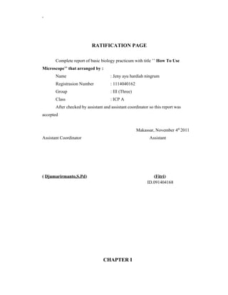
  • 1. , RATIFICATION PAGE Complete report of basic biology practicum with title ’’ How To Use Microscope’’ that arranged by : Name : Jeny ayu hardiah ningrum Registrasion Number : 1114040162 Group : III (Three) Class : ICP A After checked by assistant and assistant coordinator so this report was accepted Makassar, November 4th 2011 Assistant Coordinator Assistant ( Djumarirmanto,S.Pd) (Fitri) ID.091404168 CHAPTER I
  • 2. , INTRODUCTION A.Background Microscope ,this word not new for us,I have use when I in junior high school and senior high school,we had now practicum of microscope is one of practicum in biology department ,biology is one of branch of science wich explained about matter and the energy which has connection with human life and the process of their life,human life concist of seme cell very small,for the first time we must to know how to use the microscope because we just can see cell with use the microscope which can help us to see cell and structure of cell Do this practicum is the challenge for us because we must know components or part of microscope before we do this practicum about microscope, we can not get shadow, but my teacher help me,and my observation have succesed, and this practicum very accostumed but I always try although the tenses in report very hurd, not only present tense but past tens also. light very important for this practicum if don’t have light,I can not see cell and I can not find something This practicum is very interest and it is very challenge, so we have to try and try use it better than past time when we still studied ,I always studied in junior high school and senior high scool because I hope in future I can become a biology teacher.the reasons to do this practicum very many because it is one of the subject which we must know it.seeing happening problem, I interest to do praktikum this and to know more about microscope, therefore of that i hopes more know a lot of about microscope and part. not only sectioned but logistic and way utilizes also need at knows that deep utilize tool not false again. B.Purpose
  • 3. , The purpose of practicum is the students of University can use the biology microscope and safe to see a simple preparate. C.Benefit The benefit this practicum is the student of university more to know,part of microscope and function.the students can use microscope well more than senior high school,we can know the purpose of this practicum ,the manner to keep the microscope and the right procedure wor k to use microscope.
  • 4. , CHAPTER II PREVIEW OF LITERATURE Light microscopes is an indispensable technique for cell and molecular biologist to study cellular structures and biological processes in both living and fixed cells.this chapter provides and overview of light microscopy .describes the important parts of the microscope and goes on to explain how to set up a standard research microscope for bright field and phase contrast microscopy.there is also a short section on concfocalmicroscopy,more comprehensive descriptions of the different forms of light microscopy are found elsewhere.Microsopes are instruments that produce an enlarged image of a specimen. The eyepieces and the objectives are the main components of the magnification system of the microscope,the product of the magnification of the objective lens and the ocular give the total magnification of the microscope.the visibility of the magnifed specimen depends on contrast and resolution. Contrast is the difference in light bintensity between an object and its background. some biological samples contain coloured compouns. The key components of the compound microscope consist of : the eyepieces ,body tubes,nose pieces and objectives are part of the magnification system of the microscope.To use a microscope properly,and to get the most out of it ,it is important to understand the purpose and function of each of the microscope’s components (Harris,2006) The simple example of a microscope is a double convex lens of the type that is used as a magnifiying glass .in the late 1500s to dutch spectacle makers developed the compound microscope .their device had two convex lenses placed at either and of a tube and was capable of magnifiying an object to 10 times.its actual size today,developments in microscopy provide scientist with a wide selection of instruments with wich to view the smallest organism and even the components of individual cells.These microscopes range in complexity from the relatively simple models you will use in the laboratory today to highty sophisticated scanning and transmission electron microscopes (Helms R.Dorris ,2006)
  • 5. , A compound microscope contains two lens system .one is located in the ocular and the other in the objective ,each of the lenses magnifies the object independently .the ocular eyepiece magnification is disagnated in a similar manner,in order to determine the total magnification of acombination a two lenses multiply themanification of your microscope for both low and high power (windell,1975) There are two objectives mounted on a movable stage instead of a movable body tube,on each on side of the arm near the base you will see a large knob and represents both the coarse and fine adjustment,turning the adjustment one quarter turn in either direction represents fine adjustment ,turning it a greater amount represents coarse adjustment (windell,1957). There are two objecteves the nosepiece called the resolving nosepiece.a spring catch enganges the nose piece to hold each objective in position,the shorter objective is the low power objective and is marked 10x.the high power objective is longer and is ,arked 43x or 45x.The microscope is parfocal which means that anobject is in focus when the noce piece is rotated from one position to another .when switching from low to high power watch from the side to see whether or not the objective will touch the slide .if it does touch,lower the stage slightly before placing the objective into position (windell,1957). A microscope is an instrument used to see objects that are too small for the naked eye. The science of investigating small objects using such an instrument is called microscopy. Microscopic means invisible to the eye unless aided by a microscope. There are many types of microscopes, the most common and first to be invented is the optical microscope which uses light to image the sample. Other major types of microscopes are the electron microscope (both the transmission electron microscope and the scanning electron microscope) and the various types of scanning probe microscope(anonymous a,2011) The first microscope to be developed was the optical microscope, although the original inventor is not easy to identify. An early microscope was made in 1590 in Middelburg, Netherlands. Two eyeglass makers are variously given credit: Hans
  • 6. , Lippershey (who developed an early telescope) and Zacharias Janssen. Giovanni Faber coined the name microscope for Galileo Galilei's compound microscope in 1625 (Galileo had called it the "occhiolino" or "little eye"). The first detailed account of the interior construction of living tissue based on the use of a microscope did not appear until 1644, in Giambattista Odierna's L'occhio della mosca, or The Fly's Eye. It was not until the 1660s and 1670s that the microscope was used extensively for research in Italy, Holland and England. Marcelo Malpighi in Italy began the analysis of biological structures beginning with the lungs. Robert Hooke's Micrographia had a huge impact, largely because of its impressive illustrations. The greatest contribution came from Antoni van Leeuwenhoek who discovered red blood cells and spermatozoa and helped popularise microscopy as a technique. On 9 October 1676, Leeuwenhoek reported the discovery of micro-organisms. In 1893 August Köhler developed a key technique for sample illumination, Köhler illumination, which is central to modern light microscopy. This method of sample illumination gives rise to extremely even lighting and overcomes many limitations of older techniques of sample illumination. Further developments in sample illumination came from Fritz Zernike in 1953 and George Nomarski 1955 for their development of phase contrast and differential interference contrast illumination which allow imaging of transparent samples. Specialized techniques may exceed this magnification but the resolution is diffraction limited. The use of shorter wavelengths of light, such as the ultraviolet, is one way to improve the spatial resolution of the optical microscope, as are devices such as the near-field scanning optical microscope.Sarfus, a recent optical technique increases the sensitivity of standard optical microscope to a point it becomes possible to directly visualize nanometric films (down to 0.3 nanometre) and isolated nanoobjects (down to 2 nm-diameter). The technique is based on the use of non-reflecting substrates for cross-polarized reflected light microscopy (anonymous b,2011). CHAPTER III PRACTICUM METHOD
  • 7. , A.Time and Place Day / Date :Monday/October 25th 2011 Time :10.00 A.M until 12.30 P.M Place :Biology laboratory 3th flour at FMIPA UNM B.Tool and Material 1. Tool a. Biology microscope b. Tool box have contents: 1) Object glass 2) Cover glass 3) Petri dish 4) Tweezers 5) Hand pipette c. The equipments are served by the student of university: 1) New razor blade 2) New flannel cloth 3) Cotton cloth 4) Drawing Book 5) Tooth picks 2. Material
  • 8. , a. The materials are served by laboratory: 1) Flute water 2) Filter paper 3) Cotton or barn b. The materials are served by the student of university: 1) Hibiscus rosa-sinensis 2) Hibiscus tillaceus 3) Cucurbita moschata 4) Allium cepa C.Work produce 1. Prepared the microscope a. Put the microscope on the work table in front of you b. Cleaned up the body of microscope by using flannel eloth. Didn’t shine the lens of microscope with cloth. c. Opened toolbox than took petri dish which have object glass and cover glass. Cleaned up the object glass with cotton cloth or filter paper. d. Only there were microscope, toolbox and is contents, guide book and note book, the materials of practicum on the table, besides it put on the other place which had been served. 2. Straightened up the light for enter to the tube a. Paid attention to your practicum room condition, from where the bright light came (from front, left or right ). Directed the mirror of microscope to the light source. Opened diaphragm or revolved the slab to the medium hole position.
  • 9. , The microscope which have condenser straightened the position near the stage and used thin mirror. The microscope without condenser used concave mirror. b. Settled the revolver position until the short objective lens looked out to the stage until click sound. c. Brought the tube to down until the objective tip with the stage 5-10 mm or the tube maximize down. d. With the left eye closed the right eye looked the ocular will appeared white round area. If the light didn’t flat, moved mirror position until the light flat. If more dazzled, constricted the diaphragm or the hole of slab. If white round area still hazy, it was need the light, opened diaphragm put on the biggest lab. e. The microscope ready to used for observed the preparate. 3. The method to set the lens distance with preparate a. Revolved the macro meter to the thumb, dropped the tube, objective distance with stage become small. b. Put the object glass which have preserved preparate on the stage so that the material which observed been in the middle in the hole stage. Hold the preparate with stage clips so that it didn’t move. c. Paid attention the objective distance with the object glass not more 10 mm. If the distance loose, the hand revolved macro meter to down the tube while looked from beside objective tip approach the object glass until 5-10 mm. d. Looked from ocular while the hand revolved macro meter to up the tube slowly. Observed the while round area until the shadow appear. If that had done and the shadow had not appeared, it is had past. Repeat again from 3.3.
  • 10. , if had there was a shadow but still hazy so saw it while turned the micro meter up and down until the shadow clearly visible. e. Looked into the ocular (what increases is used?) and the objective (what increase is used?) counted the shadow increase which we see. f. Put out side the preparat when we had observed. 4. Made simple preparate a. Took the object glass which was cleaned, held everywhere. b. Sprinkled water on the middle. c. Pulped the cotton fibers with tweezers and put it on the middle drops of water. d. One of the hand held cover glass between the thumb and index finger on the contrary side. e. The side of cover glass touched on the object glass near the drops of water with 45 declivity than released it so it cover the drops of water. Reserved the surplus of water which ooze out on the glass side by using filter paper. f. Put on the preparate make on the stage and inspected it like the step 3.2, 3.3,3.4, and 3.5. 5. Changed the magnification a. The 4.6 surveillance had success, 3.4 and 3.5, increased the shadow which measles. Didn’t touch preparate position. b. Revolved until the longest of objective lens (strong) vertical on the stage and click sound (looked the increase).
  • 11. , c. Looked while turned the micro meter until maesles the shadow whish more big. Inspected the shadow. d. Failed to found the biggest shadow, upward the tube with turned the macro meter contrary direction with the thumb. Revolved again revolved to put the small objective lens on the position from the beginning. Without change preparate position, did the step 3.3, 3.4, 3.5, and continued to 5.1, 5.2, 5.3 until success. e. If would observe another object, so upward the tube. Put out preparate which was observed and cleaned object glass and cover glass. f. Made a new preparate conform with step 4.1 -4.6. g. The end of activity which used the microscope, take note of this metter. 1) Could not store preparate on the atage 2) Cleaned the wet preparate with filter paper. 3) (object glass + cover glass). Stored it in petri dish and put into the tool box. 4) Cleaned the microscope body with flannel cloth. 5) Put the microscope into the microscope box. 6) Cleaned the all epuipments were used with cotton cloth and put into its box. 7) Put each our equipments into box for used at the next practicum.
  • 12. , CHAPTER IV OBSERVATION RESULT A. Observation result We had done this practicum and got bthe result of it,The microscope,its components and its functions ,the observation result are the result of the practicum.The picture of microscope and its components:
  • 13. , The information of the picture 1. Ocular lens 2. Macro meter 3. Micro meter 4. Microscope arm 5. Mechanic activator 6. Inclination point 7. Supervisor condenser 8. Mast 9. Base 10. Mirror 11. Diapraghm 12. Condenser 13. Object table 14. Clippers 15. Objective lens 16. Revolver 17. Tube :
  • 15. , OBSERVATION PAGE Allium cepa Magnification 10 x 10 Notes: Cell wall Cytoplasm Neucleus Cell membran From internet
  • 16. , OBSERVATION PAGE Hibiscus rosa-sinensis Magnification 10 x 10 Notes: Trichome star Cell wall Cell membrane Cytoplasm Neucleus stomata From internet
  • 17. ,
  • 18. OBSERVATION PAGE Cucurbitha moschata , Magnification 10 x 10 Notes : Neucleus Cytoplasm Cell wall From internet OBSERVATION PAGE Hibiscus tilachius Magnification 10x10 Notes: Trichome star Cell wall Neucleus
  • 19. , B.Discussion Microscope is one of important tool for activity in biology laboratory,for see small things structure.it has some components .like accept a shadow,move the tube up and down,and many function other.like a 1st practicum i have many information from assistant about microscope and I have try use biology microscope.I have see cell of Hibiscus rosa-sinensis ,Hibiscus tillaceus ,Cucurbita moschata and Allium cepa and when I see in internet not difference for my research. The component and function of microscope are:The function of ocular lens is to accept a shadow from objective and magnify the shadow.mechanic activator is as the regulator of the place of the object glass on the stage .supervisor condenser is up and down the condenser. base is the place microscope standing.mirror is the captor and the reflection of the light.diaphragm is control the light that will enter to the condenser.condenser is provides a bright from the mirror which will eneter to the hole of stage and many other part.The microscope have objective amd ocular lense which have enlargement of objective lens and ocular lens CHAPTER V
  • 20. , CONCLUSION AND SUGGESTION A.Conclusion Student at university can use the biology microscope and safe to see a simple preparate,and the practicum have succes B. Suggestion 1. Suggestion for laboratory I hope for next practicum student can use microscope for each self .and the laboratory can become good place or suitable for practicum. 2. Suggestion for Assistant I hope assistant can give information and directive about practicum ,may be can give time to make the result observation 3. Suggestion for the all friends I hope all friend can hear and can see if assistant and coordinator assistant give information,so we can do practicum BIBLIOGRAPHY
  • 21. , Anonymous a.2011.Microscope,www.wikipedia.com,accesed at October 30 th 2011 Anonymous b.2011.Microscope,www.wikipedia.com,accesed at October 30 th 2011 Doris,R.doris.2006.Biology in the laboratory.city:publisher Robin,Harris.2006.cell biology protocol.Usa:john willey and soon,ltd Tim pengajar,2011.penuntun praktikum biologi dasar.Makassar UNM Windell,john.1975.Intestigations for practicing biology THE ANSWER OF QUESTION 1. The name of mechanic part of the microscop are
  • 22. , a. Ocular lens b. Objective lens c. Condenser d. Mirror 2. The name of mechanic parts of the microscop are a. Tube b. Revolver c. Stage d. Clippers e. Diaphragm f. Arm g. Mast h. Micro meter i. Macro meter j. Condenser setting k. Mechanic activator 3. The function of mechanic are a) Tube function as the place of ocular lens b) Revolver as place of objective lens c) Stage as the place of object glass
  • 23. , d) Clippers function to clip the object glass e) Diaphragm as the regulator of the light which enter to condenser f) Arm function to held when we want to move microscope g) Mast function is the place of microscope standing h) Micro meter as the tool for move up and down the tube smoothly i) Macro meter as the tool for move up and down the tube roughly j) Condenser setting if we turn can up and down the condenser k) Mechanic activator function manage the place of object glass on the table 4. When the shadow in the white round area will be shifted to the front left, we must shift the object glass to the opposite direction, because the quality of ocular lens will receive with the objective lens shadow 5. Because it can make the lens of microscope will be broken. And if the lens broken, we can not use it again.
  • 24. ,