
Recomendados
Mais conteúdo relacionado
Mais procurados
Mais procurados (12)
Destaque
Destaque (20)
Semelhante a Chapter8
Semelhante a Chapter8 (20)
Último
Último (20)
Chapter8
- 1. Chapter 8: Anatomical Considerations Part I
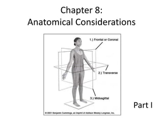
- 2. Anatomical Position • The subject is standing erect, the arms of the subject are at the sides with the palms of the hands facing the observer, the feet are together, and the subject is facing the observer.
- 3. • Anatomical Guide – • Linear Guide – • Anatomical Limit –
- 4. • Blood in veins flows OPPOSITE blood in arteries… • Therefore… • The anatomical limit and the linear guide for the veins would be the opposite of those of the respective arteries.
- 5. • The anatomical guides for arteries and veins would be the same.
- 6. Common Carotid Artery • Linear Guide –
- 7. Common Carotid Artery • Anatomical Guide –
- 8. Common Carotid Artery • Anatomical Limit – • Right – begins at the level of the right sternoclavicular articulation and extends to the superior border of the thyroid cartilage. • Left – begins at the level of the second costal cartilage and extends to the superior border of the thyroid cartilage.
- 9. Common Carotid Artery • Origins – • Right – a terminal branch of the brachiocephalic artery. • Left – is a branch off the arch of the aorta.
- 10. Common Carotid Artery • Branches – • Right – no branches of the right common carotid, except the terminal bifurcation into the right internal and external carotid arteries. • Left – no branches except the terminal bifurcation into the left internal and external carotid arteries.
- 12. Common Carotid Artery • Branches of the Right and Left External Carotid Arteries – • Ascending pharyngeal • Superior thyroid • Lingual • Facial • Occipital • Posterior auricular • Maxillary • Superficial temporal
- 13. Common Carotid Artery • Branches of the Right and Left Internal Carotid Arteries – • Branches arising within the carotid canal • Ophthalmic • Anterior cerebral • Middle cerebral • Posterior communicating • Choroidal branches
- 14. Common Carotid Artery • Relationship of the Common Carotid to the Internal Jugular Vein – • The internal jugular vein lies…
- 15. Common Carotid Artery • Contents of the Carotid Sheath – • Internal jugular vein • Vagus nerve • Common carotid artery What is the carotid sheath?
- 16. Axillary Artery • Linear Guide –
- 17. Axillary Artery • Anatomical Guide –
- 18. Axillary Artery • Anatomical Limit – • Extends from a point beginning at the lateral border of the first rib and extends to the inferior border of the tendon of the teres major muscle.
- 19. Axillary Artery • Origin – • A continuation of the subclavian artery.
- 20. Axillary Artery Branches – • Highest (supreme) thoracic artery • Thoracoacromial artery • Lateral thoracic artery • Subscapular artery • Anterior humeral circumflex artery • Posterior humeral circumflex artery
- 21. Axillary Artery • Relationship of Axillary Artery to the Axillary Vein – • The axillary artery is located…
- 22. Axillary Artery • Incision for Raising the Axillary Vessels –
- 23. Brachial Artery • Linear Guide –
- 24. Brachial Artery • Anatomical Guide –
- 25. Brachial Artery • Anatomical Limit – • Extends from a point beginning at the inferior border of the tendon of the teres major muscle and extends to a point inferior to the antecubital fossa.
- 26. Brachial Artery • Origin – • The brachial artery is a continuation of the axillary artery.
- 27. Brachial Artery • Relationship of the Brachial Artery and the Basilic Vein – • The accompanying basilic vein is located…
- 28. Brachial Artery • Location of the Incision –
- 29. Radial Artery • Linear Guide –
- 30. Radial Artery • Anatomical Guide –
- 31. Radial Artery • Anatomical Limit – • Extends from a point approximately 1 inch below an in front of the bend of the elbow to a point over the base of the thumb (thenar eminence).
- 32. Radial Artery • Origin – • Originates at the bifurcation of the brachial artery.
- 33. Radial Artery • Relationship of the Radial Artery and the Venae Comitantes – • Two small veins (venae comintantes) lie…
- 34. Ulnar Artery • Linear Guide –
- 35. Ulnar Artery • Anatomical Guide –
- 36. Ulnar Artery • Anatomical Limit – • Extends from a point approximately 1 inch below and in front of the bend of the elbow to a point over the pisiform bone (hypothenar eminence).
- 37. Ulnar Artery • Origin – • Originates at the bifurcation of the brachial artery.
- 38. Ulnar Artery • Relationship of the Ulnar Artery to the Venae Comitantes – • Two small veins (venae comitantes) lie…
- 39. Arteries of the Body Trunk • Ascending Aorta • Arch of the Aorta • Right Subclavian • Left Subclavian • Descending Thoracic Aorta • Descending Abdominal Aorta
- 40. Arteries of the Body Trunk
- 41. Ascending Aorta • Originates at the left ventricle. • Branches – • Right Coronary Artery • Left Coronary Artery
- 42. Arch of the Aorta • Center of Arterial Solution Distribution • Branches – • Brachiocephalic Artery • Left Common Carotid Artery • Left Subclavian Artery
- 43. Right Subclavian • Begins at the right sternoclavicular articulation and extends to the lateral border of the first rib. • For full autopsy (neck organs removed) – branches need to be clamped. • Braches – • Vertebral Artery • Internal Thoracic artery • Inferior Thyroid
- 44. Left Subclavian • Begins at the level of the left second costal cartilage and extends to the lateral border of the first rib.
- 45. Descending Thoracic Aorta • Branches – • Include NINE (9) pair of thoracic intercostal arteries.
- 46. Descending Abdominal Aorta • Extends from the diaphragm to the lower border of the fourth lumbar vertebra. • Branches – • Parietal (Inferior Phrenic, Superior Suprarenals, Lumbar, Middle Sacral) • Visceral unpaired (Celiac Axis, Superior Mesenteric, Inferior Mesenteric) • Visceral paired (Middle Suprarenals, Renals, Internal Spermatic, Ovarian, Common Iliacs)
- 47. External Iliac Artery and Vein • Continuation of the common iliac artery. • Common iliac is a terminal branch of the abdominal aorta. • Extends to a point under the center of the inguinal ligament. • Lies exactly at this ligament lateral to the external iliac vein.
- 48. External Iliac Artery and Vein • Used in autopsied bodies for the injection of the lower extremities. • In unautopsied bodies, it can be used for the morbidly obese. • Iliofemoral –