SlideShare uma empresa Scribd logo
1 de 8
Baixar para ler offline
AMERICAN SOCIETY OF ECHOCARDIOGRAPHY REPORT


                   American Society of Echocardiography
                 Guidelines and Standards for Performance of
                          the Fetal Echocardiogram
                     A statement of the Pediatric Council of the American Society of Echocardiography
                 represented by, Jack Rychik, MD, Nancy Ayres, MD, Bettina Cuneo, MD, Nina Gotteiner,
                      MD, Lisa Hornberger, MD, Philip J. Spevak, MD, and Mary Van Der Veld, MD


                                                                    studies suggest improved physiological state after
INTRODUCTION
                                                                    birth and improved surgical outcome for infants
Fetal echocardiography is the ultrasonic evaluation                 who have had prenatal diagnosis via fetal echocar-
                                                                    diography.2,3 In addition, accurate diagnosis via fetal
of the human fetal cardiovascular system. General                   echocardiography allows for appropriate counseling
antepartum obstetrical ultrasound has become a                      to take place and for parents to take the opportunity
standard part of gestational care and is commonly                   to learn about the cardiac anomaly. This knowledge
used for the determination of fetal age, size, gender,              can allay parental fears, improve psychological state,
or well-being and for the detection of congenital                   and bolster coping skills in dealing with the birth of
anomalies. A variety of maternal or fetal disorders
                                                                    a child with life-threatening cardiovascular illness.4
may result in abnormality of the fetal cardiovascular
                                                                       Fetal echocardiography can also contribute to
system to a degree which demands evaluation at a
                                                                    improved understanding when applied to cardiovas-
level above and beyond that attainable with standard
                                                                    cular abnormalities unrelated to congenital heart
antepartum obstetrical ultrasound. In these circum-
                                                                    disease. Such data, when presented in a multidisci-
stances, a fetal echocardiogram should be per-
                                                                    plinary format, can help elucidate the pathophysiol-
formed.
                                                                    ogy of a variety of disorders and can be used to both
   Improved operator skill amongst physicians per-
                                                                    guide and monitor disease therapy.
forming general antepartum obstetrical ultrasound,
                                                                       Performance and interpretation of fetal echocar-
in combination with increased sensitivity of present
day ultrasound systems, has resulted in improved                    diography requires a unique set of advanced skills
detection of fetal cardiovascular abnormalities and                 and knowledge. The fetal heart is of small size and
increased requirements for more detailed evalua-                    dynamic in nature. A myriad of complex anatomical
tion. Congenital heart disease is the most common                   and physiological derangements are possible. The
congenital anomaly found in the human.1 As the                      American Institute of Ultrasound in Medicine5 and
detection rates for congenital anomalies continue to                the American College of Radiology6 have recom-
increase, the demand for fetal echocardiography has                 mended performance of a 4-chamber view of the
grown. Accurate diagnosis of congenital heart dis-                  heart as part of the standard for an antepartum
ease via fetal echocardiography provides many ben-                  obstetrical ultrasound. However, studies have
efits. It allows for a smooth transition between the                shown that when using the 4-chamber view alone,
pre- and post-natal states, with the opportunity to                 important congenital heart disease may go unrecog-
provide immediate care at birth, thereby avoiding                   nized.7 Addition of right and left ventricular outflow
the onset of hemodynamic compromise. Recent                         tract and great artery visualization improves the
                                                                    yield allowing for more effective screening for con-
From The Children’s Hospital of Philadelphia (J.R.), Texas Chil-    genital heart disease,8-10 however many anomalies
dren’s Hospital, Houston (N.A.), Hope Children’s Hospital, Oak      can still be missed. Hence general antenatal obstet-
Lawn, Ill (B.C.), Children’s Memorial Hospital, Chicago (N.G.),     rical ultrasound can function as a screen for fetal
University of California San Francisco (L.H.), Johns Hopkins        cardiovascular disorders; suspicion or detection of a
Medical Center, Baltimore (P.J.S.), and Mott Children’s Hospital,
Ann Arbor, Mich (M.V.).                                             fetal cardiovascular abnormality requires referral for
Address reprint requests to the American Society of Echocardiog-
                                                                    a more comprehensive evaluation to a physician
raphy, 1500 Sunday Drive, Suite 102, Raleigh, NC 27607 (919)        with expertise in fetal echocardiography. Well-
787-5574.                                                           trained pediatric cardiologists, maternal-fetal medi-
J Am Soc Echocardiogr 2004;17:803-10.                               cine specialists, or obstetrical radiologists who have
0894-7317/$30.00                                                    acquired the appropriate knowledge base and skills
Copyright 2004 American Society of Echocardiography, Property       as outlined below may perform fetal echocardiogra-
of ASE. Reprint of these documents, beyond single use, is prohib-   phy.
ited without the prior written authorization of the ASE.               The purpose of this statement from the American
doi:10.1016/j.echo.2004.04.011                                      Society of Echocardiography is to define the distin-


                                                                                                                      803
Journal of the American Society of Echocardiography
804 Pediatric Council of the American Society of Echocardiography                                                  July 2004


guishing elements of the fetal echocardiogram from          Table 1 Examples of indications for fetal
other forms of ultrasonic evaluation and to provide         echocardiography
guidelines and standards for physician performance               Maternal indications                  Fetal indications
and interpretation of fetal echocardiography.
                                                            ● Family history of CHD          ● Abnormal obstetrical ultrasound
                                                                                               screen
                                                            ● Metabolic disorders (eg,       ● Extracardiac abnormality
                                                              diabetes, PKU)
SKILLS AND KNOWLEDGE REQUIRED
                                                            ● Exposure to teratogens         ● Chromosomal abnormality
                                                            ● Exposure to prostaglandin      ● Arrhythmia
Performance and interpretation of fetal echocardi-            synthetase inhibitors (eg,
ography requires a special set of skills and knowl-           ibuprofen, salicylic acid,
edge. The physician performing and interpreting               indomethacin)
fetal echocardiography must:                                ● Rubella infection              ● Hydrops
                                                            ● Autoimmune disease (eg,        ● Increased first trimester nuchal
  ●   be able to recognize the full spectrum of simple        SLE, Sjogren’s)
                                                                       ¨                       translucency
      and complex, acquired and congenital, heart           ● Familial inherited disorders   ● Multiple gestation and suspicion
      disease and its manifestations and natural his-         (Ellisvan Creveld, Marfan,       of twin-twin transfusion
      tory throughout gestation, and recognize the            Noonan’s, etc)                   syndrome
      limitations of fetal echocardiography in detect-      ● In vitro fertilization
      ing important associated lesions                      CHD, Congenital heart disease; PKU, phenyl ketonuria; SLE, sytemic lupus
  ●   have the skill and ability to apply all modalities    erythematosus.
      of echocardiography including 2-dimensional,
      M-mode, pulsed-wave, continuous wave, and
      Doppler color flow mapping in recognizing and         ography will also offer counseling to the family of
      evaluating both the normal and abnormal fetal         the fetus. In these circumstances, the physician
      cardiovascular state                                  offering counsel must have a thorough knowledge
  ●   have knowledge of the anatomy and physiology          base of all management strategies and be familiar
      of the cardiovascular system throughout the           with current outcomes for treatment of congenital
      stages of human development                           and acquired cardiovascular disease.
  ●   have a thorough understanding of the spectrum
      of fetal arrhythmias and the ability to utilize the
      spectrum of echocardiographic modalities for          INDICATIONS
      their assessment
  ●   be knowledgeable in the principles of biological
                                                            Indications for fetal echocardiography can be sepa-
      ultrasound instrumentation and its application
                                                            rated into maternal and fetal indications. Examples
      in human pregnancy
  ●
                                                            are listed in Table 1.
      have a thorough understanding of maternal-fetal
                                                               There are presently no strong prenatal markers
      physiology as well as maternal conditions that
                                                            available for identifying the fetus with congenital
      may affect the developing fetus
  ●
                                                            heart disease. Family history of congenital heart
      be familiar with the latest developments in
                                                            disease or the presence of a chromosomal anomaly
      obstetrical diagnostics, which include invasive
                                                            are relative risk factors. Increased nuchal translu-
      and non-invasive tests available throughout
                                                            cency present at 10 to 13 weeks gestation has been
      pregnancy
  ●
                                                            associated with an increased risk of congenital heart
      have knowledge of the growing field of invasive
                                                            disease, even in the absence of chromosomal anom-
      fetal intervention and its possible effects on the
                                                            aly.12 Recent reports indicate up to a 3-fold increase
      fetal cardiovascular system.
                                                            in the prevalence of congenital heart disease over
   Specific training requirements and maintenance           the general population in infants conceived via
of competency guidelines have been developed by             intracytoplasmic sperm injection and in-vitro fertili-
the American College of Cardiology in conjunction           zation.13
with the American Heart Association and American
Society of Echocardiography and are endorsed by
our group.11                                                THE FETAL ECHOCARDIOGRAM
   The physician performing and interpreting fetal
echocardiography should have access to a multidis-          Timing of Examination
ciplinary team with expertise in maternal-fetal med-
icine, genetics, neonatology, pediatric surgery, pedi-      The optimal timing for performance of a compre-
atric cardiology, and pediatric cardiac surgery, with       hensive transabdominal fetal echocardiogram is 18
availability for consultative services and advice. Of-      to 22 weeks gestation. Images can be more difficult
tentimes the physician performing fetal echocardi-          to obtain after 30 weeks gestation, as the ratio of
Journal of the American Society of Echocardiography
Volume 17 Number 7                                    Pediatric Council of the American Society of Echocardiography 805


fetal body mass-to-amniotic fluid increases. Acquir-             Table 2 Essential components of the fetal
ing images of the fetal heart at 15 to 18 weeks is               echocardiogram
possible; however performing a comprehensive car-                       Feature                   Essential component
diac evaluation study at this age can be difficult and
                                                                 Anatomic overview         Fetal number and position in the
may require repeat assessment at 18 to 22 weeks.
                                                                                             uterus
                                                                                           Establish stomach position and
Equipment                                                                                    abdominal situs
Ultrasound systems used for fetal echocardiography                                         Establish cardiac position
should have capabilities for performing 2-dimen-                 Biometric examination     Cardiothoracic ratio
                                                                                           Biparietal diameter
sional, M-mode, and Doppler imaging. The require-
                                                                                           Femur length
ments of fetal echocardiography are more stringent               Cardiac imaging           Four-chamber view
than for the examination of an infant or child with                views/sweeps            Four-chamber view angled
congenital or acquired heart disease. This is due to                                         towards great arteries
the increased demands for both spatial and temporal                                        (“Five-chamber” view)
resolution. Anatomic surveys require axial resolu-                                         Long-axis view (left ventricular
tion of 1 mm or less and this is particularly important                                      outflow)
given the small size of critical fetal cardiac struc-                                      Long-axis view (right ventricular
tures. Frames rates of 80 to 100 Hz are frequently                                           outflow)
needed to view important events occurring at heart                                         Short-axis sweep (cephalad angling
                                                                                             includes “3-vessel” view
rates in excess of 140 beats per minute. To meet
                                                                                           Caval long-axis view
these requirements, imaging systems need to be                                             Ductal arch view
optimally configured. In general, system settings are                                      Aortic arch view
adjusted to minimize persistence and spatial averag-             Doppler examination       Inferior and superior vena cava
ing and to increase frame rate. All modalities of                                          Pulmonary veins
Doppler including color, pulse, high pulse repeti-                                         Hepatic veins
tion frequency, and continuous wave should be                                              Ductus venosus
available. Tissue Doppler imaging has been recently                                        Foramen ovale
applied in the assessment of fetal arrhythmia.14                                           Atrioventricular valves
Harmonic imaging is useful when acoustic penetra-                                          Semilunar valves
                                                                                           Ductus arteriosus
tion is difficult such as in the presence of maternal
                                                                                           Transverse aortic arch
obesity. Phased array transducers with fundamental                                         Umbilical artery
frequencies between 4 and 12 MHz are generally                                             Umbilical vein
used. Curvilinear probes may be helpful given the                Measurement data          Atrioventricular valve diameter
wider near-field of view. High frequency transducers                                       Semilunar valve diameter
with a narrower footprint commonly used in echo-                                           Main pulmonary artery
cardiography of infants may also be helpful.                                               Ascending aorta
                                                                                           Branch pulmonary arteries
Examination Technique                                                                      Transverse aortic arch
                                                                                           Ventricular length
The essential components of the fetal echocardio-                                          Ventricular short-axis dimensions
gram are listed in Table 2. Although the goal is to              Examination of            M-mode of atrial and ventricular
achieve visualization of each of the essential compo-              rhythm and rate           wall motion
nents, not all will be visualized in every fetus at                                        Doppler examination of atrial and
every examination. Fetal position in the uterus or                                           ventricular flow patterns
increased activity may limit the ability to obtain
visualization of each of the components.
   The number of vessels in the umbilical cord is                views are described below with a brief explanation
counted and Doppler sampling of the umbilical                    of how to achieve the view and the structures
artery and umbilical vein is performed. After estab-             generally well seen. Reference sources are available,
lishing the position of the fetus and the right/left and         which illustrate these views in detail.15 Figures 1 and
anterior/posterior orientation, an initial survey of             2 demonstrate the anatomical correlates to the to-
the fetus is used to estimate the gestational age and            mographic imaging planes used for the views de-
to establish abdominal situs and cardiac position.               scribed below. The authors recognize that based on
The presence or absence of fluid in the pericardial,             operator style, alternative or additional sweeps and
pleural, or peritoneal space should be noted. The                views may be utilized to image the various struc-
position of the inferior vena cava and descending                tures of the fetal heart and still accomplish a com-
aorta at the level of the diaphragm are established.             prehensive fetal echocardiogram.
   Multiple scanning positions and sweeps are nec-                  Four-chamber view. The 4-chamber view is gen-
essary to adequately image the fetal heart. Suggested            erally easy to achieve and is useful for identifying the
Journal of the American Society of Echocardiography
806 Pediatric Council of the American Society of Echocardiography                                                        July 2004


        Figure 1 Illustration of the tomographic planes used to image the fetal cardiovascular system. Imaging
        planes displayed are in a normal human fetus. Starting at the top left, the following views are demonstrated
        in a clockwise manner: 1, apical (4-chamber) view; 2, apical (5-chamber) view angled towards the aorta;
        3, long-axis view of the left ventricular outflow tract; 4, long-axis view of the right ventricular outflow
        tract; 5, short-axis view at the level of the great vessels; 6, short-axis view with caudaud angling at the level
        of the ventricles; 7, caval long-axis view; 8, ductal arch view; 9, aortic arch view.


atria, ventricles, and respective septae (Table 3).                times, the pulmonary veins can be identified enter-
The diameters of the mitral and tricuspid valve                    ing the left atrium posteriorly.
annuli are measured. The lengths of the left and                      Short-axis view. The short-axis view is obtained
right ventricle can also be measured. Numerous                     by scanning perpendicular to the long axis of the
standards for dimensional measures based on gesta-                 heart (Table 4). It is an excellent view for identifying
tional age have been published.16-19 The view is                   the return of the pulmonary veins. In general, the
inadequate for determining the conotruncus and in                  connections of both lower lobe veins can be visual-
particular, excluding transposition of the great arter-            ized. The upper pulmonary veins may be identified
ies. From the standard 4-chamber view, one should                  as they course directly beneath the branch pulmo-
sweep posteriorly to demonstrate the coronary si-                  nary arteries. Sweeping cranially, one can measure
nus and then anteriorly to identify the aorta. Often-              right ventricular and left ventricular diastolic dimen-
Journal of the American Society of Echocardiography
Volume 17 Number 7                                    Pediatric Council of the American Society of Echocardiography 807


          Figure 2 Illustrations of the anatomical correlates for each of the designated tomographic imaging planes
          used for imaging of the fetal cardiovascular system. Each numbered view relates to the clockwise
          illustration of the fetal heart in Figure 1. Ao, Aorta; IVC, inferior vena cava; LA, left atrium; LV, left
          ventricle; MV, mitral valve; PA, pulmonary artery; PD, patent ductus; RA, right atrium; RV, right
          ventricle; SVC, superior vena cava.
Journal of the American Society of Echocardiography
808 Pediatric Council of the American Society of Echocardiography                                                  July 2004


Table 3 Structures viewed in the 4- and 5- chamber view       Table 5 Structures viewed in the cardiac long-axis sweep
● Atrial and ventricular size                                 ● Superior and inferior vena cava
● Atrial and ventricular septae                               ● Left ventricular outflow tract
● Atrioventricular size and function                          ● Ascending aorta
● Coronary sinus                                              ● Great vessel connection and size
● Ventricular function in long axis                           ● Ductus arteriosus and proximal ductal arch
● Semilunar valve function (may not, however, be optimal to
  differentiate aorta from main pulmonary artery)
● Pulmonary veins                                             Table 6 Structures viewed in the caval long-axis view
                                                              ● Superior vena cava
                                                              ● Inferior vena cava and eustachian valve
Table 4 Structures viewed in the cardiac short-axis sweep
                                                              ● Patent foramen ovale
● Pulmonary venous return                                     ● Right pulmonary artery
● Inferior vena cava and hepatic veins
● Ventricular short-axis dimensions
● Ventricular-arterial relationship                           Table 7 Structures viewed in the ductal and aortic arch
● Right ventricular outflow tract                             views
● Branch pulmonary arteries and origin
● Caval connections                                           ● Main pulmonary artery
● Innominate vein                                             ● Branch pulmonary arteries
● Ductus arteriosus                                           ● Patent ductus arteriosus and direction of flow
● Determination of arch sidedness and branching               ● Aortic arch dimension (ascending, transverse, isthmus, and
                                                                descending)
                                                              ● Direction of flow in the aortic arch

sions. These measurements should be made at the
level of the tips of the papillary muscles of the left
ventricle. Even more cranially, one can identify the          ing to the right allows visualization of both cavae
main pulmonary artery as it supplies the branch               (although these structures are better visualized in
pulmonary arteries. Identification of the branch              the caval view described below).
pulmonary arteries facilitates determination of the              Caval long-axis view. The caval long-axis view is
conotruncus. The superior vena cava can be identi-            obtained with the imaging plane parallel to the caval
fied crossing anterior to the ipsilateral branch pul-         connections to the right atrium (Table 6). Continuity
monary artery. Even more superiorly, the innomi-              of the inferior vena cava as it passes through the
nate vein is noted and as well, the branching of the          liver is established. The septum primum is seen
aortic arch and arch sidedness. Sweeping inferiorly           extending into the left atrium. Color Doppler inter-
one can demonstrate the connection of the inferior            rogation demonstrates the normal right to left flow
vena cava and hepatic veins with the right atrium. A          across the foramen ovale, which is reversed in
cross-sectional view obtained at the level of the             lesions of critical left heart hypoplasia.20
superior mediastinum provides an image of the                    Ductal view and aortic arch view. The ductal view
“3-vessel view.” In this plane the relationship and           is obtained when the imaging plane is aligned with
size differences between the superior vena cava, the          the right ventricular outflow tract and main pulmo-
ascending aorta, and main pulmonary artery can be             nary artery. The aortic arch is obtained with the
recognized. The main pulmonary artery is more                 beam aligned from anterior right of the fetal chest to
anterior and to the left of the other structures, and         posterior left of the fetal chest (Table 7). In the
tends to have a larger diameter. The ascending aorta          ductal view, the main pulmonary artery, and ductal
is the next, more rightward structure that is posi-           arch are well seen and main pulmonary artery size is
tioned slightly posterior relative to the main pulmo-         easily measured. The direction of ductal flow is
nary artery. The superior vena cava is the most               appreciated and the velocity recorded. Sweeping to
posterior and rightward vessel of the 3. Slight dis-          either side may allow visualization of the branch
crepancies in the position or size of these 3 vessels         pulmonary arteries. As the ductus connects with the
can identify subtle pathology of the outlets and              descending aorta, the isthmus is well seen and the
arches. Anterior to the 3 vessels is the thymus,              direction of flow established.21 In the aortic arch
which wraps over the anterior aspect of the superior          view, antegrade flow though the ascending aorta,
mediastinum.                                                  transverse arch, and descending aorta is established
   Cardiac long-axis view. The long-axis view is              with color Doppler interrogation.
aligned with the left ventricular outflow tract (Table           Heart rate and rhythm. The rate and mechanism
5). The continuity between the mitral and aortic              of rhythm is established by identifying mechanical
valves and the absence of sub-aortic conus is noted.          events associated with both atrial and ventricular
The size of the ascending aorta is measured. Sweep-           systole. Atrial systole is identified with either an
Journal of the American Society of Echocardiography
Volume 17 Number 7                                    Pediatric Council of the American Society of Echocardiography 809


M-mode of the lateral atrial wall or atrial append-              maximum temperature increase that may result from
age,22 pulse Doppler interrogation of the outflow                the exposure at those ultrasound system settings is
tract at a location where atrioventricular valve in-             2°C). The risk of mechanically induced ultrasound
flow is detected,23 or pulse tissue Doppler of the               damage is displayed by the mechanical index (MI),
atrial wall.14 The identification of atrioventricular            which is defined as the ratio of maximal peak
valve inflow serves as a proxy for atrial systole.               rarefractional pressure to the square root of the
Ventricular systole can be identified with M-mode of             ultrasound frequency. The risk of mechanical injury
the ventricular free wall or aortic valve or again,              rises with increasing MI.
using pulse Doppler interrogation of the outflow                    As newer modalities such as Doppler applications
tract or tissue Doppler of the ventricular myocar-               assessing tissue motion and real-time 3-dimensional
dium. Doppler flow in the outflow tract serves as a              imaging continue to develop, bioeffects on the fetus
proxy for ventricular systole. Measurement of the                will need to continue to be monitored. As there are
time interval between two successive beats allows                no strictly defined limits established, use of ultra-
calculation of rate. Simultaneous interrogation of left          sound energy in fetal echocardiography is best
ventricular inflow and outflow allows assessment of              expressed by the “ALARA” principle—as low as
atrioventricular conduction and the “mechanical”                 reasonably achievable.28
PR interval.23

                                                                 SUMMARY
ULTRASOUND SAFETY DURING PREGNANCY
                                                                 The fetal echocardiogram is a unique ultrasound
                                                                 examination, which differs from the antenatal ob-
The standard fetal echocardiographic examination                 stetrical ultrasound and from the conventional echo-
utilizes all modalities of diagnostic ultrasound in-             cardiogram in the infant, child, or adult. A unique,
cluding 2-dimensional (B-mode) imaging, Doppler,                 high level set of skills and knowledge is required in
and Doppler color flow mapping. Ultrasound energy                order to perform this test. In this statement, we
expenditures increase with each modality used and                outline the indications and essential performance
are most intense when Doppler color flow mapping                 components of the fetal echocardiogram, as well as
is applied to a small region of interest, as is com-             highlight the importance of operator cognizance of
monly the case when examining the structures of                  potential safety concerns. This statement contrib-
the fetal heart.24 Hence special consideration should            utes to the establishment of a standard for perfor-
be given to the use of ultrasound energy in the
                                                                 mance of the fetal echocardiogram, as the use of this
developing fetus. While theoretical concerns exist,
                                                                 valuable assay continues to expand in the future.
to date there have been no confirmed harmful
effects detected.25 Those performing fetal echocar-
                                                                 We wish to acknowledge Dr David Low for his artistic
diography should be aware of these effects and                   contributions to this report.
should limit power output and time of exposure to
no more than that which is absolutely necessary to
complete the examination.
   As ultrasound technology has advanced and new                 REFERENCES
modalities added, power output on newer systems
has changed. In 1985, the Food and Drug Adminis-                  1. Hoffman JI, Kaplan S. The incidence of congenital heart
tration 510(K) guide strictly limited ultrasound                     disease. J Am Coll Cardiol 2002;39:1890-900.
                                                                  2. Verheijen PM, Lisowski LA, Stoutenbeek P, Hitchcock JF,
power output on imaging systems. However, since
                                                                     Brenner JI, Cope JA, et al. Prenatal diagnosis of congenital
1992 much greater output levels have been allowed                    heart disease affects preoperative acidosis in the newborn
in conjunction with a display of power output,                       patient. J Thorac Cardiovasc Surg 2001;121:798.
thereby placing responsibility upon the user to                   3. Tworetzky W, McElhinney DB, Reddy VM, Brook MM,
make educated decisions regarding relative risk of a                 Hanley FL, Silverman NH. Improved surgical outcome after
particular modality.26 Potential bioeffects of ultra-                fetal diagnosis of hypoplastic left heart syndrome. Circulation
sound energy can be categorized as thermal, or                       2001;103:1269-73.
relating to increase in temperature in the region of              4. Sklansky M, Tang A, Levy D, Grossfeld P, Kashani I, Shaugh-
insonation, or mechanical, relating primarily to cav-                nessy R, et al. Maternal psychological impact of fetal echocar-
itation.27 Current ultrasound systems allow for dis-                 diography. J Am Soc Echocardiogr 2002;15:159-66.
                                                                  5. Standards for the Performance of the Antepartum Obstetrical
play of potential increase in temperature via the
                                                                     Ultrasound Examination. Copyright 1994, by the American
thermal index (TI) assigned for either soft-tissue                   Institute of Ultrasound in Medicine.
(TIS), or bone (TIB). The TI represents an estimate               6. American College of Radiology Standard for the Performance
of the temperature rise in the field and is approxi-                 of Antepartum Obstetrical Ultrasound.
mately proportional to the temperature increase in                7. Nelson NL, Filly RA, Goldstein RB, Callen PW. The AIUM/
degrees Celsius (eg, a TI of 2 means that the                        ACR antepartum obstetrical sonographic guidelines: expecta-
Journal of the American Society of Echocardiography
810 Pediatric Council of the American Society of Echocardiography                                                              July 2004


      tions for detection of anomalies. J Ultrasound Med 1993;4:              ography in the normal human fetus from 18 weeks to term.
      186-96.                                                                 Am J Cardiol 1992;70:1459-67.
 8.   Carvalho JS, Mavrides E, Shinebourne EA, Campbell S, Thil-        18.   Schmidt KG, Silverman NH, Van Hare GF, Hawkins JA,
      aganathan B. Improving the effectiveness of routine prenatal            Cloez JL, Rudolph AM. Two-dimensional echocardiographic
      screening for major congenital heart defects. Heart 2002;88:            determination of ventricular volumes in the fetal heart. Circu-
      387-91.                                                                 lation 1990;81:325-33.
 9.   Buskens E, Grobbee DE, Frohn-Mulder IME, Stewart PA,              19.   Phillipos EZ, Robertson MA, Still KD. The echocardio-
      Juttmann RE, Wladimiroff JW, et al. Efficacy of routine fetal           graphic assessment of the human foramen ovale. J Am Soc
      ultrasound screening for congenital heart disease in normal             Echocardiogr 1994;7:257-63.
      pregnancy. Circulation 1996;94:67-72.                             20.   Berning RA, Silverman NH, Villegas M, Sahn DJ, Martin GR,
10.   Stumpflen I, Stumpflen A, Wimmer M, Bernaschek G. Effect                Rice MJ. Reversed shunting across the ductus arteriosus or
      of detailed fetal echocardiography as part of routine prenatal          atrial septum in utero heralds severe congenital heart disease.
      ultrasonographic screening on detection of congenital heart             J Am Coll Cardiol 1996;27:481-6.
      disease. Lancet 1996;348:854-7.                                   21.   Fouron JC, Zarelli M, Drblik SP, Lessard M. Normal flow
11.   Quinones MA, Douglas PS, Foster E, Gorcsan J, Lewos JF,                 velocity profile of the fetal aortic isthmus through normal
      Pearlman AS, et al. ACC/AHA clinical competence statement
                                                                              gestation. Am J Cardiol 1994;74:483-6.
      on echocardiography: a report of the American College of
                                                                        22.   Kleinman C, Donnerstein R, Jaffe C, DeVore G, Weinstein
      Cardiology/American Heart Association/American College
                                                                              EM, Lynch DC, et al. Fetal echocardiography. A tool for
      of Physicians-American Society of Internal Medicine task force
                                                                              evaluation of in utero cardiac arrhythmias and monitoring of
      on clinical competence (committee on echocardiography).
                                                                              in utero therapy. Am J Cardiol 1983;51:237-43.
      J Am Coll Cardiol 2003;41:687-708.
                                                                        23.   Glickstein JS, Buyon J, Friedman D. Pulsed Doppler echocar-
12.   Ghi T, Huggon IC, Zosmer N, Nicolaides KH. Incidence of
                                                                              diographic assessment of the fetal PR interval. Am J Cardiol
      major structural cardiac defects associated with increased nu-
      chal translucency but normal karyotype. Ultrasound Obstet               2000;86:236-9.
      Gynecol 2001;18:610-4.                                            24.   Kurjak A. Are color and pulsed Doppler sonography safe in
13.   Hansen M, Kurinczuk JJ, Bower C, Webb S. The risk of major              early pregnancy? J Perinat Med 1999;27:423-30.
      birth defects after intracytoplasmic sperm injection and in       25.   Abramowicz JS, Kossoff G, Marsal K, Ter Haar G. Literature
      vitro fertilization. N Engl J Med 2002;346:725-30.                      review by the ISUOG bioefects and safety committee. Ultra-
14.   Rein AJ, O’Donnell C, Geva T, Nir A, Perles Z, Hashimoto I,             sound Obstet Gynecol 2002;19:318-9.
      et al. Use of tissue velocity imaging in the diagnosis of fetal   26.   Deane C, Lees C. Doppler obstetric ultrasound: a graphical
      cardiac arrhythmias. Circulation 2002;106:1827-33.                      display of temporal changes in safety indices. Ultrasound
15.   Allan L, Hornberger L, Sharland G, editors. Textbook of fetal           Obstet Gynecol 2000;15:418-23.
      cardiology. London: Greenwich Medical Media; 2000.                27.   Miller MW, Brayman AA, Abramowicz JA. Obstetric ultra-
16.   Sharland GK, Allan LD. Normal fetal cardiac measurements                sonography: a biophysical consideration of patient safety–the
      derived by cross-sectional echocardiography. Ultrasound Ob-             “rules” have changed. Am J Obstet Gynecol 1998;179:241-54.
      stet Gynecol 1992;2:175-81.                                       28.   International Society of Ultrasound in Obstetrics and Gyne-
17.   Tan J, Silverman NH, Hoffman JIE, Villegas M, Schmidt KG.               cology (ISUOG). Safety statement, 2000. Ultrasound Obstet
      Cardiac dimensions determined by cross-sectional echocardi-             Gynecol 2000;16:594-6.

Mais conteúdo relacionado

Mais procurados

Doppler ultrasound in the management of fetal growth restriction and IUGR
Doppler ultrasound in the management of fetal growth restriction and IUGRDoppler ultrasound in the management of fetal growth restriction and IUGR
Doppler ultrasound in the management of fetal growth restriction and IUGRChukwuma Onyeije, MD, FACOG
 
Fetal echocardiographic parameters and surgical outcomes in congenital left s...
Fetal echocardiographic parameters and surgical outcomes in congenital left s...Fetal echocardiographic parameters and surgical outcomes in congenital left s...
Fetal echocardiographic parameters and surgical outcomes in congenital left s...gisa_legal
 
Cardiac mdct for determining aetiology of pulmonary hypertension
Cardiac mdct for determining aetiology of  pulmonary hypertensionCardiac mdct for determining aetiology of  pulmonary hypertension
Cardiac mdct for determining aetiology of pulmonary hypertensionMuhammad Ayub
 
Prediction for rashkind procedure in fetuses with d tgvb
Prediction for rashkind procedure in fetuses with d tgvbPrediction for rashkind procedure in fetuses with d tgvb
Prediction for rashkind procedure in fetuses with d tgvbgisa_legal
 
Novo metodo prediz rashkind em tgvb
Novo metodo prediz rashkind em tgvbNovo metodo prediz rashkind em tgvb
Novo metodo prediz rashkind em tgvbgisa_legal
 
Carvalho 2014-ultrasound in-obstetrics_&_gynecology
Carvalho 2014-ultrasound in-obstetrics_&_gynecologyCarvalho 2014-ultrasound in-obstetrics_&_gynecology
Carvalho 2014-ultrasound in-obstetrics_&_gynecologygisa_legal
 
EMGuideWire's Radiology Reading Room: Stress-Induced Cardiomyopathy
EMGuideWire's Radiology Reading Room: Stress-Induced CardiomyopathyEMGuideWire's Radiology Reading Room: Stress-Induced Cardiomyopathy
EMGuideWire's Radiology Reading Room: Stress-Induced CardiomyopathySean M. Fox
 
BEGUM, JAMILA RESUME 16
BEGUM, JAMILA RESUME 16BEGUM, JAMILA RESUME 16
BEGUM, JAMILA RESUME 16jamila begum
 
Translucência nucal
Translucência nucalTranslucência nucal
Translucência nucalgisa_legal
 
Nuclear Imaging In Cardiology Cme
Nuclear Imaging In Cardiology CmeNuclear Imaging In Cardiology Cme
Nuclear Imaging In Cardiology CmeMuhammad Ayub
 
Intracranial hemorrhage newborn
Intracranial hemorrhage newbornIntracranial hemorrhage newborn
Intracranial hemorrhage newbornSingaram_Paed
 
EMGuideWire's Radiology Reading Room: Aortic Dissection
EMGuideWire's Radiology Reading Room: Aortic DissectionEMGuideWire's Radiology Reading Room: Aortic Dissection
EMGuideWire's Radiology Reading Room: Aortic DissectionSean M. Fox
 
Nuclear imaging in cardiology
Nuclear imaging in cardiologyNuclear imaging in cardiology
Nuclear imaging in cardiologyrahatul quadir
 
Functional echocardiography in the fetus with non cardiac disease
Functional echocardiography in the fetus with non cardiac diseaseFunctional echocardiography in the fetus with non cardiac disease
Functional echocardiography in the fetus with non cardiac diseaseLuis Carlos Murillo Valencia
 
Functional echocardiography in the fetus with non cardiac disease
Functional echocardiography in the  fetus with non cardiac diseaseFunctional echocardiography in the  fetus with non cardiac disease
Functional echocardiography in the fetus with non cardiac diseasegisa_legal
 

Mais procurados (20)

Doppler ultrasound in the management of fetal growth restriction and IUGR
Doppler ultrasound in the management of fetal growth restriction and IUGRDoppler ultrasound in the management of fetal growth restriction and IUGR
Doppler ultrasound in the management of fetal growth restriction and IUGR
 
Fetal echocardiographic parameters and surgical outcomes in congenital left s...
Fetal echocardiographic parameters and surgical outcomes in congenital left s...Fetal echocardiographic parameters and surgical outcomes in congenital left s...
Fetal echocardiographic parameters and surgical outcomes in congenital left s...
 
Cardiac mdct for determining aetiology of pulmonary hypertension
Cardiac mdct for determining aetiology of  pulmonary hypertensionCardiac mdct for determining aetiology of  pulmonary hypertension
Cardiac mdct for determining aetiology of pulmonary hypertension
 
Prediction for rashkind procedure in fetuses with d tgvb
Prediction for rashkind procedure in fetuses with d tgvbPrediction for rashkind procedure in fetuses with d tgvb
Prediction for rashkind procedure in fetuses with d tgvb
 
Novo metodo prediz rashkind em tgvb
Novo metodo prediz rashkind em tgvbNovo metodo prediz rashkind em tgvb
Novo metodo prediz rashkind em tgvb
 
Carvalho 2014-ultrasound in-obstetrics_&_gynecology
Carvalho 2014-ultrasound in-obstetrics_&_gynecologyCarvalho 2014-ultrasound in-obstetrics_&_gynecology
Carvalho 2014-ultrasound in-obstetrics_&_gynecology
 
Coronary CTA
Coronary CTACoronary CTA
Coronary CTA
 
EMGuideWire's Radiology Reading Room: Stress-Induced Cardiomyopathy
EMGuideWire's Radiology Reading Room: Stress-Induced CardiomyopathyEMGuideWire's Radiology Reading Room: Stress-Induced Cardiomyopathy
EMGuideWire's Radiology Reading Room: Stress-Induced Cardiomyopathy
 
Arco ao à d
Arco ao à dArco ao à d
Arco ao à d
 
BEGUM, JAMILA RESUME 16
BEGUM, JAMILA RESUME 16BEGUM, JAMILA RESUME 16
BEGUM, JAMILA RESUME 16
 
Translucência nucal
Translucência nucalTranslucência nucal
Translucência nucal
 
Poster Houston
Poster HoustonPoster Houston
Poster Houston
 
CSI
CSICSI
CSI
 
Nuclear Imaging In Cardiology Cme
Nuclear Imaging In Cardiology CmeNuclear Imaging In Cardiology Cme
Nuclear Imaging In Cardiology Cme
 
Intracranial hemorrhage newborn
Intracranial hemorrhage newbornIntracranial hemorrhage newborn
Intracranial hemorrhage newborn
 
EMGuideWire's Radiology Reading Room: Aortic Dissection
EMGuideWire's Radiology Reading Room: Aortic DissectionEMGuideWire's Radiology Reading Room: Aortic Dissection
EMGuideWire's Radiology Reading Room: Aortic Dissection
 
Europace 2016 acosta
Europace 2016 acostaEuropace 2016 acosta
Europace 2016 acosta
 
Nuclear imaging in cardiology
Nuclear imaging in cardiologyNuclear imaging in cardiology
Nuclear imaging in cardiology
 
Functional echocardiography in the fetus with non cardiac disease
Functional echocardiography in the fetus with non cardiac diseaseFunctional echocardiography in the fetus with non cardiac disease
Functional echocardiography in the fetus with non cardiac disease
 
Functional echocardiography in the fetus with non cardiac disease
Functional echocardiography in the  fetus with non cardiac diseaseFunctional echocardiography in the  fetus with non cardiac disease
Functional echocardiography in the fetus with non cardiac disease
 

Semelhante a Guidelines and Standards for Performance of Fetal Echocardiography

ISUOG consensus statement: what constitutes a fetal echocardiogram?
ISUOG consensus statement: what constitutes a fetal echocardiogram?ISUOG consensus statement: what constitutes a fetal echocardiogram?
ISUOG consensus statement: what constitutes a fetal echocardiogram?Tony Terrones
 
Lindsay impacto
Lindsay impactoLindsay impacto
Lindsay impactogisa_legal
 
Artigo civ e eco fetal
Artigo civ e eco fetalArtigo civ e eco fetal
Artigo civ e eco fetalgisa_legal
 
Cmwh 8-2015-039
Cmwh 8-2015-039Cmwh 8-2015-039
Cmwh 8-2015-039gisa_legal
 
ANTENATAL_SCREENING_IN_FIRST_AND_SECOND_TRIMESTER.ppt
ANTENATAL_SCREENING_IN_FIRST_AND_SECOND_TRIMESTER.pptANTENATAL_SCREENING_IN_FIRST_AND_SECOND_TRIMESTER.ppt
ANTENATAL_SCREENING_IN_FIRST_AND_SECOND_TRIMESTER.ppt9459654457
 
Diagnosisoftetralogyoffallotanditsvariantsinthe 130818165058-phpapp01
Diagnosisoftetralogyoffallotanditsvariantsinthe 130818165058-phpapp01Diagnosisoftetralogyoffallotanditsvariantsinthe 130818165058-phpapp01
Diagnosisoftetralogyoffallotanditsvariantsinthe 130818165058-phpapp01accoll
 
Diagnosis of tetralogy of fallot and its variants in the
Diagnosis of tetralogy of fallot and its variants in theDiagnosis of tetralogy of fallot and its variants in the
Diagnosis of tetralogy of fallot and its variants in thegisa_legal
 
2004 pediat. motivos encaminhamento
2004 pediat. motivos encaminhamento 2004 pediat. motivos encaminhamento
2004 pediat. motivos encaminhamento gisa_legal
 
Fetal growth restriction:Evidence based management 2018
Fetal growth restriction:Evidence based management 2018Fetal growth restriction:Evidence based management 2018
Fetal growth restriction:Evidence based management 2018Lifecare Centre
 
Drs. Potter and Richardson's CMC Pediatric X-Ray Mastery September Cases
Drs. Potter and Richardson's CMC Pediatric X-Ray Mastery September CasesDrs. Potter and Richardson's CMC Pediatric X-Ray Mastery September Cases
Drs. Potter and Richardson's CMC Pediatric X-Ray Mastery September CasesSean M. Fox
 
Fetal echocardiographic screening in twins for
Fetal echocardiographic screening in twins forFetal echocardiographic screening in twins for
Fetal echocardiographic screening in twins forgisa_legal
 
Pulmonary outflow tract obstruction in fetuses with complex
Pulmonary outflow tract obstruction in fetuses with complexPulmonary outflow tract obstruction in fetuses with complex
Pulmonary outflow tract obstruction in fetuses with complexgisa_legal
 
20150918 E. Pompilii - Microarray in diagnosi prenatale: la complessità della...
20150918 E. Pompilii - Microarray in diagnosi prenatale: la complessità della...20150918 E. Pompilii - Microarray in diagnosi prenatale: la complessità della...
20150918 E. Pompilii - Microarray in diagnosi prenatale: la complessità della...Roberto Scarafia
 
Genetic counselling 7 march13-Dr.Gourav
Genetic counselling 7 march13-Dr.GouravGenetic counselling 7 march13-Dr.Gourav
Genetic counselling 7 march13-Dr.GouravGourav Thakre
 
Rn com ccc eco fetal
Rn com ccc   eco fetalRn com ccc   eco fetal
Rn com ccc eco fetalgisa_legal
 
Ann pediatrcard eco fetal no diag precoce
Ann pediatrcard eco fetal no diag precoceAnn pediatrcard eco fetal no diag precoce
Ann pediatrcard eco fetal no diag precocegisa_legal
 

Semelhante a Guidelines and Standards for Performance of Fetal Echocardiography (20)

ISUOG consensus statement: what constitutes a fetal echocardiogram?
ISUOG consensus statement: what constitutes a fetal echocardiogram?ISUOG consensus statement: what constitutes a fetal echocardiogram?
ISUOG consensus statement: what constitutes a fetal echocardiogram?
 
Presentation1
Presentation1Presentation1
Presentation1
 
Lindsay impacto
Lindsay impactoLindsay impacto
Lindsay impacto
 
Artigo civ e eco fetal
Artigo civ e eco fetalArtigo civ e eco fetal
Artigo civ e eco fetal
 
Cmwh 8-2015-039
Cmwh 8-2015-039Cmwh 8-2015-039
Cmwh 8-2015-039
 
ANTENATAL_SCREENING_IN_FIRST_AND_SECOND_TRIMESTER.ppt
ANTENATAL_SCREENING_IN_FIRST_AND_SECOND_TRIMESTER.pptANTENATAL_SCREENING_IN_FIRST_AND_SECOND_TRIMESTER.ppt
ANTENATAL_SCREENING_IN_FIRST_AND_SECOND_TRIMESTER.ppt
 
Diagnosisoftetralogyoffallotanditsvariantsinthe 130818165058-phpapp01
Diagnosisoftetralogyoffallotanditsvariantsinthe 130818165058-phpapp01Diagnosisoftetralogyoffallotanditsvariantsinthe 130818165058-phpapp01
Diagnosisoftetralogyoffallotanditsvariantsinthe 130818165058-phpapp01
 
Diagnosis of tetralogy of fallot and its variants in the
Diagnosis of tetralogy of fallot and its variants in theDiagnosis of tetralogy of fallot and its variants in the
Diagnosis of tetralogy of fallot and its variants in the
 
2004 pediat. motivos encaminhamento
2004 pediat. motivos encaminhamento 2004 pediat. motivos encaminhamento
2004 pediat. motivos encaminhamento
 
Pediatric radiology
Pediatric radiologyPediatric radiology
Pediatric radiology
 
Fetal growth restriction:Evidence based management 2018
Fetal growth restriction:Evidence based management 2018Fetal growth restriction:Evidence based management 2018
Fetal growth restriction:Evidence based management 2018
 
Drs. Potter and Richardson's CMC Pediatric X-Ray Mastery September Cases
Drs. Potter and Richardson's CMC Pediatric X-Ray Mastery September CasesDrs. Potter and Richardson's CMC Pediatric X-Ray Mastery September Cases
Drs. Potter and Richardson's CMC Pediatric X-Ray Mastery September Cases
 
Fetal echocardiographic screening in twins for
Fetal echocardiographic screening in twins forFetal echocardiographic screening in twins for
Fetal echocardiographic screening in twins for
 
Pulmonary outflow tract obstruction in fetuses with complex
Pulmonary outflow tract obstruction in fetuses with complexPulmonary outflow tract obstruction in fetuses with complex
Pulmonary outflow tract obstruction in fetuses with complex
 
20150918 E. Pompilii - Microarray in diagnosi prenatale: la complessità della...
20150918 E. Pompilii - Microarray in diagnosi prenatale: la complessità della...20150918 E. Pompilii - Microarray in diagnosi prenatale: la complessità della...
20150918 E. Pompilii - Microarray in diagnosi prenatale: la complessità della...
 
Genetic counselling 7 march13-Dr.Gourav
Genetic counselling 7 march13-Dr.GouravGenetic counselling 7 march13-Dr.Gourav
Genetic counselling 7 march13-Dr.Gourav
 
Lindsay geral
Lindsay geralLindsay geral
Lindsay geral
 
Ecocardio y neo
Ecocardio y neoEcocardio y neo
Ecocardio y neo
 
Rn com ccc eco fetal
Rn com ccc   eco fetalRn com ccc   eco fetal
Rn com ccc eco fetal
 
Ann pediatrcard eco fetal no diag precoce
Ann pediatrcard eco fetal no diag precoceAnn pediatrcard eco fetal no diag precoce
Ann pediatrcard eco fetal no diag precoce
 

Mais de Tony Terrones

MCOD Infecciones Perinatales.pdf
MCOD Infecciones Perinatales.pdfMCOD Infecciones Perinatales.pdf
MCOD Infecciones Perinatales.pdfTony Terrones
 
MCOD Iniciación.pdf
MCOD Iniciación.pdfMCOD Iniciación.pdf
MCOD Iniciación.pdfTony Terrones
 
MCOD Anexos Ovulares.pdf
MCOD Anexos Ovulares.pdfMCOD Anexos Ovulares.pdf
MCOD Anexos Ovulares.pdfTony Terrones
 
MCOD Consensos Eco Gine.pdf
MCOD Consensos Eco Gine.pdfMCOD Consensos Eco Gine.pdf
MCOD Consensos Eco Gine.pdfTony Terrones
 
MCOD Anatomía Fetal.pdf
MCOD Anatomía Fetal.pdfMCOD Anatomía Fetal.pdf
MCOD Anatomía Fetal.pdfTony Terrones
 
Trayecto Formativo en Ecocardiografía Fetal
Trayecto Formativo en Ecocardiografía FetalTrayecto Formativo en Ecocardiografía Fetal
Trayecto Formativo en Ecocardiografía FetalTony Terrones
 
Curso de Ecocardiografía Fetal
Curso de Ecocardiografía FetalCurso de Ecocardiografía Fetal
Curso de Ecocardiografía FetalTony Terrones
 
Deterioro laboral en Diagnóstico Maipú
Deterioro laboral en Diagnóstico MaipúDeterioro laboral en Diagnóstico Maipú
Deterioro laboral en Diagnóstico MaipúTony Terrones
 
Taller de Clínica Ecográfica
Taller de Clínica EcográficaTaller de Clínica Ecográfica
Taller de Clínica EcográficaTony Terrones
 
IVE en Pcia de Bs As. Anexo
IVE en Pcia de Bs As. AnexoIVE en Pcia de Bs As. Anexo
IVE en Pcia de Bs As. AnexoTony Terrones
 
Ecógrafo Philips HD3
Ecógrafo Philips HD3Ecógrafo Philips HD3
Ecógrafo Philips HD3Tony Terrones
 
Curso e-learning Universitario de Ecografía TV
Curso e-learning Universitario de Ecografía TVCurso e-learning Universitario de Ecografía TV
Curso e-learning Universitario de Ecografía TVTony Terrones
 

Mais de Tony Terrones (20)

MCOD Urgencias.pdf
MCOD Urgencias.pdfMCOD Urgencias.pdf
MCOD Urgencias.pdf
 
MCOD Infecciones Perinatales.pdf
MCOD Infecciones Perinatales.pdfMCOD Infecciones Perinatales.pdf
MCOD Infecciones Perinatales.pdf
 
MCOD Iniciación.pdf
MCOD Iniciación.pdfMCOD Iniciación.pdf
MCOD Iniciación.pdf
 
MCOD Anexos Ovulares.pdf
MCOD Anexos Ovulares.pdfMCOD Anexos Ovulares.pdf
MCOD Anexos Ovulares.pdf
 
MCOD TN y ADNf.pdf
MCOD TN y ADNf.pdfMCOD TN y ADNf.pdf
MCOD TN y ADNf.pdf
 
MCOD Consensos Eco Gine.pdf
MCOD Consensos Eco Gine.pdfMCOD Consensos Eco Gine.pdf
MCOD Consensos Eco Gine.pdf
 
MCOD CCF.pdf
MCOD CCF.pdfMCOD CCF.pdf
MCOD CCF.pdf
 
MCOD Anatomía Fetal.pdf
MCOD Anatomía Fetal.pdfMCOD Anatomía Fetal.pdf
MCOD Anatomía Fetal.pdf
 
MCOD 3D.pdf
MCOD 3D.pdfMCOD 3D.pdf
MCOD 3D.pdf
 
MCOD 2T.pdf
MCOD 2T.pdfMCOD 2T.pdf
MCOD 2T.pdf
 
MCOD 1T.pdf
MCOD 1T.pdfMCOD 1T.pdf
MCOD 1T.pdf
 
Trayecto Formativo en Ecocardiografía Fetal
Trayecto Formativo en Ecocardiografía FetalTrayecto Formativo en Ecocardiografía Fetal
Trayecto Formativo en Ecocardiografía Fetal
 
Curso de Ecocardiografía Fetal
Curso de Ecocardiografía FetalCurso de Ecocardiografía Fetal
Curso de Ecocardiografía Fetal
 
Deterioro laboral en Diagnóstico Maipú
Deterioro laboral en Diagnóstico MaipúDeterioro laboral en Diagnóstico Maipú
Deterioro laboral en Diagnóstico Maipú
 
COVID-19
COVID-19COVID-19
COVID-19
 
Taller de Clínica Ecográfica
Taller de Clínica EcográficaTaller de Clínica Ecográfica
Taller de Clínica Ecográfica
 
IVE en Pcia de Bs As. Anexo
IVE en Pcia de Bs As. AnexoIVE en Pcia de Bs As. Anexo
IVE en Pcia de Bs As. Anexo
 
Ecógrafo Philips HD3
Ecógrafo Philips HD3Ecógrafo Philips HD3
Ecógrafo Philips HD3
 
Diplomaturas
DiplomaturasDiplomaturas
Diplomaturas
 
Curso e-learning Universitario de Ecografía TV
Curso e-learning Universitario de Ecografía TVCurso e-learning Universitario de Ecografía TV
Curso e-learning Universitario de Ecografía TV
 

Último

Sports & Fitness Value Added Course FY..
Sports & Fitness Value Added Course FY..Sports & Fitness Value Added Course FY..
Sports & Fitness Value Added Course FY..Disha Kariya
 
Measures of Central Tendency: Mean, Median and Mode
Measures of Central Tendency: Mean, Median and ModeMeasures of Central Tendency: Mean, Median and Mode
Measures of Central Tendency: Mean, Median and ModeThiyagu K
 
Russian Call Girls in Andheri Airport Mumbai WhatsApp 9167673311 💞 Full Nigh...
Russian Call Girls in Andheri Airport Mumbai WhatsApp  9167673311 💞 Full Nigh...Russian Call Girls in Andheri Airport Mumbai WhatsApp  9167673311 💞 Full Nigh...
Russian Call Girls in Andheri Airport Mumbai WhatsApp 9167673311 💞 Full Nigh...Pooja Nehwal
 
1029 - Danh muc Sach Giao Khoa 10 . pdf
1029 -  Danh muc Sach Giao Khoa 10 . pdf1029 -  Danh muc Sach Giao Khoa 10 . pdf
1029 - Danh muc Sach Giao Khoa 10 . pdfQucHHunhnh
 
JAPAN: ORGANISATION OF PMDA, PHARMACEUTICAL LAWS & REGULATIONS, TYPES OF REGI...
JAPAN: ORGANISATION OF PMDA, PHARMACEUTICAL LAWS & REGULATIONS, TYPES OF REGI...JAPAN: ORGANISATION OF PMDA, PHARMACEUTICAL LAWS & REGULATIONS, TYPES OF REGI...
JAPAN: ORGANISATION OF PMDA, PHARMACEUTICAL LAWS & REGULATIONS, TYPES OF REGI...anjaliyadav012327
 
Presentation by Andreas Schleicher Tackling the School Absenteeism Crisis 30 ...
Presentation by Andreas Schleicher Tackling the School Absenteeism Crisis 30 ...Presentation by Andreas Schleicher Tackling the School Absenteeism Crisis 30 ...
Presentation by Andreas Schleicher Tackling the School Absenteeism Crisis 30 ...EduSkills OECD
 
Activity 01 - Artificial Culture (1).pdf
Activity 01 - Artificial Culture (1).pdfActivity 01 - Artificial Culture (1).pdf
Activity 01 - Artificial Culture (1).pdfciinovamais
 
Introduction to Nonprofit Accounting: The Basics
Introduction to Nonprofit Accounting: The BasicsIntroduction to Nonprofit Accounting: The Basics
Introduction to Nonprofit Accounting: The BasicsTechSoup
 
The Most Excellent Way | 1 Corinthians 13
The Most Excellent Way | 1 Corinthians 13The Most Excellent Way | 1 Corinthians 13
The Most Excellent Way | 1 Corinthians 13Steve Thomason
 
social pharmacy d-pharm 1st year by Pragati K. Mahajan
social pharmacy d-pharm 1st year by Pragati K. Mahajansocial pharmacy d-pharm 1st year by Pragati K. Mahajan
social pharmacy d-pharm 1st year by Pragati K. Mahajanpragatimahajan3
 
Mastering the Unannounced Regulatory Inspection
Mastering the Unannounced Regulatory InspectionMastering the Unannounced Regulatory Inspection
Mastering the Unannounced Regulatory InspectionSafetyChain Software
 
1029-Danh muc Sach Giao Khoa khoi 6.pdf
1029-Danh muc Sach Giao Khoa khoi  6.pdf1029-Danh muc Sach Giao Khoa khoi  6.pdf
1029-Danh muc Sach Giao Khoa khoi 6.pdfQucHHunhnh
 
Beyond the EU: DORA and NIS 2 Directive's Global Impact
Beyond the EU: DORA and NIS 2 Directive's Global ImpactBeyond the EU: DORA and NIS 2 Directive's Global Impact
Beyond the EU: DORA and NIS 2 Directive's Global ImpactPECB
 
Z Score,T Score, Percential Rank and Box Plot Graph
Z Score,T Score, Percential Rank and Box Plot GraphZ Score,T Score, Percential Rank and Box Plot Graph
Z Score,T Score, Percential Rank and Box Plot GraphThiyagu K
 
Web & Social Media Analytics Previous Year Question Paper.pdf
Web & Social Media Analytics Previous Year Question Paper.pdfWeb & Social Media Analytics Previous Year Question Paper.pdf
Web & Social Media Analytics Previous Year Question Paper.pdfJayanti Pande
 
APM Welcome, APM North West Network Conference, Synergies Across Sectors
APM Welcome, APM North West Network Conference, Synergies Across SectorsAPM Welcome, APM North West Network Conference, Synergies Across Sectors
APM Welcome, APM North West Network Conference, Synergies Across SectorsAssociation for Project Management
 
Disha NEET Physics Guide for classes 11 and 12.pdf
Disha NEET Physics Guide for classes 11 and 12.pdfDisha NEET Physics Guide for classes 11 and 12.pdf
Disha NEET Physics Guide for classes 11 and 12.pdfchloefrazer622
 

Último (20)

Sports & Fitness Value Added Course FY..
Sports & Fitness Value Added Course FY..Sports & Fitness Value Added Course FY..
Sports & Fitness Value Added Course FY..
 
Measures of Central Tendency: Mean, Median and Mode
Measures of Central Tendency: Mean, Median and ModeMeasures of Central Tendency: Mean, Median and Mode
Measures of Central Tendency: Mean, Median and Mode
 
Russian Call Girls in Andheri Airport Mumbai WhatsApp 9167673311 💞 Full Nigh...
Russian Call Girls in Andheri Airport Mumbai WhatsApp  9167673311 💞 Full Nigh...Russian Call Girls in Andheri Airport Mumbai WhatsApp  9167673311 💞 Full Nigh...
Russian Call Girls in Andheri Airport Mumbai WhatsApp 9167673311 💞 Full Nigh...
 
1029 - Danh muc Sach Giao Khoa 10 . pdf
1029 -  Danh muc Sach Giao Khoa 10 . pdf1029 -  Danh muc Sach Giao Khoa 10 . pdf
1029 - Danh muc Sach Giao Khoa 10 . pdf
 
JAPAN: ORGANISATION OF PMDA, PHARMACEUTICAL LAWS & REGULATIONS, TYPES OF REGI...
JAPAN: ORGANISATION OF PMDA, PHARMACEUTICAL LAWS & REGULATIONS, TYPES OF REGI...JAPAN: ORGANISATION OF PMDA, PHARMACEUTICAL LAWS & REGULATIONS, TYPES OF REGI...
JAPAN: ORGANISATION OF PMDA, PHARMACEUTICAL LAWS & REGULATIONS, TYPES OF REGI...
 
Presentation by Andreas Schleicher Tackling the School Absenteeism Crisis 30 ...
Presentation by Andreas Schleicher Tackling the School Absenteeism Crisis 30 ...Presentation by Andreas Schleicher Tackling the School Absenteeism Crisis 30 ...
Presentation by Andreas Schleicher Tackling the School Absenteeism Crisis 30 ...
 
Activity 01 - Artificial Culture (1).pdf
Activity 01 - Artificial Culture (1).pdfActivity 01 - Artificial Culture (1).pdf
Activity 01 - Artificial Culture (1).pdf
 
Introduction to Nonprofit Accounting: The Basics
Introduction to Nonprofit Accounting: The BasicsIntroduction to Nonprofit Accounting: The Basics
Introduction to Nonprofit Accounting: The Basics
 
The Most Excellent Way | 1 Corinthians 13
The Most Excellent Way | 1 Corinthians 13The Most Excellent Way | 1 Corinthians 13
The Most Excellent Way | 1 Corinthians 13
 
social pharmacy d-pharm 1st year by Pragati K. Mahajan
social pharmacy d-pharm 1st year by Pragati K. Mahajansocial pharmacy d-pharm 1st year by Pragati K. Mahajan
social pharmacy d-pharm 1st year by Pragati K. Mahajan
 
Mastering the Unannounced Regulatory Inspection
Mastering the Unannounced Regulatory InspectionMastering the Unannounced Regulatory Inspection
Mastering the Unannounced Regulatory Inspection
 
1029-Danh muc Sach Giao Khoa khoi 6.pdf
1029-Danh muc Sach Giao Khoa khoi  6.pdf1029-Danh muc Sach Giao Khoa khoi  6.pdf
1029-Danh muc Sach Giao Khoa khoi 6.pdf
 
INDIA QUIZ 2024 RLAC DELHI UNIVERSITY.pptx
INDIA QUIZ 2024 RLAC DELHI UNIVERSITY.pptxINDIA QUIZ 2024 RLAC DELHI UNIVERSITY.pptx
INDIA QUIZ 2024 RLAC DELHI UNIVERSITY.pptx
 
Beyond the EU: DORA and NIS 2 Directive's Global Impact
Beyond the EU: DORA and NIS 2 Directive's Global ImpactBeyond the EU: DORA and NIS 2 Directive's Global Impact
Beyond the EU: DORA and NIS 2 Directive's Global Impact
 
Z Score,T Score, Percential Rank and Box Plot Graph
Z Score,T Score, Percential Rank and Box Plot GraphZ Score,T Score, Percential Rank and Box Plot Graph
Z Score,T Score, Percential Rank and Box Plot Graph
 
Web & Social Media Analytics Previous Year Question Paper.pdf
Web & Social Media Analytics Previous Year Question Paper.pdfWeb & Social Media Analytics Previous Year Question Paper.pdf
Web & Social Media Analytics Previous Year Question Paper.pdf
 
Mattingly "AI & Prompt Design: The Basics of Prompt Design"
Mattingly "AI & Prompt Design: The Basics of Prompt Design"Mattingly "AI & Prompt Design: The Basics of Prompt Design"
Mattingly "AI & Prompt Design: The Basics of Prompt Design"
 
Código Creativo y Arte de Software | Unidad 1
Código Creativo y Arte de Software | Unidad 1Código Creativo y Arte de Software | Unidad 1
Código Creativo y Arte de Software | Unidad 1
 
APM Welcome, APM North West Network Conference, Synergies Across Sectors
APM Welcome, APM North West Network Conference, Synergies Across SectorsAPM Welcome, APM North West Network Conference, Synergies Across Sectors
APM Welcome, APM North West Network Conference, Synergies Across Sectors
 
Disha NEET Physics Guide for classes 11 and 12.pdf
Disha NEET Physics Guide for classes 11 and 12.pdfDisha NEET Physics Guide for classes 11 and 12.pdf
Disha NEET Physics Guide for classes 11 and 12.pdf
 

Guidelines and Standards for Performance of Fetal Echocardiography

  • 1. AMERICAN SOCIETY OF ECHOCARDIOGRAPHY REPORT American Society of Echocardiography Guidelines and Standards for Performance of the Fetal Echocardiogram A statement of the Pediatric Council of the American Society of Echocardiography represented by, Jack Rychik, MD, Nancy Ayres, MD, Bettina Cuneo, MD, Nina Gotteiner, MD, Lisa Hornberger, MD, Philip J. Spevak, MD, and Mary Van Der Veld, MD studies suggest improved physiological state after INTRODUCTION birth and improved surgical outcome for infants Fetal echocardiography is the ultrasonic evaluation who have had prenatal diagnosis via fetal echocar- diography.2,3 In addition, accurate diagnosis via fetal of the human fetal cardiovascular system. General echocardiography allows for appropriate counseling antepartum obstetrical ultrasound has become a to take place and for parents to take the opportunity standard part of gestational care and is commonly to learn about the cardiac anomaly. This knowledge used for the determination of fetal age, size, gender, can allay parental fears, improve psychological state, or well-being and for the detection of congenital and bolster coping skills in dealing with the birth of anomalies. A variety of maternal or fetal disorders a child with life-threatening cardiovascular illness.4 may result in abnormality of the fetal cardiovascular Fetal echocardiography can also contribute to system to a degree which demands evaluation at a improved understanding when applied to cardiovas- level above and beyond that attainable with standard cular abnormalities unrelated to congenital heart antepartum obstetrical ultrasound. In these circum- disease. Such data, when presented in a multidisci- stances, a fetal echocardiogram should be per- plinary format, can help elucidate the pathophysiol- formed. ogy of a variety of disorders and can be used to both Improved operator skill amongst physicians per- guide and monitor disease therapy. forming general antepartum obstetrical ultrasound, Performance and interpretation of fetal echocar- in combination with increased sensitivity of present day ultrasound systems, has resulted in improved diography requires a unique set of advanced skills detection of fetal cardiovascular abnormalities and and knowledge. The fetal heart is of small size and increased requirements for more detailed evalua- dynamic in nature. A myriad of complex anatomical tion. Congenital heart disease is the most common and physiological derangements are possible. The congenital anomaly found in the human.1 As the American Institute of Ultrasound in Medicine5 and detection rates for congenital anomalies continue to the American College of Radiology6 have recom- increase, the demand for fetal echocardiography has mended performance of a 4-chamber view of the grown. Accurate diagnosis of congenital heart dis- heart as part of the standard for an antepartum ease via fetal echocardiography provides many ben- obstetrical ultrasound. However, studies have efits. It allows for a smooth transition between the shown that when using the 4-chamber view alone, pre- and post-natal states, with the opportunity to important congenital heart disease may go unrecog- provide immediate care at birth, thereby avoiding nized.7 Addition of right and left ventricular outflow the onset of hemodynamic compromise. Recent tract and great artery visualization improves the yield allowing for more effective screening for con- From The Children’s Hospital of Philadelphia (J.R.), Texas Chil- genital heart disease,8-10 however many anomalies dren’s Hospital, Houston (N.A.), Hope Children’s Hospital, Oak can still be missed. Hence general antenatal obstet- Lawn, Ill (B.C.), Children’s Memorial Hospital, Chicago (N.G.), rical ultrasound can function as a screen for fetal University of California San Francisco (L.H.), Johns Hopkins cardiovascular disorders; suspicion or detection of a Medical Center, Baltimore (P.J.S.), and Mott Children’s Hospital, Ann Arbor, Mich (M.V.). fetal cardiovascular abnormality requires referral for Address reprint requests to the American Society of Echocardiog- a more comprehensive evaluation to a physician raphy, 1500 Sunday Drive, Suite 102, Raleigh, NC 27607 (919) with expertise in fetal echocardiography. Well- 787-5574. trained pediatric cardiologists, maternal-fetal medi- J Am Soc Echocardiogr 2004;17:803-10. cine specialists, or obstetrical radiologists who have 0894-7317/$30.00 acquired the appropriate knowledge base and skills Copyright 2004 American Society of Echocardiography, Property as outlined below may perform fetal echocardiogra- of ASE. Reprint of these documents, beyond single use, is prohib- phy. ited without the prior written authorization of the ASE. The purpose of this statement from the American doi:10.1016/j.echo.2004.04.011 Society of Echocardiography is to define the distin- 803
  • 2. Journal of the American Society of Echocardiography 804 Pediatric Council of the American Society of Echocardiography July 2004 guishing elements of the fetal echocardiogram from Table 1 Examples of indications for fetal other forms of ultrasonic evaluation and to provide echocardiography guidelines and standards for physician performance Maternal indications Fetal indications and interpretation of fetal echocardiography. ● Family history of CHD ● Abnormal obstetrical ultrasound screen ● Metabolic disorders (eg, ● Extracardiac abnormality diabetes, PKU) SKILLS AND KNOWLEDGE REQUIRED ● Exposure to teratogens ● Chromosomal abnormality ● Exposure to prostaglandin ● Arrhythmia Performance and interpretation of fetal echocardi- synthetase inhibitors (eg, ography requires a special set of skills and knowl- ibuprofen, salicylic acid, edge. The physician performing and interpreting indomethacin) fetal echocardiography must: ● Rubella infection ● Hydrops ● Autoimmune disease (eg, ● Increased first trimester nuchal ● be able to recognize the full spectrum of simple SLE, Sjogren’s) ¨ translucency and complex, acquired and congenital, heart ● Familial inherited disorders ● Multiple gestation and suspicion disease and its manifestations and natural his- (Ellisvan Creveld, Marfan, of twin-twin transfusion tory throughout gestation, and recognize the Noonan’s, etc) syndrome limitations of fetal echocardiography in detect- ● In vitro fertilization ing important associated lesions CHD, Congenital heart disease; PKU, phenyl ketonuria; SLE, sytemic lupus ● have the skill and ability to apply all modalities erythematosus. of echocardiography including 2-dimensional, M-mode, pulsed-wave, continuous wave, and Doppler color flow mapping in recognizing and ography will also offer counseling to the family of evaluating both the normal and abnormal fetal the fetus. In these circumstances, the physician cardiovascular state offering counsel must have a thorough knowledge ● have knowledge of the anatomy and physiology base of all management strategies and be familiar of the cardiovascular system throughout the with current outcomes for treatment of congenital stages of human development and acquired cardiovascular disease. ● have a thorough understanding of the spectrum of fetal arrhythmias and the ability to utilize the spectrum of echocardiographic modalities for INDICATIONS their assessment ● be knowledgeable in the principles of biological Indications for fetal echocardiography can be sepa- ultrasound instrumentation and its application rated into maternal and fetal indications. Examples in human pregnancy ● are listed in Table 1. have a thorough understanding of maternal-fetal There are presently no strong prenatal markers physiology as well as maternal conditions that available for identifying the fetus with congenital may affect the developing fetus ● heart disease. Family history of congenital heart be familiar with the latest developments in disease or the presence of a chromosomal anomaly obstetrical diagnostics, which include invasive are relative risk factors. Increased nuchal translu- and non-invasive tests available throughout cency present at 10 to 13 weeks gestation has been pregnancy ● associated with an increased risk of congenital heart have knowledge of the growing field of invasive disease, even in the absence of chromosomal anom- fetal intervention and its possible effects on the aly.12 Recent reports indicate up to a 3-fold increase fetal cardiovascular system. in the prevalence of congenital heart disease over Specific training requirements and maintenance the general population in infants conceived via of competency guidelines have been developed by intracytoplasmic sperm injection and in-vitro fertili- the American College of Cardiology in conjunction zation.13 with the American Heart Association and American Society of Echocardiography and are endorsed by our group.11 THE FETAL ECHOCARDIOGRAM The physician performing and interpreting fetal echocardiography should have access to a multidis- Timing of Examination ciplinary team with expertise in maternal-fetal med- icine, genetics, neonatology, pediatric surgery, pedi- The optimal timing for performance of a compre- atric cardiology, and pediatric cardiac surgery, with hensive transabdominal fetal echocardiogram is 18 availability for consultative services and advice. Of- to 22 weeks gestation. Images can be more difficult tentimes the physician performing fetal echocardi- to obtain after 30 weeks gestation, as the ratio of
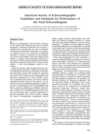
  • 3. Journal of the American Society of Echocardiography Volume 17 Number 7 Pediatric Council of the American Society of Echocardiography 805 fetal body mass-to-amniotic fluid increases. Acquir- Table 2 Essential components of the fetal ing images of the fetal heart at 15 to 18 weeks is echocardiogram possible; however performing a comprehensive car- Feature Essential component diac evaluation study at this age can be difficult and Anatomic overview Fetal number and position in the may require repeat assessment at 18 to 22 weeks. uterus Establish stomach position and Equipment abdominal situs Ultrasound systems used for fetal echocardiography Establish cardiac position should have capabilities for performing 2-dimen- Biometric examination Cardiothoracic ratio Biparietal diameter sional, M-mode, and Doppler imaging. The require- Femur length ments of fetal echocardiography are more stringent Cardiac imaging Four-chamber view than for the examination of an infant or child with views/sweeps Four-chamber view angled congenital or acquired heart disease. This is due to towards great arteries the increased demands for both spatial and temporal (“Five-chamber” view) resolution. Anatomic surveys require axial resolu- Long-axis view (left ventricular tion of 1 mm or less and this is particularly important outflow) given the small size of critical fetal cardiac struc- Long-axis view (right ventricular tures. Frames rates of 80 to 100 Hz are frequently outflow) needed to view important events occurring at heart Short-axis sweep (cephalad angling includes “3-vessel” view rates in excess of 140 beats per minute. To meet Caval long-axis view these requirements, imaging systems need to be Ductal arch view optimally configured. In general, system settings are Aortic arch view adjusted to minimize persistence and spatial averag- Doppler examination Inferior and superior vena cava ing and to increase frame rate. All modalities of Pulmonary veins Doppler including color, pulse, high pulse repeti- Hepatic veins tion frequency, and continuous wave should be Ductus venosus available. Tissue Doppler imaging has been recently Foramen ovale applied in the assessment of fetal arrhythmia.14 Atrioventricular valves Harmonic imaging is useful when acoustic penetra- Semilunar valves Ductus arteriosus tion is difficult such as in the presence of maternal Transverse aortic arch obesity. Phased array transducers with fundamental Umbilical artery frequencies between 4 and 12 MHz are generally Umbilical vein used. Curvilinear probes may be helpful given the Measurement data Atrioventricular valve diameter wider near-field of view. High frequency transducers Semilunar valve diameter with a narrower footprint commonly used in echo- Main pulmonary artery cardiography of infants may also be helpful. Ascending aorta Branch pulmonary arteries Examination Technique Transverse aortic arch Ventricular length The essential components of the fetal echocardio- Ventricular short-axis dimensions gram are listed in Table 2. Although the goal is to Examination of M-mode of atrial and ventricular achieve visualization of each of the essential compo- rhythm and rate wall motion nents, not all will be visualized in every fetus at Doppler examination of atrial and every examination. Fetal position in the uterus or ventricular flow patterns increased activity may limit the ability to obtain visualization of each of the components. The number of vessels in the umbilical cord is views are described below with a brief explanation counted and Doppler sampling of the umbilical of how to achieve the view and the structures artery and umbilical vein is performed. After estab- generally well seen. Reference sources are available, lishing the position of the fetus and the right/left and which illustrate these views in detail.15 Figures 1 and anterior/posterior orientation, an initial survey of 2 demonstrate the anatomical correlates to the to- the fetus is used to estimate the gestational age and mographic imaging planes used for the views de- to establish abdominal situs and cardiac position. scribed below. The authors recognize that based on The presence or absence of fluid in the pericardial, operator style, alternative or additional sweeps and pleural, or peritoneal space should be noted. The views may be utilized to image the various struc- position of the inferior vena cava and descending tures of the fetal heart and still accomplish a com- aorta at the level of the diaphragm are established. prehensive fetal echocardiogram. Multiple scanning positions and sweeps are nec- Four-chamber view. The 4-chamber view is gen- essary to adequately image the fetal heart. Suggested erally easy to achieve and is useful for identifying the
  • 4. Journal of the American Society of Echocardiography 806 Pediatric Council of the American Society of Echocardiography July 2004 Figure 1 Illustration of the tomographic planes used to image the fetal cardiovascular system. Imaging planes displayed are in a normal human fetus. Starting at the top left, the following views are demonstrated in a clockwise manner: 1, apical (4-chamber) view; 2, apical (5-chamber) view angled towards the aorta; 3, long-axis view of the left ventricular outflow tract; 4, long-axis view of the right ventricular outflow tract; 5, short-axis view at the level of the great vessels; 6, short-axis view with caudaud angling at the level of the ventricles; 7, caval long-axis view; 8, ductal arch view; 9, aortic arch view. atria, ventricles, and respective septae (Table 3). times, the pulmonary veins can be identified enter- The diameters of the mitral and tricuspid valve ing the left atrium posteriorly. annuli are measured. The lengths of the left and Short-axis view. The short-axis view is obtained right ventricle can also be measured. Numerous by scanning perpendicular to the long axis of the standards for dimensional measures based on gesta- heart (Table 4). It is an excellent view for identifying tional age have been published.16-19 The view is the return of the pulmonary veins. In general, the inadequate for determining the conotruncus and in connections of both lower lobe veins can be visual- particular, excluding transposition of the great arter- ized. The upper pulmonary veins may be identified ies. From the standard 4-chamber view, one should as they course directly beneath the branch pulmo- sweep posteriorly to demonstrate the coronary si- nary arteries. Sweeping cranially, one can measure nus and then anteriorly to identify the aorta. Often- right ventricular and left ventricular diastolic dimen-
  • 5. Journal of the American Society of Echocardiography Volume 17 Number 7 Pediatric Council of the American Society of Echocardiography 807 Figure 2 Illustrations of the anatomical correlates for each of the designated tomographic imaging planes used for imaging of the fetal cardiovascular system. Each numbered view relates to the clockwise illustration of the fetal heart in Figure 1. Ao, Aorta; IVC, inferior vena cava; LA, left atrium; LV, left ventricle; MV, mitral valve; PA, pulmonary artery; PD, patent ductus; RA, right atrium; RV, right ventricle; SVC, superior vena cava.
  • 6. Journal of the American Society of Echocardiography 808 Pediatric Council of the American Society of Echocardiography July 2004 Table 3 Structures viewed in the 4- and 5- chamber view Table 5 Structures viewed in the cardiac long-axis sweep ● Atrial and ventricular size ● Superior and inferior vena cava ● Atrial and ventricular septae ● Left ventricular outflow tract ● Atrioventricular size and function ● Ascending aorta ● Coronary sinus ● Great vessel connection and size ● Ventricular function in long axis ● Ductus arteriosus and proximal ductal arch ● Semilunar valve function (may not, however, be optimal to differentiate aorta from main pulmonary artery) ● Pulmonary veins Table 6 Structures viewed in the caval long-axis view ● Superior vena cava ● Inferior vena cava and eustachian valve Table 4 Structures viewed in the cardiac short-axis sweep ● Patent foramen ovale ● Pulmonary venous return ● Right pulmonary artery ● Inferior vena cava and hepatic veins ● Ventricular short-axis dimensions ● Ventricular-arterial relationship Table 7 Structures viewed in the ductal and aortic arch ● Right ventricular outflow tract views ● Branch pulmonary arteries and origin ● Caval connections ● Main pulmonary artery ● Innominate vein ● Branch pulmonary arteries ● Ductus arteriosus ● Patent ductus arteriosus and direction of flow ● Determination of arch sidedness and branching ● Aortic arch dimension (ascending, transverse, isthmus, and descending) ● Direction of flow in the aortic arch sions. These measurements should be made at the level of the tips of the papillary muscles of the left ventricle. Even more cranially, one can identify the ing to the right allows visualization of both cavae main pulmonary artery as it supplies the branch (although these structures are better visualized in pulmonary arteries. Identification of the branch the caval view described below). pulmonary arteries facilitates determination of the Caval long-axis view. The caval long-axis view is conotruncus. The superior vena cava can be identi- obtained with the imaging plane parallel to the caval fied crossing anterior to the ipsilateral branch pul- connections to the right atrium (Table 6). Continuity monary artery. Even more superiorly, the innomi- of the inferior vena cava as it passes through the nate vein is noted and as well, the branching of the liver is established. The septum primum is seen aortic arch and arch sidedness. Sweeping inferiorly extending into the left atrium. Color Doppler inter- one can demonstrate the connection of the inferior rogation demonstrates the normal right to left flow vena cava and hepatic veins with the right atrium. A across the foramen ovale, which is reversed in cross-sectional view obtained at the level of the lesions of critical left heart hypoplasia.20 superior mediastinum provides an image of the Ductal view and aortic arch view. The ductal view “3-vessel view.” In this plane the relationship and is obtained when the imaging plane is aligned with size differences between the superior vena cava, the the right ventricular outflow tract and main pulmo- ascending aorta, and main pulmonary artery can be nary artery. The aortic arch is obtained with the recognized. The main pulmonary artery is more beam aligned from anterior right of the fetal chest to anterior and to the left of the other structures, and posterior left of the fetal chest (Table 7). In the tends to have a larger diameter. The ascending aorta ductal view, the main pulmonary artery, and ductal is the next, more rightward structure that is posi- arch are well seen and main pulmonary artery size is tioned slightly posterior relative to the main pulmo- easily measured. The direction of ductal flow is nary artery. The superior vena cava is the most appreciated and the velocity recorded. Sweeping to posterior and rightward vessel of the 3. Slight dis- either side may allow visualization of the branch crepancies in the position or size of these 3 vessels pulmonary arteries. As the ductus connects with the can identify subtle pathology of the outlets and descending aorta, the isthmus is well seen and the arches. Anterior to the 3 vessels is the thymus, direction of flow established.21 In the aortic arch which wraps over the anterior aspect of the superior view, antegrade flow though the ascending aorta, mediastinum. transverse arch, and descending aorta is established Cardiac long-axis view. The long-axis view is with color Doppler interrogation. aligned with the left ventricular outflow tract (Table Heart rate and rhythm. The rate and mechanism 5). The continuity between the mitral and aortic of rhythm is established by identifying mechanical valves and the absence of sub-aortic conus is noted. events associated with both atrial and ventricular The size of the ascending aorta is measured. Sweep- systole. Atrial systole is identified with either an
  • 7. Journal of the American Society of Echocardiography Volume 17 Number 7 Pediatric Council of the American Society of Echocardiography 809 M-mode of the lateral atrial wall or atrial append- maximum temperature increase that may result from age,22 pulse Doppler interrogation of the outflow the exposure at those ultrasound system settings is tract at a location where atrioventricular valve in- 2°C). The risk of mechanically induced ultrasound flow is detected,23 or pulse tissue Doppler of the damage is displayed by the mechanical index (MI), atrial wall.14 The identification of atrioventricular which is defined as the ratio of maximal peak valve inflow serves as a proxy for atrial systole. rarefractional pressure to the square root of the Ventricular systole can be identified with M-mode of ultrasound frequency. The risk of mechanical injury the ventricular free wall or aortic valve or again, rises with increasing MI. using pulse Doppler interrogation of the outflow As newer modalities such as Doppler applications tract or tissue Doppler of the ventricular myocar- assessing tissue motion and real-time 3-dimensional dium. Doppler flow in the outflow tract serves as a imaging continue to develop, bioeffects on the fetus proxy for ventricular systole. Measurement of the will need to continue to be monitored. As there are time interval between two successive beats allows no strictly defined limits established, use of ultra- calculation of rate. Simultaneous interrogation of left sound energy in fetal echocardiography is best ventricular inflow and outflow allows assessment of expressed by the “ALARA” principle—as low as atrioventricular conduction and the “mechanical” reasonably achievable.28 PR interval.23 SUMMARY ULTRASOUND SAFETY DURING PREGNANCY The fetal echocardiogram is a unique ultrasound examination, which differs from the antenatal ob- The standard fetal echocardiographic examination stetrical ultrasound and from the conventional echo- utilizes all modalities of diagnostic ultrasound in- cardiogram in the infant, child, or adult. A unique, cluding 2-dimensional (B-mode) imaging, Doppler, high level set of skills and knowledge is required in and Doppler color flow mapping. Ultrasound energy order to perform this test. In this statement, we expenditures increase with each modality used and outline the indications and essential performance are most intense when Doppler color flow mapping components of the fetal echocardiogram, as well as is applied to a small region of interest, as is com- highlight the importance of operator cognizance of monly the case when examining the structures of potential safety concerns. This statement contrib- the fetal heart.24 Hence special consideration should utes to the establishment of a standard for perfor- be given to the use of ultrasound energy in the mance of the fetal echocardiogram, as the use of this developing fetus. While theoretical concerns exist, valuable assay continues to expand in the future. to date there have been no confirmed harmful effects detected.25 Those performing fetal echocar- We wish to acknowledge Dr David Low for his artistic diography should be aware of these effects and contributions to this report. should limit power output and time of exposure to no more than that which is absolutely necessary to complete the examination. As ultrasound technology has advanced and new REFERENCES modalities added, power output on newer systems has changed. In 1985, the Food and Drug Adminis- 1. Hoffman JI, Kaplan S. The incidence of congenital heart tration 510(K) guide strictly limited ultrasound disease. J Am Coll Cardiol 2002;39:1890-900. 2. Verheijen PM, Lisowski LA, Stoutenbeek P, Hitchcock JF, power output on imaging systems. However, since Brenner JI, Cope JA, et al. Prenatal diagnosis of congenital 1992 much greater output levels have been allowed heart disease affects preoperative acidosis in the newborn in conjunction with a display of power output, patient. J Thorac Cardiovasc Surg 2001;121:798. thereby placing responsibility upon the user to 3. Tworetzky W, McElhinney DB, Reddy VM, Brook MM, make educated decisions regarding relative risk of a Hanley FL, Silverman NH. Improved surgical outcome after particular modality.26 Potential bioeffects of ultra- fetal diagnosis of hypoplastic left heart syndrome. Circulation sound energy can be categorized as thermal, or 2001;103:1269-73. relating to increase in temperature in the region of 4. Sklansky M, Tang A, Levy D, Grossfeld P, Kashani I, Shaugh- insonation, or mechanical, relating primarily to cav- nessy R, et al. Maternal psychological impact of fetal echocar- itation.27 Current ultrasound systems allow for dis- diography. J Am Soc Echocardiogr 2002;15:159-66. 5. Standards for the Performance of the Antepartum Obstetrical play of potential increase in temperature via the Ultrasound Examination. Copyright 1994, by the American thermal index (TI) assigned for either soft-tissue Institute of Ultrasound in Medicine. (TIS), or bone (TIB). The TI represents an estimate 6. American College of Radiology Standard for the Performance of the temperature rise in the field and is approxi- of Antepartum Obstetrical Ultrasound. mately proportional to the temperature increase in 7. Nelson NL, Filly RA, Goldstein RB, Callen PW. The AIUM/ degrees Celsius (eg, a TI of 2 means that the ACR antepartum obstetrical sonographic guidelines: expecta-
  • 8. Journal of the American Society of Echocardiography 810 Pediatric Council of the American Society of Echocardiography July 2004 tions for detection of anomalies. J Ultrasound Med 1993;4: ography in the normal human fetus from 18 weeks to term. 186-96. Am J Cardiol 1992;70:1459-67. 8. Carvalho JS, Mavrides E, Shinebourne EA, Campbell S, Thil- 18. Schmidt KG, Silverman NH, Van Hare GF, Hawkins JA, aganathan B. Improving the effectiveness of routine prenatal Cloez JL, Rudolph AM. Two-dimensional echocardiographic screening for major congenital heart defects. Heart 2002;88: determination of ventricular volumes in the fetal heart. Circu- 387-91. lation 1990;81:325-33. 9. Buskens E, Grobbee DE, Frohn-Mulder IME, Stewart PA, 19. Phillipos EZ, Robertson MA, Still KD. The echocardio- Juttmann RE, Wladimiroff JW, et al. Efficacy of routine fetal graphic assessment of the human foramen ovale. J Am Soc ultrasound screening for congenital heart disease in normal Echocardiogr 1994;7:257-63. pregnancy. Circulation 1996;94:67-72. 20. Berning RA, Silverman NH, Villegas M, Sahn DJ, Martin GR, 10. Stumpflen I, Stumpflen A, Wimmer M, Bernaschek G. Effect Rice MJ. Reversed shunting across the ductus arteriosus or of detailed fetal echocardiography as part of routine prenatal atrial septum in utero heralds severe congenital heart disease. ultrasonographic screening on detection of congenital heart J Am Coll Cardiol 1996;27:481-6. disease. Lancet 1996;348:854-7. 21. Fouron JC, Zarelli M, Drblik SP, Lessard M. Normal flow 11. Quinones MA, Douglas PS, Foster E, Gorcsan J, Lewos JF, velocity profile of the fetal aortic isthmus through normal Pearlman AS, et al. ACC/AHA clinical competence statement gestation. Am J Cardiol 1994;74:483-6. on echocardiography: a report of the American College of 22. Kleinman C, Donnerstein R, Jaffe C, DeVore G, Weinstein Cardiology/American Heart Association/American College EM, Lynch DC, et al. Fetal echocardiography. A tool for of Physicians-American Society of Internal Medicine task force evaluation of in utero cardiac arrhythmias and monitoring of on clinical competence (committee on echocardiography). in utero therapy. Am J Cardiol 1983;51:237-43. J Am Coll Cardiol 2003;41:687-708. 23. Glickstein JS, Buyon J, Friedman D. Pulsed Doppler echocar- 12. Ghi T, Huggon IC, Zosmer N, Nicolaides KH. Incidence of diographic assessment of the fetal PR interval. Am J Cardiol major structural cardiac defects associated with increased nu- chal translucency but normal karyotype. Ultrasound Obstet 2000;86:236-9. Gynecol 2001;18:610-4. 24. Kurjak A. Are color and pulsed Doppler sonography safe in 13. Hansen M, Kurinczuk JJ, Bower C, Webb S. The risk of major early pregnancy? J Perinat Med 1999;27:423-30. birth defects after intracytoplasmic sperm injection and in 25. Abramowicz JS, Kossoff G, Marsal K, Ter Haar G. Literature vitro fertilization. N Engl J Med 2002;346:725-30. review by the ISUOG bioefects and safety committee. Ultra- 14. Rein AJ, O’Donnell C, Geva T, Nir A, Perles Z, Hashimoto I, sound Obstet Gynecol 2002;19:318-9. et al. Use of tissue velocity imaging in the diagnosis of fetal 26. Deane C, Lees C. Doppler obstetric ultrasound: a graphical cardiac arrhythmias. Circulation 2002;106:1827-33. display of temporal changes in safety indices. Ultrasound 15. Allan L, Hornberger L, Sharland G, editors. Textbook of fetal Obstet Gynecol 2000;15:418-23. cardiology. London: Greenwich Medical Media; 2000. 27. Miller MW, Brayman AA, Abramowicz JA. Obstetric ultra- 16. Sharland GK, Allan LD. Normal fetal cardiac measurements sonography: a biophysical consideration of patient safety–the derived by cross-sectional echocardiography. Ultrasound Ob- “rules” have changed. Am J Obstet Gynecol 1998;179:241-54. stet Gynecol 1992;2:175-81. 28. International Society of Ultrasound in Obstetrics and Gyne- 17. Tan J, Silverman NH, Hoffman JIE, Villegas M, Schmidt KG. cology (ISUOG). Safety statement, 2000. Ultrasound Obstet Cardiac dimensions determined by cross-sectional echocardi- Gynecol 2000;16:594-6.