Top Rated Hyderabad Call Girls Erragadda ⟟ 9332606886 ⟟ Call Me For Genuine ...
Blood and tissue flagellates
1. BLOOD AND TISSUE FLAGELLATES
Trypanosoma cruzi
DISEASE: Chagas’ Disease or American Trypanosomiasis
the ONLY trypanosome that has an intracellular
amastigote stage
VERTEBRATE HOST: humans, domesticated and wild
animals
VECTOR/INTERMEDIATE HOST: Reduviid bug/ Kissing
bug/Triatomid bug/Assasin bug
Triatoma infestans
Triatoma sordida
Panstrongylus megistus
Rhodnius prolixus
GEOGRAPHIC DISTRIBUTION
Southern part of United States, Mexico, Brazil
Central America, South America
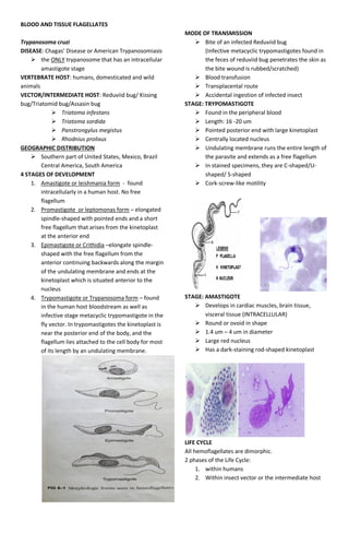
4 STAGES OF DEVELOPMENT
1. Amastigote or leishmania form - found
intracellularly in a human host. No free
flagellum
2. Promastigote or leptomonas form – elongated
spindle-shaped with pointed ends and a short
free flagellum that arises from the kinetoplast
at the anterior end
3. Epimastigote or Crithidia –elongate spindle-
shaped with the free flagellum from the
anterior continuing backwards along the margin
of the undulating membrane and ends at the
kinetoplast which is situated anterior to the
nucleus
4. Trypomastigote or Trypanosoma form – found
in the human host bloodstream as well as
infective stage metacyclic trypomastigote in the
fly vector. In trypomastigotes the kinetoplast is
near the posterior end of the body, and the
flagellum lies attached to the cell body for most
of its length by an undulating membrane.
MODE OF TRANSMISSION
Bite of an infected Reduviid bug
(Infective metacyclic trypomastigotes found in
the feces of reduviid bug penetrates the skin as
the bite wound is rubbed/scratched)
Blood transfusion
Transplacental route
Accidental ingestion of infected insect
STAGE: TRYPOMASTIGOTE
Found in the peripheral blood
Length: 16 -20 um
Pointed posterior end with large kinetoplast
Centrally located nucleus
Undulating membrane runs the entire length of
the parasite and extends as a free flagellum
In stained specimens, they are C-shaped/U-
shaped/ S-shaped
Cork-screw-like motility
STAGE: AMASTIGOTE
Develops in cardiac muscles, brain tissue,
visceral tissue (INTRACELLULAR)
Round or ovoid in shape
1.4 um – 4 um in diameter
Large red nucleus
Has a dark-staining rod-shaped kinetoplast
LIFE CYCLE
All hemoflagellates are dimorphic.
2 phases of the Life Cycle:
1. within humans
2. Within insect vector or the intermediate host
2. LIFE CYCLE IN MAN
Infected reduviid bug partakes a mealdefecates
passing out metacyclic trypomastigotepenetrates the
skin via mucus membranestrypomastigotes engulfed
by histiocytesdevelops into amastigote
intracellularly multiply by binary fissionmay
transitionally pass through a promastigote
stageepimastigotetrypomastigoteinfected
histiocyte ruptures in 4-5 days releasing
trypomastigotes in the bloodstreaminfects new
histiocytes and replicate again as
amastigotestrypomastigote in bloodstream is
ingested by reduviid bug as it takes a blood meal
LIFE CYCLE IN THE INSECT VECTOR
Trypanosomes pass through the posterior portion of the
midgut of reduviid bug develop into
epimastigotemultiplies by longitudinal binary
fissiondevelop into infective metacyclic
trypomastigoteappears in the insect’s rectum 8-10
days after infectionexcreted in the bug’s
fecesenters human host via scratch on skin or
through mucous membranes that are rubbed with
fingers contaminated with bug’s feces
INFECTED CELLS
Frequently infects:
1. Reticuloendothelial cells of spleen, liver, cardiac
muscles, smooth and skeletal muscles
2. Skin
3. Gonads
4. Intestinal mucosa
5. Placenta
PATHOGENESIS AND CLINICAL MANIFESTION
Chagoma – localized inflammation at the site of
infection (usually in the face)
Small, painful, reddish nodule that takes
2-3 months to resolve
Trypomastigotes and amastigotes may
be aspirated from Chagoma
ACUTE FORM
4 days – 2 wks after the insect bite
High fever, lymphadenopathy, swelling of the
entire body
Severe symptoms are seen in young children
with CNS involvement early in the infection.
Amastigotes are seen in the meningeal tissues.
Leads to death within a few days or weeks
Meningoencephalitis in neonates
Romaña’s Sign – conjunctivitis and unilateral
edema of the eyes
Some patients will experience complete
recovery after the acute stage but most will
progress to Chronic Chagas’ Disease
CHRONIC FORM
More common in adults
More common than the acute form
Cardiac damage– most common serious form of
the disease
CHF (Congestive Heart Failure)
Mega syndrome
Megaesophagus
Megacolon
Cardiomegaly
CNS involvement - signs of agitation,
disorentiation, aphasia, comadeath if left
untreated
DIAGNOSIS
Patient history – being in an endemic area
Clinical Presentation: Febrile episodes,
enlargement of the lymph nodes, Chagoma,
Romaña’s sign, myocarditis, CNS and digestive
problems
Demonstration of parasites in blood, CSF, and
tissue Giemsa staining
Centrifugation – examination of the buffy coat
layer
Blood cultures – if parasites are scanty
Animal inoculation
Xenodiagnosis – successful even in the later
part of the disease when blood films are
negative
3. Xenodiagnosis
SEROLOGIC TESTS
Complement fixation
Direct and Indirect Hemagglutination
Indirect Immunofluorescent Antibody Test
(IFAT)
PCR
ELISA – directs T. cruzi antigen in urine
TREATMENT
Nifurtimox (aka Lampit or Bayer 2502) – DOC
Allupurinol
Benznidazole
Megaesophagus and Megacolon –surgical
intervention
PREVENTION
Education of the endemic population to raise
awareness about the disease
Insect control – application of insecticide on the
roof and walls
Housing improvement to eliminate cracks and
crevices where the insect resides
Screening of blood for transfusion
Routine addition of gentian violet dye
to blood bottles in final concentration
of 0.025% to kill T. cruzi
______________________________________________
Trypanosoma brucei complex
Both sp. are morphologically indistinguishable
Same life cycle
Both are pathogenic for humans in Africa
“brucei-gambiense-rhodesiense complex”
Disease: African Sleeping Sickness
STAGES OF DEVELOPMENT
Polymorphic trypanosomes – seen in blood and
CSF
Epimastigotes – seen in vector
Trypanosoma brucei gambiense
Disease: West African Sleeping Sickness/
Gambian Trypanosomiasis
Has a chronic course which ends with
CNS involvement leading to death after
several years
Parasite can be found in the wet lowlands and
rainforest of West and Central Africa
MODE OF TRANSMISSION
Bite of an infected Tsetse fly (Intermediate host
and vector)
(Riverine tsetse flies of the palpalis group)
Glossina palpalis
Glossina tachinoides (tsetse fly)
Glossina fuscipes
Day biters
Blood transfusion
Organ transplant
Transplacental route
LIFE CYCLE
Trypomastigotes are ingested by tsetse fly when it takes
a blood meal on infected humans develop into
epimastigotemultiplies within the fly in the gut and
salivary glands. As tsetse fly takes another blood meal,
saliva containing metacyclic trypomastigotes is
transmitted to the human hosttrypomastigotes
multiply in the bloodstream (may be ingested by
fly)goes to CNS causing sleeping sickness
CLINICAL MANIFESTATIONS AND PATHOGENESIS
1st
Stage
Asymptomatic incubation period
Trypanosomal chancre at the insect bite
2nd
Stage
Trypomastigotes found in the bloodstream and
lymphatic system
1st
distinct symptom: fever followed by afebrile
periods
Headache, malaise, anorexia, night sweats,
weakness, joint and muscle pain, tachycardia
Lymphadenopathy and glandular enlargement
Winterbottom’s sign – enlargement of the post-
cervical chain of lymph nodes
4. Erythematous (red )rash, pruritus, edema
Kerandel’s sign – delayed sensation to pain
3RD
Stage
Takes 6 mos. – 1 yr after the onset of first
symptoms
Meningoencephalitic stage
Increased fatigue, mental dullness, apathy,
diminished motor control
Somnolence (excessive sleepiness)
Demonstration of trypomastigotes in px’s CSF
Sleepiness progresses to coma death
LABORATORY DIAGNOSIS
Demonstration of trypomastigotes in:
blood, lymph node aspirates, bone
marrow – early stage
CSF – late stage
Direct wet mounts examined for motile
trypanosomes
Concentration techniques - Trypanosomes can
be found in the buffy coat layer of blood after
centrifugation
Serologic Tests: Card Agglutination
Trypanosomiasis Test (CATT)
TREATMENT
Pentamidine –2nd
stage
Suramin – for 1st
and 2nd
stage
more toxic than pentamidine
May be prescribed during pregnancy
Melarsoprol – 3rd
stage; CNS involvement
Eflornithine - the resurrection drug
PREVENTION
Control, management, and avoidance of insect
vector
Wearing protective clothing against tsetse fly
Application of repellents
Clearing of vegetation where tsetse fly breeds
When traveling to endemic areas, wear khaki or
olive drab clothing
Trypanosoma brucei rhodesiense
East African Sleeping Sickness/Rhodesian
Trypanosomiasis
Geographical distribution: Central and Eastern
Africa
Pathology is similar to Gambian form but more
severe and fatal (terminating within 1 yr)
More common in males than in females
Trypomastigotes are found in the peripheral
blood during febrile periods
Glomerulonephritis
Myocarditis
Somnolence – indicates CNS involvement
Entire course of the disease takes 9-12 months
MODE OF TRANSMISSION
Bite of an infected tsetse fly
Glossina pallidipes
Glossina morsitans
Glossina swynnertoni
Reservoir host: antelopes, game animals, domesticated
cattle, waterbuck, impala, and warhog
LABORATORY DX
Same as gambian trypanosomiasis
______________________________________________
Leishmania spp.
Basis of differentiation of Leishmania sp:
Geographic distribution
Pathogenesis
Stages of Development:
Promastigoteamastigote
Serologic tests to differentiate the species:
Kinetoplast DNA (kDNA)
DNA hydridization
______________________________________________
Leishmania tropica complex
L. tropica
L. aethiopica
L. major
Vector
Sand fly (Stomoxys calcitrans)
Infective Stage
Promastigote
Diagnostic Stage
Amastigote (intracellular in mononuclear
phagocytic cells)
MODE OF INFECTION
Bite of infected sand fly
Blood transfusion
DISEASE
Old World Cutaneous Leishmaniasis
Recidivans
Chronic Relapsing Cutaneous Leishmaniasis
Oriental Sore
Aleppo or Baghdad or Delhi boil
Dry or Urban Cutaneous Leishmaniasis
EPIDEMIOLOGY
Areas bordering Mediterranean, Middle East, Republic
of Georgia, and India
LIFE CYCLE IN THE VECTOR
sand fly ingests amastigote from infected human after
taking a blood meal within the insect, amastigote
transforms into promastigotemultiply
promastigotes migrate to the pharynx of sand
flyTransmits the promastigote to humans
LIFE CYCLE IN HUMAN HOSTS
Promastigotes are engulfed by phagocytestransform
into amastigotesmultiply inside cellcell ruptures
releasing amastigotes which invade other macrophages
5. then multiply intracellularly leading to tissue
destruction
CLINICAL MANIFESTATIONS AND PATHOGENESIS
Incubation period: 2-24 months
First Sign/Early Lesion: Oriental sore
Late Lesion: multiple on exposed surface of the
body
Healing occurs over 1-2 yrs without treatment
leaving a depigmented scar
Diffuse Cutaneous Leishmaniasis occurs in
patients with impaired immunity
Lesions are loaded with parasites
COMPLICATIONS
Secondary bacterial infections
Leishmania recidiva – due to an exaggerated
delayed hypersensitivity response to parasite
antigen
Facial lesions with scanty parasites
LABORATORY DX
Characteristic feature of lesion: elevated and
indurated margin of the ulcer
Demonstration of amastigotes from lesions or
biopsy
Giemsa or Wright’s stain
Presence of promastigotes from specimen
cultured on NNN media
Montenegro (Leishmanin) Skin Test – delayed
hypersensitivity reaction
Promastigotes are administered
intradermally
(+) result: induration and erythema of
4-5 mm or more in diameter
SEROLOGIC TESTS
Indirect fluorescent Antibody Assay (IFAT)
Complement Fixation
Immunoperoxidase test
Direct/Indirect Hemagglutination
TREATMENT
Sodium stibogluconate (Pentostam) – DOC
Meglumine antimonate (Glucantime)
Amphoterecin B and ketoconazole
Prompt TX for individuals with active lesions to
prevent autoinfection
PREVENTION
Vector and reservoir control
______________________________________________
Leishmania mexicana complex
Leishmania brasiliensis, L. panamensis, L. peruviana, L.
guyanensis
STAGES OF DEVELOPMENT
Amastigote
Promastigote
DISEASE
New World Cutaneous Leishmaniasis
American Leishmaniasis
Espundia
Uta – form of the disease with mucosal features
Mucocutaneous Leishmaniasis
Pain bois (forest yaws)
VECTOR: Lutzomyia sandfly
MODE OF TRANSMISSION
Contamination of the bite wound
By contact
GEOGRAPHIC DISTRIBUTION
From Mexico to Argentina
CLINICAL MANIFESTATIONS
Mucocutaneous lesions - Growth of polyp-like
appendages in the nasal cavity
Development of ulcers on or around the oral
and nasal mucosa
Needs to be differentiated with lepromatous
leprosy
Tapir nose – destruction of oropharyngeal
mucosa
Chiclero ulcer erosion of the pinna of ear of
forest workers
LABORATORY DX
Same for Leishmania sp.
TREATMENT
Pentostam – DOC
Camolar and Amphoterecin B
______________________________________________
Leishmania donovani complex
L. donovani, L. infantum, L. chagasi
MORPHOLOGY
In Mammalian tissues
- Amastigotes (leishmania stage)
In the gut of sandflies or in cultures
- promastigote form (leptomonas stage)
DISEASE (fatal if not treated)
Visceral Leishmaniasis
Kala azar or Black Disease
Dum-dum fever
Death Fever
Ponos
Tropical splenomegaly
6. GEOGRAPHIC DISTRIBUTION
India, Pakistan, Thailand, parts of Africa, China
VECTOR
Phlebotomus sandflies
Lutzomyia
RESERVOIR HOST
Humans, dog, wild animals
CLINICAL MANIFESTATIONS
More common in males
Affects the spleen, liver, bone marrow,
peripheral blood, lymphatics
Most prominent symptoms:
Fever
Splenomegaly
Cachexia
Anemia
Recovery from kala-azar leads to lasting
immunity
Butterfly rash
LABORATORY DIAGNOSIS
History and PE
Demonstration of the parasites from the blood
and tissues
Culture using Novy-MacNeal-Nicolle Medium –
promastigotes
Bone marrow aspirate and splenic puncture
SEROLOGIC TESTS
Indirect hemagglutination test
ELISA
Formol-gel aldehyde test of Napier
Direct Agglutination Test
TREATMENT
Pentostam and Lomidine – DOC
Amphoterecin B
Allopurinol or gamma intervention + Pentostam
PREVENTION
Avoid and destroy infected dogs
Destroy breeding places
Use repellents
Vector control
Health education