![ARTICLE RESEARCH
O2 consumption rate O2 consumption rate O2 consumption rate
a 0 b (μmol cm–3 d–1) (μmol cm–3 d–1) (μmol cm–3 d–1)
0 20 40 60 0 20 40 60 0 20 40 60
O2 concentration (μM) O2 concentration (μM) O2 concentration (μM)
0 100 200 300 400 0 100 200 300 400 0 100 200 300 400
5 a b c
0 0 0
1 1 1
2 2 2
10
Depth (mm)
O2
Depth (mm)
Before cutting 10–60 minutes after cutting 1 day after cutting
3 3 3
pH
5 6 7 8
ΣH2S 5
pH
5
15
c 10 O2 consumption rate 10
O2 concentration
15 pH 15
ΣH2 S concentration
Sediment surfaces
20 200 μm 20 Cut line 20
5 6 7 8 5 6 7 8
pH pH
0 10 20 30 40 50 0 10 20 30 40 50
25 ΣH2S (μM) ΣH2S (μM)
6 7 8
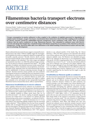
pH Figure 2 | Biogeochemical impacts of filament cutting. a–c, Microprofiles of
oxygen, sulphide and pH measured in undisturbed sediment cores (a),
0 100 200 300
10–60 min (b) and 1 day (c) after passing a thin tungsten wire (50 mm diameter)
horizontally through the sediment near the oxic–anoxic interface. Data are
[O2] or ΣH2S (μM) presented as mean values 6 s.e.m. (oxygen and pH, n 5 9; sulphide, n 5 6).
5 μm Volume-specific rates of oxygen consumption rates (grey areas) are calculated
from numerical modelling of the measured oxygen concentration profiles.
Structure of Desulfobulbaceae filaments
d Desulfobulbaceae DSB706
The multicellular Desulfobulbaceae filaments showed a unique struc-
ture with uniform ridges running along their entire length (Fig. 5a).
Desulfocapsa sulfexigens Transmission electron microscopy of thin sections showed two types
Desulfonema Desulfurivibrio alkaliphilus of filaments that either had 15 ridges and were about 400 nm wide or
magnum
Desulfobacter
Desulfobulbus japonicus
had 17 ridges and were about 700 nm wide (Fig. 5b, c). Each ridge
postgatei contained a filled, 70–100 nm wide channel between the cytoplasmic
Desulfobulbus propionicus
and the outer membrane. The adjacent cells within the filaments were
Desulfovibrio Filament DNA separated by 200 nm wide gaps bridged by the ridge filling and tightly
desulfuricans Filament RNA
Sediment RNA wrapped up by the outer membrane, which seemed to encase the
0.10 Glass microsphere DNA entire filament (Fig. 5d, e). A similar collective outer membrane of
ELF645 the filamentous multicellular cyanobacterium Anabaena sp. has been
suggested to ease exchange of nutrients between cells without leakage
Figure 1 | Filamentous Desulfobulbaceae in current-producing sediments. to the surroundings6. It is possible that the outer membrane of the
a, Microprofiles of O2, pH and SH2S (SH2S 5 ([H2S] 1 [HS2] 1 [S22]).
Desulfobulbaceae filaments has a similar function with respect to
b, Tuft of filamentous Desulfobulbaceae collected from the sulphide-free zone.
c, Filamentous Desulfobulbaceae identified by fluorescence in situ electrons. On the basis of all the morphological data, we propose that
hybridization targeting 16S rRNA. Filament cells appear yellow from overlay of
O2 consumption rate O2 consumption rate O2 consumption rate
images obtained with probe DSB706 (labelled green) and probe ELF654 (μmol cm–3 d–1) (μmol cm–3 d–1) (μmol cm–3 d–1)
(labelled red); other cells appear blue from DNA-staining with 49,6-diamidino- 0 20 40 60 0 20 40 60 0 20 40 60
2-phenylindole (DAPI). d, Phylogenetic affiliation (by maximum likelihood) of O2 concentration (μM) O2 concentration (μM) O2 concentration (μM)
the filaments based on 16S rRNA sequences. Scale bar, 10% estimated sequence 0 100 200 300 400 0 100 200 300 400 0 100 200 300 400
divergence; filled and open circles show bootstrap support .80% and .60%,
respectively (by maximum parsimony; 1,000 iterations); the specificity of the a b c
0 0 0
probes used for FISH is indicated by the green and yellow shading. Accession
numbers are given in Supplementary Table 1.
Depth (mm)
1 1 1
oxygen consumption rates (Tukey’s test under ANOVA; P , 0.001; 2 2 2
alpha 5 0.05; n 5 18) and absence of pH peaks in the oxic zone of Pore size: 2.0 μm Pore size: 0.8 μm Pore size: 0.22 μm
3 3 3
these cores. This observation confirms that passage of bacteria-sized
objects and not only dissolved or colloidal compounds were required. 5 6
pH
7 8 5 6
pH
7 8 5 6
pH
7 8
Sediment particles were not essential as mediators for the electron
transport either, because manifestations of long-distance electron Figure 3 | Biogeochemical effects of filter pore size. a–c, Depth
transport also developed in incubations where a 5-mm sediment layer distributions of oxygen concentrations (red circles) and pH (blue circles)
measured after 20 days of incubation in sediment cores containing
had been replaced by non-conductive glass microspheres (Fig. 4). The
polycarbonate filters with different pore sizes: 2.0 mm (a), 0.8 mm (b) and
filamentous Desulfobulbaceae were abundant in the glass micro- 0.22 mm (c). The filter position is indicated by the dashed line. Volume-specific
sphere layer as confirmed by FISH and 16S rRNA gene sequencing rates of oxygen consumption (grey bars) were estimated from numerical
(Figs 1c and 4b), and no other connecting structures were observed by modelling of the oxygen concentration profiles. Data are presented as mean
light and scanning electron microscopy (SEM) inspection. values 6 s.e.m. (n 5 6).
8 NO V E M B E R 2 0 1 2 | VO L 4 9 1 | N AT U R E | 2 1 9
©2012 Macmillan Publishers Limited. All rights reserved](data:image/gif;base64,R0lGODlhAQABAIAAAAAAAP///yH5BAEAAAAALAAAAAABAAEAAAIBRAA7)
Recomendados
Recomendados
Mais conteúdo relacionado
Mais procurados
Mais procurados (20)
Semelhante a Bacterial cable
Semelhante a Bacterial cable (20)
Bacterial cable
- 1. ARTICLE doi:10.1038/nature11586 Filamentous bacteria transport electrons over centimetre distances Christian Pfeffer1, Steffen Larsen2, Jie Song3, Mingdong Dong3, Flemming Besenbacher3, Rikke Louise Meyer2,3, Kasper Urup Kjeldsen1, Lars Schreiber1, Yuri A. Gorby4, Mohamed Y. El-Naggar5, Kar Man Leung4,5, Andreas Schramm1,2, Nils Risgaard-Petersen1 & Lars Peter Nielsen1,2 Oxygen consumption in marine sediments is often coupled to the oxidation of sulphide generated by degradation of organic matter in deeper, oxygen-free layers. Geochemical observations have shown that this coupling can be mediated by electric currents carried by unidentified electron transporters across centimetre-wide zones. Here we present evidence that the native conductors are long, filamentous bacteria. They abounded in sediment zones with electric currents and along their length they contained strings with distinct properties in accordance with a function as electron transporters. Living, electrical cables add a new dimension to the understanding of interactions in nature and may find use in technology development. Marine sediments become anoxic because oxygen is consumed by micro- identity to any cultured member of this family (Fig. 1d). Almost bial processes at the surface. Without available oxygen the microorgan- identical (. 99%) 16S rRNA sequences were detected in the suboxic isms living below the surface are supposed to depend on energetically less zones of three replicate cores (Fig. 1d), whereas they were absent in the favourable, anaerobic processes1. Recently, however, electric currents subjacent sulphidic zones. The filaments were identified and quan- have been found to directly connect oxygen reduction at the surface with tified in the sediments by fluorescence in situ hybridization (FISH) sulphide oxidation in the subsurface2, even when oxygen and sulphide with specific 16S rRNA-targeting probes (Fig. 1c). The length density are separated by more than 1 cm. Half of the sediment oxygen consump- of filaments was at least 117 m cm23 in the oxic and suboxic sediment tion can be driven by electrons transported from below2,3. The spatial zone and no filaments were detected in the deeper sulphidic zone. separation of oxidation and reduction processes invokes steep pH gra- With an average cell length of 3 mm, this length density corresponded dients leading to distinct dissolutions and precipitations of minerals3. to 4 3 107 cells cm23. Individual filaments were difficult to dissect out Microbial activity apparently drives the electrochemical half-reactions without breaking them, however, fragments up to 1.5 cm long were and the establishment of electron-conducting structures through the recorded, supporting that the bacteria had the length to span the sediment2. Bacterial nanowires, humus particles and semi-conductive entire suboxic zone. mineral grains are known to conduct electrons over nanometre to micro- metre distances, and alone or in combination they have been proposed to Desulfobulbaceae filaments qualify as conductors facilitate electron transport over centimetre distances in marine sedi- We demonstrated experimentally that electron transport through the ment2,4,5. Experimental tests and observations have failed to confirm suboxic zone was mediated by a coherent structure like a bacterial these proposals so far, and instead we have unexpectedly found long, filament and not by diffusive electron shuttles or casual contact filamentous bacteria structured like electric cables as reported below. between conductive elements. Evidence for solid conductors was found by passing a very thin tungsten wire (50 mm diameter) hori- Filamentous Desulfobulbaceae in electric sediment zontally through sediment 1–2 mm below the oxic–anoxic interface, When sulphidic defaunated marine sediment was incubated in the thus transiently interrupting the sediment continuum (see Sup- dark with overlying oxic sea water, the porewater chemistry gradually plementary Information for details). The cut resulted in an immediate developed in accordance with the establishment of an electron trans- and lasting halt of the electron transport, as indicated by a significant port mechanism that coupled oxygen reduction at the sediment sur- drop in oxygen consumption (Tukey’s test under analysis of variance face to sulphide oxidation in deeper anoxic layers (Fig. 1a). As (ANOVA); P , 0.001; alpha 5 0.05; n 5 27), attenuation of the pH described previously2,3, the electric coupling of spatially segregated peak in the oxic zone and contraction of the suboxic zone (Fig. 2), the processes was evident from (1) the presence of a distinct pH peak latter being similar to the response to momentary oxygen removal demonstrating aerobic proton consumption—an indicator of electro- observed in a previous study2. The role of bacteria in establishing a chemical oxygen reduction2,3—and (2) the formation of a 12–15-mm centimetre-long electron transport mechanism was further investi- deep suboxic zone separating sulphide oxidation from the associated gated in incubations, where filters with defined pore sizes were oxygen reduction2,3. When sediment from the top 20 mm was gently inserted into sediment cores to selectively exclude or permit vertical washed, tufts of entangled filamentous bacteria appeared (Fig. 1b). growth or migration of bacteria. Manifestations of long-distance elec- Reverse transcription, cloning and sequencing of 16S ribosomal RNA tron transport appeared in cores containing filter barriers with pore from dissected filaments identified them as novel members of the sizes of 2.0 mm (Fig. 3), but did not appear in cores containing filter deltaproteobacterial family Desulfobulbaceae with , 92% sequence barriers with pore sizes # 0.8 mm as indicated from significantly lower 1 Center for Geomicrobiology, Department of Bioscience, Aarhus University, 8000 Aarhus C, Denmark. 2Section for Microbiology, Department of Bioscience, Aarhus University, 8000 Aarhus C, Denmark. 3 Centre for DNA Nanotechnology (CDNA), Interdisciplinary Nanoscience Center (iNANO), Aarhus University, 8000 Aarhus C, Denmark. 4Department of Biological Sciences, University of Southern California, Los Angeles, California 90089, USA. 5Department of Physics and Astronomy, University of Southern California, Los Angeles, California 90089, USA. 2 1 8 | N AT U R E | VO L 4 9 1 | 8 NO V E M B E R 2 0 1 2 ©2012 Macmillan Publishers Limited. All rights reserved
- 2. ARTICLE RESEARCH O2 consumption rate O2 consumption rate O2 consumption rate a 0 b (μmol cm–3 d–1) (μmol cm–3 d–1) (μmol cm–3 d–1) 0 20 40 60 0 20 40 60 0 20 40 60 O2 concentration (μM) O2 concentration (μM) O2 concentration (μM) 0 100 200 300 400 0 100 200 300 400 0 100 200 300 400 5 a b c 0 0 0 1 1 1 2 2 2 10 Depth (mm) O2 Depth (mm) Before cutting 10–60 minutes after cutting 1 day after cutting 3 3 3 pH 5 6 7 8 ΣH2S 5 pH 5 15 c 10 O2 consumption rate 10 O2 concentration 15 pH 15 ΣH2 S concentration Sediment surfaces 20 200 μm 20 Cut line 20 5 6 7 8 5 6 7 8 pH pH 0 10 20 30 40 50 0 10 20 30 40 50 25 ΣH2S (μM) ΣH2S (μM) 6 7 8 pH Figure 2 | Biogeochemical impacts of filament cutting. a–c, Microprofiles of oxygen, sulphide and pH measured in undisturbed sediment cores (a), 0 100 200 300 10–60 min (b) and 1 day (c) after passing a thin tungsten wire (50 mm diameter) horizontally through the sediment near the oxic–anoxic interface. Data are [O2] or ΣH2S (μM) presented as mean values 6 s.e.m. (oxygen and pH, n 5 9; sulphide, n 5 6). 5 μm Volume-specific rates of oxygen consumption rates (grey areas) are calculated from numerical modelling of the measured oxygen concentration profiles. Structure of Desulfobulbaceae filaments d Desulfobulbaceae DSB706 The multicellular Desulfobulbaceae filaments showed a unique struc- ture with uniform ridges running along their entire length (Fig. 5a). Desulfocapsa sulfexigens Transmission electron microscopy of thin sections showed two types Desulfonema Desulfurivibrio alkaliphilus of filaments that either had 15 ridges and were about 400 nm wide or magnum Desulfobacter Desulfobulbus japonicus had 17 ridges and were about 700 nm wide (Fig. 5b, c). Each ridge postgatei contained a filled, 70–100 nm wide channel between the cytoplasmic Desulfobulbus propionicus and the outer membrane. The adjacent cells within the filaments were Desulfovibrio Filament DNA separated by 200 nm wide gaps bridged by the ridge filling and tightly desulfuricans Filament RNA Sediment RNA wrapped up by the outer membrane, which seemed to encase the 0.10 Glass microsphere DNA entire filament (Fig. 5d, e). A similar collective outer membrane of ELF645 the filamentous multicellular cyanobacterium Anabaena sp. has been suggested to ease exchange of nutrients between cells without leakage Figure 1 | Filamentous Desulfobulbaceae in current-producing sediments. to the surroundings6. It is possible that the outer membrane of the a, Microprofiles of O2, pH and SH2S (SH2S 5 ([H2S] 1 [HS2] 1 [S22]). Desulfobulbaceae filaments has a similar function with respect to b, Tuft of filamentous Desulfobulbaceae collected from the sulphide-free zone. c, Filamentous Desulfobulbaceae identified by fluorescence in situ electrons. On the basis of all the morphological data, we propose that hybridization targeting 16S rRNA. Filament cells appear yellow from overlay of O2 consumption rate O2 consumption rate O2 consumption rate images obtained with probe DSB706 (labelled green) and probe ELF654 (μmol cm–3 d–1) (μmol cm–3 d–1) (μmol cm–3 d–1) (labelled red); other cells appear blue from DNA-staining with 49,6-diamidino- 0 20 40 60 0 20 40 60 0 20 40 60 2-phenylindole (DAPI). d, Phylogenetic affiliation (by maximum likelihood) of O2 concentration (μM) O2 concentration (μM) O2 concentration (μM) the filaments based on 16S rRNA sequences. Scale bar, 10% estimated sequence 0 100 200 300 400 0 100 200 300 400 0 100 200 300 400 divergence; filled and open circles show bootstrap support .80% and .60%, respectively (by maximum parsimony; 1,000 iterations); the specificity of the a b c 0 0 0 probes used for FISH is indicated by the green and yellow shading. Accession numbers are given in Supplementary Table 1. Depth (mm) 1 1 1 oxygen consumption rates (Tukey’s test under ANOVA; P , 0.001; 2 2 2 alpha 5 0.05; n 5 18) and absence of pH peaks in the oxic zone of Pore size: 2.0 μm Pore size: 0.8 μm Pore size: 0.22 μm 3 3 3 these cores. This observation confirms that passage of bacteria-sized objects and not only dissolved or colloidal compounds were required. 5 6 pH 7 8 5 6 pH 7 8 5 6 pH 7 8 Sediment particles were not essential as mediators for the electron transport either, because manifestations of long-distance electron Figure 3 | Biogeochemical effects of filter pore size. a–c, Depth transport also developed in incubations where a 5-mm sediment layer distributions of oxygen concentrations (red circles) and pH (blue circles) measured after 20 days of incubation in sediment cores containing had been replaced by non-conductive glass microspheres (Fig. 4). The polycarbonate filters with different pore sizes: 2.0 mm (a), 0.8 mm (b) and filamentous Desulfobulbaceae were abundant in the glass micro- 0.22 mm (c). The filter position is indicated by the dashed line. Volume-specific sphere layer as confirmed by FISH and 16S rRNA gene sequencing rates of oxygen consumption (grey bars) were estimated from numerical (Figs 1c and 4b), and no other connecting structures were observed by modelling of the oxygen concentration profiles. Data are presented as mean light and scanning electron microscopy (SEM) inspection. values 6 s.e.m. (n 5 6). 8 NO V E M B E R 2 0 1 2 | VO L 4 9 1 | N AT U R E | 2 1 9 ©2012 Macmillan Publishers Limited. All rights reserved
- 3. RESEARCH ARTICLE O2 concentration (μM) a 200 nm 0 100 200 300 400 a b 0 2 4 Depth (mm) 6 8 200 nm 200 nm 10 12 14 16 18 5 μm 6 7 8 b d pH c e 0 20 40 60 80 100 ΣH2S (μM) Figure 4 | Effect of layer of glass beads intercalated in the sediment. a, Oxygen concentrations (red lines), pH (blue lines) and sulphide concentrations (black lines) in three sediment cores with an intercalated layer of electrically inert glass microspheres reaching from 3 mm down to 8 mm sediment depth (hatched area). b, Micrograph showing filamentous Desulfobulbaceae extracted from a glass bead section and hybridized with the specific ELF654 FISH probe. DAPI-stained chromosomes are visible in some cells of the filament. the ridges are strings transporting electrons along the filament inside 200 nm 200 nm a continuous periplasmic space, with the collective outer membrane serving as electrical insulation from the external medium. To explore f g the conductive properties of the strings, electrostatic force microscopy (EFM)7 was applied, as this technique does not require direct contact d and may therefore trace electric properties of insulated materials. A distinct elevation of electrostatic force on the ridges as compared to the intermittent areas (Fig. 5g) indicates that the underlying strings possess significant polarizability or charge storage capacity along their length as well as across cell–cell junctions. The highly organized con- 500 nm trast pattern excludes significant contribution from random leakage of polymers like extracellular DNA. These results are consistent with the proposed charge distribution role. Furthermore, we tested the hypothesis using a nanofabrication approach, similar to that pre- viously used to demonstrate charge transfer along the length of a bacterial nanowire8. Current–voltage measurements were performed –1 V 0 1V on individual filaments that were bridging two gold electrodes and Figure 5 | AFM, SEM, TEM and EFM micrographs of the filamentous fixed with platinum leads. In contrast to what should be observed if Desulfobulbaceae. a, SEM image of four cells. b, c, Thin-section TEM images the conductive structures were exposed to the surface, sweeping the of filament cross sections. d, e, Longitudinal section including a cell–cell voltage from 210 V to 10 V was not accompanied by any measurable junction (d) and oblique section of cell–cell junction (e). f, Height AFM image electrical currents. This observation supports the hypothesis that con- of cell–cell junction with inserted topographic curve along 500 nm line with the ductive structures present in the periplasmic continuum are electric- same scaling of x, y and z axes. g, 1 3 1 mm EFM image above cell–cell junction with lighter contrast closely following the ridge topography mapped in the ally insulated by the collective outer membrane. preceding AFM scan (not shown). Discussion Many prokaryotes are known to perform extracellular electron ex- Taken together, the data presented here strongly indicate that long- change as a method to control metabolic interactions with external distance electron transport from sulphide to oxygen in the sediment is solids or cells. Electrons are exchanged with insoluble electron accep- mediated by living micro-cables in the form of long filamentous bac- tors and donors like iron and manganese minerals9 or electrodes10. teria of the Desulfobulbaceae family. As the simplest metabolic model Furthermore, syntrophic interspecies electron exchange via nano- consistent with our data, we propose that the micro-cables are multi- wires and magnetite grains has recently been confirmed4,11. External cellular, aerobic, sulphide oxidizers where electrons generated by deposition of electrons preventing internal accumulation of insoluble sulphide oxidation in cells at one end can be passed through internal, and potentially harmful metabolites has also been found12. However, insulated wires to cells at the other end and here are consumed by different from these prokaryotes using external electron transport, the oxygen reduction. Charge transport by ions in the surrounding envir- competitive advantage of micro-cables seems to be their ability to sepa- onment balances the internal electron transport, thereby completing rate soluble electron acceptors and donors in space, thereby enabling the electric circuit and retaining charge balance2,3. them to monopolise major energy sources. This is done by transport of 2 2 0 | N AT U R E | VO L 4 9 1 | 8 NO V E M B E R 2 0 1 2 ©2012 Macmillan Publishers Limited. All rights reserved
- 4. ARTICLE RESEARCH electrons from donors to acceptors over long distances via insulated 2. Nielsen, L. P., Risgaard-Petersen, N., Fossing, H., Christensen, P. B. & Sayama, M. Electric currents couple spatially separated biogeochemical processes in marine internal wires with no need for electron transfer across the outer mem- sediment. Nature 463, 1071–1074 (2010). brane. Oxidation of sulphide is a major driver of oxygen consumption 3. Risgaard-Petersen, N., Revil, A., Meister, P. & Nielsen, L. P. Sulfur, iron-, and calcium in marine sediments, and the question arises how successful the fila- cycling associated with natural electric currents running through marine sediment. Geochim. Cosmochim. Acta 92, 1–13 (2012). mentous bacteria are in competing with other sulphide oxidizing bac- 4. Kato, S., Hashimoto, K. & Watanabe, K. Microbial interspecies electron transfer via teria and in controlling the process. Separation of oxygen and sulphide electric currents through conductive minerals. Proc. Natl Acad. Sci. USA 109, is common in marine sediment and conventionally explained by iron 10042–10046 (2012). 5. Roden, E. E. et al. Extracellular electron transfer through microbial reduction of and manganese biogeochemistry driven by particle mixing or by bac- solid-phase humic substances. Nature Geosci. 3, 417–421 (2010). terial nitrate transport13–16. Few studies have however directly quan- 6. Mariscal, V., Herrero, A. & Flores, E. Continuous periplasm in a filamentous, tified these pathways. heterocyst-forming cyanobacterium. Mol. Microbiol. 65, 1139–1145 (2007). 7. Lu, W., Wang, D. & Chen, L. W. Near-static dielectric polarization of individual Bacterial micro-cables represent a hitherto unknown lifestyle, which carbon nanotubes. Nano Lett. 7, 2729–2733 (2007). immediately raises many intriguing questions for further research: How 8. El-Naggar, M. Y. et al. Electrical transport along bacterial nanowires from are energy conservation and growth allocated among the cells? What is Shewanella oneidensis MR-1. Proc. Natl Acad. Sci. USA 107, 18127–18131 (2010). their genetic and metabolic diversity? How are filament division and 9. Reguera, G. et al. Extracellular electron transfer via microbial nanowires. Nature 435, 1098–1101 (2005). dispersal controlled? What is the molecular and electronic basis of the 10. Holmes, D. E., Bond, D. R. & Lovley, D. R. Electron transfer by Desulfobulbus electron transport? How widespread are they in nature? Transmis- propionicus to Fe(III) and graphite electrodes. Appl. Environ. Microbiol. 70, sion improvement and control of electric currents have been major 1234–1237 (2004). 11. Summers, Z. M. et al. Direct exchange of electrons within aggregates of an evolved drivers for electronic innovation. It appears that biological evolution syntrophic coculture of anaerobic bacteria. Science 330, 1413–1415 (2010). has worked successfully in the same direction. 12. Cologgi, D. L., Lampa-Pastirk, S., Speers, A. M., Kelly, S. D. & Reguera, G. Extracellular reduction of uranium via Geobacter conductive pili as a protective cellular mechanism. Proc. Natl Acad. Sci. USA 108, 15248–15252 (2011). METHODS SUMMARY 13. Canfield, D. E. Reactive iron in marine-sediments. Geochim. Cosmochim. Acta 53, Microbiological and geochemical data were obtained from sulphidic sediment 619–632 (1989). sampled in Aarhus Bay, Denmark. The sediment was sieved, transferred to glass 14. Fossing, H. et al. Concentration and transport of nitrate by the mat-forming sulphur core liners and incubated for three to four weeks in aquaria with circulating air- bacterium Thioploca. Nature 374, 713–715 (1994). 15. Preisler, A. et al. Biological and chemical sulfide oxidation in a Beggiatoa inhabited saturated sea water as described previously2. Oxygen, sulphide and pH micro- marine sediment. ISME J. 1, 341–353 (2007). profiles were measured as described previously17–19. 16. Sayama, M., Risgaard-Petersen, N., Nielsen, L. P., Fossing, H. & Christensen, P. B. Nucleic acids were extracted from sediment, glass microspheres and from 2 Impact of bacterial NO3 transport on sediment biogeochemistry. Appl. Environ. single filaments collected by micromanipulation. RNA was reverse-transcribed, Microbiol. 71, 7575–7577 (2005). and 16S rRNA complementary DNA fragments were PCR-amplified, cloned and 17. Jeroschewski, P., Steuckart, C. & Kuhl, M. An amperometric microsensor for the sequenced. Oligonucleotide probes for FISH were designed specific for the determination of H2S in aquatic environments. Anal. Chem. 68, 4351–4357 (1996). 18. Revsbech, N. P. An oxygen microsensor with a guard cathode. Limnol. Oceanogr. retrieved sequences and used for microscopic identification and quantification 34, 474–478 (1989). of the filamentous Desulfobulbaceae by FISH in fixed sediment samples. 19. Revsbech, N. P. & Jorgensen, B. B. Microelectrodes: their use in microbial ecology. AFM images of rinsed air-dried cells were obtained on a Nanowizard II (JPK Adv. Microb. Ecol. 9, 293–352 (1986). Instruments) in intermittent contact mode under ambient conditions with an 20. Cherniavskaya, O., Chen, L. W., Weng, V., Yuditsky, L. & Brus, L. E. Quantitative OMCL-AC160TN cantilever (Olympus). noncontact electrostatic force imaging of nanocrystal polarizability. J. Phys. Chem. B 107, 1525–1531 (2003). EFM images of rinsed air-dried cells were performed on a Bruker Dimension 3100 (Bruker Corporation) equipped with a Pt/Ir-coated tip (SCMPIC, Veeco Supplementary Information is available in the online version of the paper. Metrology) using the two-pass scan technique20. On the second pass, the probe Acknowledgements The authors thank P. G. Sørensen and L. B. Pedersen for was lifted at 5 nm with respect to the sample topography, while an oscillating bias construction of microsensors. Thanks to K. E. Thomsen for operating the TEM. potential V 5 Vdc 1 Vacsin(vt) was applied between the tip and the substrate B. B. Jørgensen is thanked for his general support. This research was financially (Vdc, direct current voltage; Vac, alternate current voltage; v, modulation fre- supported by the European Research Council (L.P.N.), the Danish National Research quency (500 Hz)). The electrostatic force can be represented by volts. Foundation (N.R.-P., C.P., K.U.K., L.S., M.D.), the Danish Council for Independent Research j Natural Sciences (FNU) (L.P.N.), the German Max Planck Society (N.R.-P., SEM of air dried cells were obtained on a NanoSEM (FEI, Nova 600 NanoSEM) C.P., K.U.K., L.S.) and the Villum Foundation (M.D.). operated in low-vacuum (60 Pa) and low-voltage (3 kV) mode to apply the charge contrast imaging data. Author Contributions This study was conceived by L.P.N., N.R.-P. and A.S. Experimental TEM of 40–60 nm cell sections were obtained on an FEI CM100 (FEI CM100). work: C.P. FISH and molecular phylogeny: S.L. Single-cell identification: K.U.K. and L.S. AFM: R.L.M., J.S. and M.D. SEM and EFM: J.S., M.D. and F.B. TEM: L.P.N. and J.S. Electric conductivity of the cell surface was addressed with nano-fabricated electrodes. Conductivity measurements using nanofabricated electro discs: J.S., Y.A.G., M.Y.E.-N., A full description of the methodology applied in the present study is given in K.M.L. and C.P. All authors contributed to discussions of the data and preparation of the the Supplementary Information. manuscript. Author Information All sequences are deposited in GenBank/EMBL/DDBJ under Received 6 July; accepted 17 September 2012. accession numbers JX091023–JX091073. Reprints and permissions information is Published online 24 October 2012. available at www.nature.com/reprints. The authors declare no competing financial interests. Readers are welcome to comment on the online version of the paper. 1. Jørgensen, B. B. in Marine Geochemistry (eds Schulz, H. D. & Zabel, M.) 173–207 Correspondence and requests for materials should be addressed to N.R.-P. (Springer, 2000). (nils.risgaard-petersen@biology.au.dk) or L.P.N. (biolpn@biology.au.dk). 8 NO V E M B E R 2 0 1 2 | VO L 4 9 1 | N AT U R E | 2 2 1 ©2012 Macmillan Publishers Limited. All rights reserved