SlideShare a Scribd company logo
1 of 70
Download to read offline
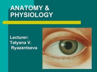
ANATOMY &
PHYSIOLOGY
Lecturer:
Tatyana V.
Ryazantseva
Outer eye:
Eyelids
The eyelids fulfill two main functions:
 protection of the eyeball
 secretion, distribution and drainage of
tears
Lid movement
 The levator extends from an attachment at the orbital
apex to attachments at the tarsal plate and skin.
● The lids are securely attached at either end to the
bony orbital margin by the medial and lateral
palpebral ligaments.
 Trauma to the medial ligament causes the lid to
flop forward and laterally, impairing function and
cosmesis.
Innervation
- Sensory innervation is from the trigeminal
(fifth) cranial nerve, via the ophthalmic
division (upper lid) and maxillary division
(lower lid).
- The orbicularis oculi is innervated by the
facial (seventh) nerve.
- The levator muscle in the upper lid is
supplied by the oculomotor (third) nerve.
Blood supply and lymphatics
The eyelids are supplied by an extensive
network of blood vessels which form an
anastomosis between branches derived
from the external carotid artery via the
face and from the internal carotid artery
via the orbit.
Blood supply and lymphatics
 Lymphatic fluid drains into the preauricular
and submandibular nodes.
 Preauricular lymphadenopathy is a useful
sign of infective eyelid swelling (especially
viral).
● Superior fornix of
conjunctiva
 Levator muscle
 Sclera
 Grey line
 Conjunctiva
 Orbicularis oculi muscle
 Inferior fornix of
conjunctiva
 Extraocular muscle
 Optic nerve
Eyelids and eyeball
Eyelid structure
Conjunctiva
 The conjunctiva is a mucous membrane
lining the eyelids and covering the anterior
eyeball up to the edge of the cornea.
 At the upper and lower reflections between
eyeball and eyelid the conjunctiva forms two
sacs, the superior and inferior fornices .
Cornea and sclera
The cornea and sclera form a spherical
shell which makes up the outer wall of the
eyeball.
Cornea and sclera
 The sclera is :
- principally collagenous,
- avascular (apart from some vessels on its surface)
- relatively acellular.
It is perforated posteriorly by the optic nerve, and by
sensory and motor nerves and blood vessels to the
eyeball.
The cornea and sclera merge at the corneal edge
(the limbus).
The chief functions of the cornea
 are protection against invasion of
microorganisms into the eye, and the
transmission and focusing (refraction) of
light.
Cornea
 Epithelium
 Bowman membrane
 Stroma
 Descemet
membrane
(posterior limiting
layer of cornea)
 Endothelium
Cornea and sclera
 Retraction of light occurs because of the
curved shape of the cornea and its greater
refractive index compared with air.
 The cornea is transparent because of the
specialised arrangement of the collagen
fibrils within the stroma, which must be kept
in a state of relative dehydration.
Sagittal section of the eye
 Fibrous tunic of
eye
 Choroid
 Retina
Sagittal section of the eye
Tear production and drainage
 The lacrimal gland secretes most of the
aqueous component of the tear film .
 It lies in the superotemporal part of the
anterior orbit Its anterior lobe can sometimes
be seen in the upper conjunctival fornix.
 It is innervated by parasympametic fibres
carried by the facial nerve.
Tear production and drainage
 Tears collect in a meniscus on the lower lid margin,
are spread across the ocular surfaces by blinking,
and drain into the superior and inferior puncta at the
nasal end of the eyelids. Single canaliculi from each
punctum unite in a common canaliculus which ends
in the lacrimal sac This is in a bony fossa crossed
anteriorly by the horizontally directed medial
palpebral ligament . Finally, tears pass down the
nasolacrimal duct and reach the nasopharyngeal
cavity via the inferior meatus. This accounts for the
unpleasant taste which follows administration of
certain eyedrops.
Tear production and drainage
 At birth, the nasolacrimal duct may not be
fully developed, causing a watery eye.
 Acquired obstruction of the nasolacrimal duct
is a common cause of a watery eye in adults.
It may lead to an acute infection of the sac.
!!!Anatomy and physiology: outer eye
Eyelids
 protect the eyeball and distribute tears across the
cornea
 closure by contraction of orbicularis ocuii muscle
(facial nerve)
 opening by levator muscle (oculomotor nerve)
 lid margin comprises a row of lashes in front of a row
of meibomian
gland orifices separated by the grey line
Conjunctiva
 a mucous membrane which contributes to
tear production and
resistance to infection.
Cornea
a highly specialised tissue
 main function is refraction and transmission of light
 structure is an outer epithelium, an avascular
hypocellular stroma
and a non-replicating endothelial monolayer
 the endothelium pumps water out of the stroma into
the anterior
chamber; failure leads to loss of transparency
Tears
● oily lipid layer secreted by meibomian glands;
aqueous layer by
lacrimal and conjunctival glands;
- mucin by conjunctival goblet cells
 drain into the puncta, then canaliculi, then
lacrimal sac, then into nose via nasolacrimal
duct.
Sensation
 trigeminal nerve, mostly via the ophthalmic
division
(maxillary to the lower lid).
Anatomy and physiology:
inner eye
BASIC PRINCIPLES
Inner eye
 The uvea comprises the iris and ciliary body
anteriorly and the choroid posteriorly .
UVEA
Iris
The iris largely consists of connective tissue
containing muscle fibres, blood vessels and pigment
cells.
Its posterior surface is lined by a layer of pigment
cells. At its centre is an aperture, the pupil.
The chief functions of the iris are to control light
entry to the retina and to reduce intraocular light
scatter.
Pupil dilation is caused by contraction of radial
smooth muscle fibres innervated by the sympathetic
nervous system.
Радужка, вид снизу
Ciliary body
 The ciliary body is a specialised structure
uniting the iris with the choroid.
 It makes aqueous humour and anchors
the lens via the zonules, through which it
modulates lens convexity.
Ciliary Body
 This is called accommodation and is
controlled by parasympathetic fibres in the
oculomotor nerve.
 The posterior part of the ciliary body merges
into the retina at the ora serrata.
Choroid
 The choroid, consisting of blood vessels,
connective tissue and pigment cells, is
sandwiched between the retina and the
sclera.
 It provides oxygen and nutrition to the
outer retinal layers.
Lens
 The discus-like lens comprises a mass of
long cells known as fibres. At the centre
these fibres are compacted into a hard
nucleus surrounded by less dense fibres, the
cortex.
 The lens is relatively dehydrated and its
fibres contain special proteins. This is why it
is transparent.
Lens
Aqueous humour
fills the anterior and posterior chambers.
The anterior chamber is the space between
the cornea and the iris.
Behind the iris and in front of the lens is the
posterior chamber.
They are connected by the pupil.
Formation
 The ciliary body forms aqueous
humour by ultrafiltration and
active secretion.
 Retina
 Choroid
 Sclera
 Optic cup
 Optic disc
 Subarachnoid space
 Anterior chamber angle
 Trabecular meshwork
 Canal of Schlemm
 Zonules
 Lenscapsule
 Lens cortex
 Lens nucleus
 Fovea at centre of macula
 Lamina cribrosa
 Path of central retinal artery and
vein
Sagittal section of the eye
●Ganglion cells
●Connecting and
processing cells
●Photoreceptor rods and
cones
●Retinal pigment epithelium
Layers of the retina
Drainage
Aqueous circulates from the posterior to the
anterior chamber through the pupil, leaving
the eye through the trabecular meshwork.
This is a specialised tissue in the anterior
chamber angle between the iris and the
cornea. From here aqueous drains into
Schlemm's canal.
Vitreous
 The vitreous body is 99% water but, vitally,
also contains collagen fibrils and hyaluronan,
which impart cohesion and a gel-like
consistency.
 The vitreous is adherent to the retina at
certain points, particularly at the optic disc
and at the ora serrata.
Retina
 The retina converts focused light images into
nerve impulses. Light has to pass through the
inner retina to reach the photoreceptors, the rods
and cones, which convert light energy into
electrical energy .
 Macula provides for central vision. At its centre is
a specialised area, the fovea, which is for high
quality vision.
 The rest of the retina is for peripheral vision.
Retina
 Cones, concentrated at the macula, are
responsible for fine vision (acuity) and
colour appreciation.
 Rods are for vision in low light levels and
the detection of movement.
Diagram of retina
Retina
 The blood supply of the retina is derived from
the central retinal artery and vein, and from
the choroid.
 The retinal vessels enter and leave the eye
through the optic nerve and run in the nerve
fibre layer. A major arterial and venous
branch, forming an 'arcade', supplies each of
the retinal quadrants .
RETINA
Optic nerve
 The ganglion cell axons in the retinal nerve
fibre layer make a right-angled turn into the
optic nerve at the optic disc, which has no
photoreceptor and corresponds to the
physiological blind spot.
Optic Nerve
 Behind the eyeball these axons become
myelinated.
 Here the optic nerve is surrounded by
cerebrospinal fluid in an anterior extension of
the subarachnoid space and is protected by
the same membranous layers as the brain.
!!!Anatomy and physiology: inner eye
Iris
 constriction is parasympathetic
 dilation sympathetic
Ciliary body
 forms aqueous humour;
 mediates accommodation
(active-parasympathetic).
Lens
 consists of a hard nucleus and
softer cortex, bounded by a capsule
and held in place by the zonules.
Aqueous drains
 Aqueous drains
through the
trabecular
meshwork in the
anterior chamber
angle between the iris
and cornea.
Retina
 Requires intact retinal and choroidal vasculature and
retinal
pigment epithelial layer for normal function.
 Photoreceptors convert light energy into electrical;
transmit to
ganglion cells via connector neurones.
 Ganglion cell axons pass across the surface of the
retina and leave the eye at the optic disc.
 Cone photoreceptors are concentrated at the macula
for high
quality colour vision.
The Orbit
 The optic nerves from each eye are
coordinated intra-cranially and connected
with other areas of the cerebral cortex.
 A set of motor centres, cranial nerve nuclei
and connections harness the two eyes
together to maintain binocular vision without
diplopia .
The Orbit
The bony walls of the orbit form a pyramidal
structure. They are the frontal, maxillary,
zygomatic, ethmoid, lacrimal and sphenoid
bones . The medial wall and the floor of the
orbit are thin.
 At the apex of the orbit, the optic foramen
conveys the optic nerve backwards to the
intracranial optic chiasm, and the ophthalmic
artery forward into the orbit .
Lateral to the foramen are two
fissures:
 The superior orbital fissure provides passage
for the lacrimal, frontal and nasociliary
nerves (ophthalmic division of the fifth cranial
nerve), the third, fourth and sixth cranial
nerves, and the superior ophthalmic vein
passing to the cavernous sinus.
The Orbit
 The inferior orbital fissure permits exit of the
inferior ophthalmic vein from the orbit and
entry of the maxillary division of the fifth
cranial nerve (thus a fracture of the floor
of the orbit can cause abnormal sensation on
the cheek).
The Orbit
 The four extraocular rectus muscles form a
cone within which are the sensory and
autonomic nerves and arteries to the eyeball,
including the optic nerve and the motor
nerves to all the extraocular muscles
including the levator but excluding the
superior oblique.
Extraocular muscles
 The four rectus muscles have a common
posterior attachment to a ring of connective
tissue which surrounds the optic foramen.
They pass forwards around the eyeball to
insert 5-7 mm behind the limbus .
Levator muscle
 Levator muscle (third nerve) passes forward
and widens into a broad aponeurosis,
attaching to the superior tarsal plate and
eyelid skin.
Nerves of the Orbit
 In addition to the motor nerves to the
extraocular muscles the orbit contains
sensory and autonomic nerves .
 The chief sensory nerve is of course the
optic nerve. It is enclosed within a sheath
continuous with the intracranial meninges, so
that the subarachnoid space extends right up
to the globe.
Nerves of the Orbit
 Branches of the ophthalmic division of the trigeminal
(fifth) nerve are sensory to the eyeball (especially the
cornea), the conjunctiva and the skin of the eyelids
extending up across the forehead and back towards
the occiput Hence.
 The nasociliary nerve is the branch to the eyeball,
but it does not terminate there. The nerve passes on
into the medial orbital wall and emerges on the side
of the nose.
Nerves of the Orbit
 Parasympathetic fibres to the lacrimal gland
pursue a complex course, passing with the
facial nerve and then following the maxillary
division of the trigeminal.
 The sensory and parasympathetic nerve
fibres reach the eyeball via the short and
long ciliary nerves which pierce the sclera
posteriorly.
The Visual Pathways
 The two optic nerves unite at the optic
chiasm above the sella turcica of the
sphenoid bone.
 The pituitary gland projects down
immediately behind the chiasm.
 Nerve fibres from the nasal retina
(temporal or lateral field of vision) cross
over to the opposite side in the chiasm, so
that the post-chiasmal fibres on the left
subserve the field of vision on the right
(and vice versa).
!!! Relation and connections: orbit and
visual pathways
 The bony walls of the orbit are thin inferiorly and
medially.
 The superior orbital fissure transmits the sensory
nerves to the eye and orbit and the 3rd, 4th and 6th
cranial nerves.
 The optic nerve and the ophthalmic artery pass
through the optic foramen.
 The 3rd nerve supplies levator; superior, inferior and
medial rectus; inferior oblique; accommodation; pupil
constriction.
!!! Relation and connections: orbit and
visual pathways
 The 6th nerve supplies lateral rectus.
 The 4th nerve supplies superior oblique.
 Extraocular muscle function: depends on the direction of
gaze:
- medial rectus: horizontal towards the nose
- lateral rectus: horizontal laterally
- obliques primarily rotate the eye
- vertical recti primarily elevate/depress the eye
 Visual pathways
- optic nerve to chiasm (fibres from nasal retina cross over) to
optic tract
- synapse in lateral geniculate body
- optic radiation to occipital cortex.
THANK YOU !
THANK YOU!

More Related Content

What's hot

What's hot (20)

Visual pathways
Visual pathwaysVisual pathways
Visual pathways
 
Anatomy of the lacrimal apparatus
Anatomy of the lacrimal apparatusAnatomy of the lacrimal apparatus
Anatomy of the lacrimal apparatus
 
Anatomy of eye
Anatomy of eyeAnatomy of eye
Anatomy of eye
 
Anatomy of the eye and Layers of eyeball
Anatomy of the eye and Layers of eyeball Anatomy of the eye and Layers of eyeball
Anatomy of the eye and Layers of eyeball
 
anatomy of eye
anatomy of eyeanatomy of eye
anatomy of eye
 
#ANATOMY OF UVEA (IRIS, CILLIARY BODY, CHOROID)
#ANATOMY OF UVEA (IRIS, CILLIARY BODY, CHOROID) #ANATOMY OF UVEA (IRIS, CILLIARY BODY, CHOROID)
#ANATOMY OF UVEA (IRIS, CILLIARY BODY, CHOROID)
 
Orbital blood supply
Orbital blood supplyOrbital blood supply
Orbital blood supply
 
Orbit anatomy
Orbit   anatomyOrbit   anatomy
Orbit anatomy
 
Extraocular muscles
Extraocular musclesExtraocular muscles
Extraocular muscles
 
Anatomy of optic nerve and its clinical significance
Anatomy of optic nerve and its clinical significanceAnatomy of optic nerve and its clinical significance
Anatomy of optic nerve and its clinical significance
 
RETINA - anatomy & physiology
RETINA - anatomy & physiologyRETINA - anatomy & physiology
RETINA - anatomy & physiology
 
Conjunctiva
ConjunctivaConjunctiva
Conjunctiva
 
Anatomy of orbit
Anatomy of orbitAnatomy of orbit
Anatomy of orbit
 
BLOOD SUPPLY OF VISUAL PATHWAY
BLOOD SUPPLY OF VISUAL PATHWAYBLOOD SUPPLY OF VISUAL PATHWAY
BLOOD SUPPLY OF VISUAL PATHWAY
 
Anatomy and Physiology of Lacrimal system
Anatomy and Physiology of Lacrimal system Anatomy and Physiology of Lacrimal system
Anatomy and Physiology of Lacrimal system
 
Visual pathway
Visual pathwayVisual pathway
Visual pathway
 
Anatomy of Eyelids & Its Clinical Correlations
Anatomy of Eyelids & Its Clinical CorrelationsAnatomy of Eyelids & Its Clinical Correlations
Anatomy of Eyelids & Its Clinical Correlations
 
Anatomy of Eyelids
Anatomy of EyelidsAnatomy of Eyelids
Anatomy of Eyelids
 
Trochlear nerve
Trochlear nerveTrochlear nerve
Trochlear nerve
 
The Orbit
The OrbitThe Orbit
The Orbit
 

Similar to Anatomy of the Eye and Vision

Anatomy of external as well internal eye
Anatomy of external as well internal eye Anatomy of external as well internal eye
Anatomy of external as well internal eye D-lip Raj Gupta
 
د احمد اسامه هاشم عيون ophthalmology
د احمد اسامه هاشم عيون ophthalmologyد احمد اسامه هاشم عيون ophthalmology
د احمد اسامه هاشم عيون ophthalmologyAhmed Osama Hashem
 
Anatomy of the eye for studentss
Anatomy of the eye for studentssAnatomy of the eye for studentss
Anatomy of the eye for studentssSana Anwari
 
01- Anatomy and Physiology of the ey ppt
01- Anatomy and Physiology of the ey ppt01- Anatomy and Physiology of the ey ppt
01- Anatomy and Physiology of the ey pptaamrutha180
 
Anatomy and Physiology.ppt of eye uploaded
Anatomy and Physiology.ppt of eye uploadedAnatomy and Physiology.ppt of eye uploaded
Anatomy and Physiology.ppt of eye uploadedPrabithaManjeshwar
 
Eyes Anatomy & Physiology with Diagnostic investigation.
Eyes Anatomy & Physiology with Diagnostic investigation.Eyes Anatomy & Physiology with Diagnostic investigation.
Eyes Anatomy & Physiology with Diagnostic investigation.DR .PALLAVI PATHANIA
 
Anatomy of the Human Eye By Mohammed Shurrab
Anatomy of the Human Eye By Mohammed ShurrabAnatomy of the Human Eye By Mohammed Shurrab
Anatomy of the Human Eye By Mohammed ShurrabMohammed Shurrab
 
vision.pptx
vision.pptxvision.pptx
vision.pptxLuzSan3
 
anatomy and physiology of eye.ppt.anatomy and physiology of eyex
anatomy and physiology of eye.ppt.anatomy and physiology of eyexanatomy and physiology of eye.ppt.anatomy and physiology of eyex
anatomy and physiology of eye.ppt.anatomy and physiology of eyexAnjuAnnMani1
 
Functional Anatomy of Eye.pptx
Functional Anatomy of Eye.pptxFunctional Anatomy of Eye.pptx
Functional Anatomy of Eye.pptxNoorin Bhimani
 

Similar to Anatomy of the Eye and Vision (20)

Anatomy of eye
Anatomy of eyeAnatomy of eye
Anatomy of eye
 
Anatomy of external as well internal eye
Anatomy of external as well internal eye Anatomy of external as well internal eye
Anatomy of external as well internal eye
 
Eye by meher
Eye by meherEye by meher
Eye by meher
 
Orbital region
Orbital regionOrbital region
Orbital region
 
د احمد اسامه هاشم عيون ophthalmology
د احمد اسامه هاشم عيون ophthalmologyد احمد اسامه هاشم عيون ophthalmology
د احمد اسامه هاشم عيون ophthalmology
 
Anatomy of the eye for studentss
Anatomy of the eye for studentssAnatomy of the eye for studentss
Anatomy of the eye for studentss
 
01- Anatomy and Physiology of the ey ppt
01- Anatomy and Physiology of the ey ppt01- Anatomy and Physiology of the ey ppt
01- Anatomy and Physiology of the ey ppt
 
Anatomy and Physiology.ppt of eye uploaded
Anatomy and Physiology.ppt of eye uploadedAnatomy and Physiology.ppt of eye uploaded
Anatomy and Physiology.ppt of eye uploaded
 
VISION.pdf
VISION.pdfVISION.pdf
VISION.pdf
 
Eyes Anatomy & Physiology with Diagnostic investigation.
Eyes Anatomy & Physiology with Diagnostic investigation.Eyes Anatomy & Physiology with Diagnostic investigation.
Eyes Anatomy & Physiology with Diagnostic investigation.
 
Eye vision
Eye visionEye vision
Eye vision
 
Anatomy of the Human Eye By Mohammed Shurrab
Anatomy of the Human Eye By Mohammed ShurrabAnatomy of the Human Eye By Mohammed Shurrab
Anatomy of the Human Eye By Mohammed Shurrab
 
Eye
EyeEye
Eye
 
Eye
EyeEye
Eye
 
The lacrimal appratus
The lacrimal appratusThe lacrimal appratus
The lacrimal appratus
 
vision.pptx
vision.pptxvision.pptx
vision.pptx
 
anatomy and physiology of eye.ppt.anatomy and physiology of eyex
anatomy and physiology of eye.ppt.anatomy and physiology of eyexanatomy and physiology of eye.ppt.anatomy and physiology of eyex
anatomy and physiology of eye.ppt.anatomy and physiology of eyex
 
anatomy.ppt
anatomy.pptanatomy.ppt
anatomy.ppt
 
anatomy And Physiology of lacrimal secretions
anatomy And Physiology of lacrimal secretions anatomy And Physiology of lacrimal secretions
anatomy And Physiology of lacrimal secretions
 
Functional Anatomy of Eye.pptx
Functional Anatomy of Eye.pptxFunctional Anatomy of Eye.pptx
Functional Anatomy of Eye.pptx
 

More from C L GUPTA EYE INSTITUTE MORADABAD UTTER PRADESH

More from C L GUPTA EYE INSTITUTE MORADABAD UTTER PRADESH (20)

Astigmatism
Astigmatism Astigmatism
Astigmatism
 
Tonometry ppt
Tonometry pptTonometry ppt
Tonometry ppt
 
Slit lump biomicroscope
Slit lump biomicroscopeSlit lump biomicroscope
Slit lump biomicroscope
 
anatomy of sclera
anatomy of sclera anatomy of sclera
anatomy of sclera
 
Pupil ppt
Pupil  pptPupil  ppt
Pupil ppt
 
anatomy And Physiology of tear film
anatomy And Physiology of tear film anatomy And Physiology of tear film
anatomy And Physiology of tear film
 
cornea and layers
cornea and layers cornea and layers
cornea and layers
 
Limbus
LimbusLimbus
Limbus
 
extra ocular muscles
extra ocular muscles extra ocular muscles
extra ocular muscles
 
Diseases of sclera
Diseases of sclera Diseases of sclera
Diseases of sclera
 
Corneal transparency
Corneal transparency Corneal transparency
Corneal transparency
 
Conjunctiva anatomy and physiology
Conjunctiva anatomy and physiology Conjunctiva anatomy and physiology
Conjunctiva anatomy and physiology
 
Color vision deficiency and ishiharas test
Color vision deficiency and ishiharas test Color vision deficiency and ishiharas test
Color vision deficiency and ishiharas test
 
Anatomy and physiology of aqueous humor
Anatomy and physiology of aqueous humor Anatomy and physiology of aqueous humor
Anatomy and physiology of aqueous humor
 
Vitreous humour
Vitreous humourVitreous humour
Vitreous humour
 
Vitamins all
Vitamins allVitamins all
Vitamins all
 
Tear film
Tear filmTear film
Tear film
 
Tca cycle
Tca cycleTca cycle
Tca cycle
 
Synthesis
SynthesisSynthesis
Synthesis
 
Retina
RetinaRetina
Retina
 

Anatomy of the Eye and Vision

  • 2. Outer eye: Eyelids The eyelids fulfill two main functions:  protection of the eyeball  secretion, distribution and drainage of tears
  • 3. Lid movement  The levator extends from an attachment at the orbital apex to attachments at the tarsal plate and skin. ● The lids are securely attached at either end to the bony orbital margin by the medial and lateral palpebral ligaments.  Trauma to the medial ligament causes the lid to flop forward and laterally, impairing function and cosmesis.
  • 4. Innervation - Sensory innervation is from the trigeminal (fifth) cranial nerve, via the ophthalmic division (upper lid) and maxillary division (lower lid). - The orbicularis oculi is innervated by the facial (seventh) nerve. - The levator muscle in the upper lid is supplied by the oculomotor (third) nerve.
  • 5. Blood supply and lymphatics The eyelids are supplied by an extensive network of blood vessels which form an anastomosis between branches derived from the external carotid artery via the face and from the internal carotid artery via the orbit.
  • 6. Blood supply and lymphatics  Lymphatic fluid drains into the preauricular and submandibular nodes.  Preauricular lymphadenopathy is a useful sign of infective eyelid swelling (especially viral).
  • 7. ● Superior fornix of conjunctiva  Levator muscle  Sclera  Grey line  Conjunctiva  Orbicularis oculi muscle  Inferior fornix of conjunctiva  Extraocular muscle  Optic nerve Eyelids and eyeball
  • 9. Conjunctiva  The conjunctiva is a mucous membrane lining the eyelids and covering the anterior eyeball up to the edge of the cornea.  At the upper and lower reflections between eyeball and eyelid the conjunctiva forms two sacs, the superior and inferior fornices .
  • 10. Cornea and sclera The cornea and sclera form a spherical shell which makes up the outer wall of the eyeball.
  • 11. Cornea and sclera  The sclera is : - principally collagenous, - avascular (apart from some vessels on its surface) - relatively acellular. It is perforated posteriorly by the optic nerve, and by sensory and motor nerves and blood vessels to the eyeball. The cornea and sclera merge at the corneal edge (the limbus).
  • 12. The chief functions of the cornea  are protection against invasion of microorganisms into the eye, and the transmission and focusing (refraction) of light.
  • 13. Cornea  Epithelium  Bowman membrane  Stroma  Descemet membrane (posterior limiting layer of cornea)  Endothelium
  • 14. Cornea and sclera  Retraction of light occurs because of the curved shape of the cornea and its greater refractive index compared with air.  The cornea is transparent because of the specialised arrangement of the collagen fibrils within the stroma, which must be kept in a state of relative dehydration.
  • 15. Sagittal section of the eye  Fibrous tunic of eye  Choroid  Retina
  • 17. Tear production and drainage  The lacrimal gland secretes most of the aqueous component of the tear film .  It lies in the superotemporal part of the anterior orbit Its anterior lobe can sometimes be seen in the upper conjunctival fornix.  It is innervated by parasympametic fibres carried by the facial nerve.
  • 18. Tear production and drainage  Tears collect in a meniscus on the lower lid margin, are spread across the ocular surfaces by blinking, and drain into the superior and inferior puncta at the nasal end of the eyelids. Single canaliculi from each punctum unite in a common canaliculus which ends in the lacrimal sac This is in a bony fossa crossed anteriorly by the horizontally directed medial palpebral ligament . Finally, tears pass down the nasolacrimal duct and reach the nasopharyngeal cavity via the inferior meatus. This accounts for the unpleasant taste which follows administration of certain eyedrops.
  • 19. Tear production and drainage  At birth, the nasolacrimal duct may not be fully developed, causing a watery eye.  Acquired obstruction of the nasolacrimal duct is a common cause of a watery eye in adults. It may lead to an acute infection of the sac.
  • 20.
  • 21. !!!Anatomy and physiology: outer eye Eyelids  protect the eyeball and distribute tears across the cornea  closure by contraction of orbicularis ocuii muscle (facial nerve)  opening by levator muscle (oculomotor nerve)  lid margin comprises a row of lashes in front of a row of meibomian gland orifices separated by the grey line
  • 22. Conjunctiva  a mucous membrane which contributes to tear production and resistance to infection.
  • 23. Cornea a highly specialised tissue  main function is refraction and transmission of light  structure is an outer epithelium, an avascular hypocellular stroma and a non-replicating endothelial monolayer  the endothelium pumps water out of the stroma into the anterior chamber; failure leads to loss of transparency
  • 24. Tears ● oily lipid layer secreted by meibomian glands; aqueous layer by lacrimal and conjunctival glands; - mucin by conjunctival goblet cells  drain into the puncta, then canaliculi, then lacrimal sac, then into nose via nasolacrimal duct.
  • 25. Sensation  trigeminal nerve, mostly via the ophthalmic division (maxillary to the lower lid).
  • 26. Anatomy and physiology: inner eye BASIC PRINCIPLES
  • 28.  The uvea comprises the iris and ciliary body anteriorly and the choroid posteriorly . UVEA
  • 29. Iris The iris largely consists of connective tissue containing muscle fibres, blood vessels and pigment cells. Its posterior surface is lined by a layer of pigment cells. At its centre is an aperture, the pupil. The chief functions of the iris are to control light entry to the retina and to reduce intraocular light scatter. Pupil dilation is caused by contraction of radial smooth muscle fibres innervated by the sympathetic nervous system.
  • 31. Ciliary body  The ciliary body is a specialised structure uniting the iris with the choroid.  It makes aqueous humour and anchors the lens via the zonules, through which it modulates lens convexity.
  • 32. Ciliary Body  This is called accommodation and is controlled by parasympathetic fibres in the oculomotor nerve.  The posterior part of the ciliary body merges into the retina at the ora serrata.
  • 33. Choroid  The choroid, consisting of blood vessels, connective tissue and pigment cells, is sandwiched between the retina and the sclera.  It provides oxygen and nutrition to the outer retinal layers.
  • 34. Lens  The discus-like lens comprises a mass of long cells known as fibres. At the centre these fibres are compacted into a hard nucleus surrounded by less dense fibres, the cortex.  The lens is relatively dehydrated and its fibres contain special proteins. This is why it is transparent.
  • 35. Lens
  • 36. Aqueous humour fills the anterior and posterior chambers. The anterior chamber is the space between the cornea and the iris. Behind the iris and in front of the lens is the posterior chamber. They are connected by the pupil.
  • 37. Formation  The ciliary body forms aqueous humour by ultrafiltration and active secretion.
  • 38.  Retina  Choroid  Sclera  Optic cup  Optic disc  Subarachnoid space  Anterior chamber angle  Trabecular meshwork  Canal of Schlemm  Zonules  Lenscapsule  Lens cortex  Lens nucleus  Fovea at centre of macula  Lamina cribrosa  Path of central retinal artery and vein Sagittal section of the eye
  • 39. ●Ganglion cells ●Connecting and processing cells ●Photoreceptor rods and cones ●Retinal pigment epithelium Layers of the retina
  • 40. Drainage Aqueous circulates from the posterior to the anterior chamber through the pupil, leaving the eye through the trabecular meshwork. This is a specialised tissue in the anterior chamber angle between the iris and the cornea. From here aqueous drains into Schlemm's canal.
  • 41. Vitreous  The vitreous body is 99% water but, vitally, also contains collagen fibrils and hyaluronan, which impart cohesion and a gel-like consistency.  The vitreous is adherent to the retina at certain points, particularly at the optic disc and at the ora serrata.
  • 42. Retina  The retina converts focused light images into nerve impulses. Light has to pass through the inner retina to reach the photoreceptors, the rods and cones, which convert light energy into electrical energy .  Macula provides for central vision. At its centre is a specialised area, the fovea, which is for high quality vision.  The rest of the retina is for peripheral vision.
  • 43. Retina  Cones, concentrated at the macula, are responsible for fine vision (acuity) and colour appreciation.  Rods are for vision in low light levels and the detection of movement.
  • 45. Retina  The blood supply of the retina is derived from the central retinal artery and vein, and from the choroid.  The retinal vessels enter and leave the eye through the optic nerve and run in the nerve fibre layer. A major arterial and venous branch, forming an 'arcade', supplies each of the retinal quadrants .
  • 47. Optic nerve  The ganglion cell axons in the retinal nerve fibre layer make a right-angled turn into the optic nerve at the optic disc, which has no photoreceptor and corresponds to the physiological blind spot.
  • 48. Optic Nerve  Behind the eyeball these axons become myelinated.  Here the optic nerve is surrounded by cerebrospinal fluid in an anterior extension of the subarachnoid space and is protected by the same membranous layers as the brain.
  • 49. !!!Anatomy and physiology: inner eye Iris  constriction is parasympathetic  dilation sympathetic
  • 50. Ciliary body  forms aqueous humour;  mediates accommodation (active-parasympathetic).
  • 51. Lens  consists of a hard nucleus and softer cortex, bounded by a capsule and held in place by the zonules.
  • 52. Aqueous drains  Aqueous drains through the trabecular meshwork in the anterior chamber angle between the iris and cornea.
  • 53. Retina  Requires intact retinal and choroidal vasculature and retinal pigment epithelial layer for normal function.  Photoreceptors convert light energy into electrical; transmit to ganglion cells via connector neurones.  Ganglion cell axons pass across the surface of the retina and leave the eye at the optic disc.  Cone photoreceptors are concentrated at the macula for high quality colour vision.
  • 54. The Orbit  The optic nerves from each eye are coordinated intra-cranially and connected with other areas of the cerebral cortex.  A set of motor centres, cranial nerve nuclei and connections harness the two eyes together to maintain binocular vision without diplopia .
  • 55. The Orbit The bony walls of the orbit form a pyramidal structure. They are the frontal, maxillary, zygomatic, ethmoid, lacrimal and sphenoid bones . The medial wall and the floor of the orbit are thin.  At the apex of the orbit, the optic foramen conveys the optic nerve backwards to the intracranial optic chiasm, and the ophthalmic artery forward into the orbit .
  • 56. Lateral to the foramen are two fissures:  The superior orbital fissure provides passage for the lacrimal, frontal and nasociliary nerves (ophthalmic division of the fifth cranial nerve), the third, fourth and sixth cranial nerves, and the superior ophthalmic vein passing to the cavernous sinus.
  • 57. The Orbit  The inferior orbital fissure permits exit of the inferior ophthalmic vein from the orbit and entry of the maxillary division of the fifth cranial nerve (thus a fracture of the floor of the orbit can cause abnormal sensation on the cheek).
  • 58. The Orbit  The four extraocular rectus muscles form a cone within which are the sensory and autonomic nerves and arteries to the eyeball, including the optic nerve and the motor nerves to all the extraocular muscles including the levator but excluding the superior oblique.
  • 59.
  • 60. Extraocular muscles  The four rectus muscles have a common posterior attachment to a ring of connective tissue which surrounds the optic foramen. They pass forwards around the eyeball to insert 5-7 mm behind the limbus .
  • 61. Levator muscle  Levator muscle (third nerve) passes forward and widens into a broad aponeurosis, attaching to the superior tarsal plate and eyelid skin.
  • 62. Nerves of the Orbit  In addition to the motor nerves to the extraocular muscles the orbit contains sensory and autonomic nerves .  The chief sensory nerve is of course the optic nerve. It is enclosed within a sheath continuous with the intracranial meninges, so that the subarachnoid space extends right up to the globe.
  • 63. Nerves of the Orbit  Branches of the ophthalmic division of the trigeminal (fifth) nerve are sensory to the eyeball (especially the cornea), the conjunctiva and the skin of the eyelids extending up across the forehead and back towards the occiput Hence.  The nasociliary nerve is the branch to the eyeball, but it does not terminate there. The nerve passes on into the medial orbital wall and emerges on the side of the nose.
  • 64.
  • 65. Nerves of the Orbit  Parasympathetic fibres to the lacrimal gland pursue a complex course, passing with the facial nerve and then following the maxillary division of the trigeminal.  The sensory and parasympathetic nerve fibres reach the eyeball via the short and long ciliary nerves which pierce the sclera posteriorly.
  • 66.
  • 67. The Visual Pathways  The two optic nerves unite at the optic chiasm above the sella turcica of the sphenoid bone.  The pituitary gland projects down immediately behind the chiasm.  Nerve fibres from the nasal retina (temporal or lateral field of vision) cross over to the opposite side in the chiasm, so that the post-chiasmal fibres on the left subserve the field of vision on the right (and vice versa).
  • 68. !!! Relation and connections: orbit and visual pathways  The bony walls of the orbit are thin inferiorly and medially.  The superior orbital fissure transmits the sensory nerves to the eye and orbit and the 3rd, 4th and 6th cranial nerves.  The optic nerve and the ophthalmic artery pass through the optic foramen.  The 3rd nerve supplies levator; superior, inferior and medial rectus; inferior oblique; accommodation; pupil constriction.
  • 69. !!! Relation and connections: orbit and visual pathways  The 6th nerve supplies lateral rectus.  The 4th nerve supplies superior oblique.  Extraocular muscle function: depends on the direction of gaze: - medial rectus: horizontal towards the nose - lateral rectus: horizontal laterally - obliques primarily rotate the eye - vertical recti primarily elevate/depress the eye  Visual pathways - optic nerve to chiasm (fibres from nasal retina cross over) to optic tract - synapse in lateral geniculate body - optic radiation to occipital cortex.