SlideShare uma empresa Scribd logo
1 de 44
Baixar para ler offline
Pulmonary Embolism
DR ZAHEER SHAH
PGR G.MED
Perspective



A Common disorder and potentially
deadly



650,000 cases occurring annually



Highest incidence in hospitalized
patients



Autopsy reports suggest it is commonly
“missed” diagnosed
Perspective


Presentation is often “atypical”



Signs and symptoms are frequently
vague and nonspecific and rarely
“classic”



Untreated mortality rate of 20% 30%, plummets to 5% with timely
intervention
3
So What Do We Do ???

Confusing

for Emergency

Physician
Do we under diagnose/over
diagnose?
4
Pathophysiology
Rudolph Virchow, 1858
Triad:


Hypercoagulability



Stasis to flow



Vessel injury

5
Venous Thromboembolism
“ The detachment of larger or smaller fragments from the
end of the softening thrombus are carried along by the
current of blood and driven in remote vessels. This
gives rise to the very frequent process upon which I
have bestowed the name EMBOLIA.”
Vessel Injury
Acquired

Stasis

Hyper-coagulability
Inherited
Risk Factors
Hypercoagulability
Malignancy
Nonmalignant thrombophilia
Pregnancy
Postpartum status (<4wk)
Estrogen/ OCP’s
Genetic mutations (Factor V Leiden, Protein C & S deficiency, Prothrombin
mutations, anti-thrombin III deficiency)

Venous Statis

Bedrest > 24 hr
Recent cast or external fixator
Long-distance travel or prolong automobile travel
Obesity
Stroke

Venous Injury

Recent surgery requiring endotracheal intubation
Recent trauma (especially the lower extremities and pelvis)
Clinical Presentation



NOTORIOUSLY DIFFICULT
1)SIZE 0f clot and pre-existing cardiopulmonary
status
2) COMMON s/s of P.E are not specific to it



The Classic Triad: (Hemoptysis, Dyspnea, Pleuritic Pain)






Not very common!



Occurs in less than 20% of patients with documented PE
Clinical Features
Symptoms in Patients with Angio Proven
PTE
Symptom

Percent

Dyspnea

84

Chest Pain, pleuritic

74

Anxiety

59

Cough

53

Hemoptysis

30

Sweating

27

Chest Pain, nonpleuritic

14

Syncope

13

9
Clinical Features
Signs with Angiographically Proven PE
Sign

Percent

Tachypnea > 20/min
Rales
Accentuated P2
Tachycardia >100/min
Fever > 37.8
Diaphoresis
S3 or S4 gallop
Thrombophebitis
Lower extremity edema

92
58
53
44
43
36
34
32
24
10
IMPORTANT DEFINITIONS
Provoked

VTE

VTE which occurred in the presence of
an antecedent (within 3 months) and transient major clinical risk
factor for VTE (for example surgery, trauma, significant immobility
and pregnancy or puerperium). The GDG also considered VTE that
occurred in association with hormonal therapy (oral contraceptive or
hormone replacement therapy) to be provoked as it

UNPROVOKED

DVT or PE in a patient with no antecedent
major clinical risk factor for VTE who is not having hormonal therapy
(oral contraceptive or hormone replacement therapy). Patients with
active cancer, thrombophilia or a family history of VTE should also be
considered as having an unprovoked episode because these
underlying risks will remain unchanged in the patient
11
Who do we work up?

- Pretest Probability


Definition: “The probability of the target disorder (PE)
before a diagnostic test result is known”.



Used to decide how to proceed with diagnostic testing
and final disposition



“Gestalt”


This is really what it boils down to!

12
WELL’S CRITERIA FOR P.E
Clinical feature
Clinical

Points

signs and symptoms of DVT (minimum of leg swelling and

pain with palpation of the deep veins)
An

3

alternative diagnosis is less likely than PE

3

Heart

rate greater than 100 beats per minute

Immobilisation
Previous

1.5

(for more than 3 days) or surgery in the previous four weeks 1.5

DVT/PE

1.5

Haemoptysis
Malignancy
Clinical

(on treatment, treated in the last 6 months, or palliative)

probability simplified score

PE likely More than 4 points
PE unlikely 4 points or less

1
1
Diagnostic Test


Imaging Studies
 CXR
 V/Q Scans
 Spiral Chest CT
 Pulmonary Angiography
 Echocardiograpy



Laboratory Analysis






CBC, ESR, Hgb/Hct,
D-Dimer
ABG’s

Ancillary Testing



EKG
Pulse Oximetry

14
Diagnostic Testing
- CXR’s
Chest X-Ray Myth:
“You have to do a chest x-ray so you can
find Hampton’s hump or a Westermark
sign.”
Reality:
Most chest x-rays in patients with PE are
nonspecific and insensitive
15
Chest X-ray Eponyms of PE


Westermark's sign




A dilation of the pulmonary vessels
proximal to the embolism along with
collapse of distal vessels, sometimes with
a sharp cutoff.

Hampton’s Hump


A triangular or rounded pleural-based
infiltrate with the apex toward the hilum,
usually located adjacent to the hilum.
16
Radiographic Eponyms
- Hampton’s Hump, Westermark’s Sign

Westermark’s
Sign
Hampton’s Hump

17
Diagnostic Testing
– EKG’s


EKG


Most Common Findings:




Tachycardia or nonspecific ST/T-wave changes

Acute cor pulmonale or right strain patterns





Tall peaked P-waves in lead II (P pulmonale)
Right axis deviation
RBBB
S1-Q3-T3 (occurs in only 20% of PE patients)
Diagnostic Testing

- Pulse Oximetry


The Pulse Oximetry Myth:




“ You must do a pulse oximetry reading, since
patients with pulmonary embolism are
hypoxemic!”

Reality:


Most patients with a PE have a normal pulse
oximetry, and most patients with an abnormal
pulse oximetry will not have a PE.
19
Diagnostic Testing
- ABG’s


The ABG/ A-a Gradient myth:




“You must do an arterial blood gas and calculate
the alveolar-arterial gradient. Normal A-a
gradient virtually rules out PE”.

Reality:


The A-a gradient is a better measure of gas
exchange than the pO2, but it is nonspecific and
insensitive in ruling out PE.
20
Diagnostic Testing
 Echocardiography
 Consider

in every patient with a documented
pulmonary embolism
 EKG

 Early

maybe helpful in demonstrating right heart strain

fibrinolysis can reduce mortality by 50%!
21
Ancillary Test


WBC


Poor sensitivity and nonspecific
 Can



Hgb/Hct




be as high as 20,000 in some patients

PTE does not alter count but if extreme,
consider polycythemia, a known risk factor

ESR


Don’t get one, terrible test in regard to
any predictive value
22
D-dimer Test


Fibrin split product



Circulating half-life of 4-6 hours



Quantitative test have 80-85% sensitivity, and 93-100% negative
predictive value



False Positives:
Pregnant Patients
Malignancy
Advanced age > 80 years
Hemmorrhage
AMI
Hepatic Impairment

Post-partum < 1 week
Surgery within 1 week
Sepsis
CVA
23
Collagen Vascular Diseases
Diagnostic Testing


D-dimer


Qualitative


Bed side RBC agglutination test




“SimpliRED D-dimer”

Quantitative


Enzyme linked immunosorbent asssay “Dimertest”



Positive assay is > 500ng/ml



VIDAS D-dimer, 2nd generation ELISA test
Ventilation/Perfusion Scan
- “V/Q Scan”
A


common modality to image the lung

And its use still stems from the PIOPED study.

 Relatively

noninvasive and sadly most often
nondiagnostic

 In

many centers remains the initial test of choice
V/Q Scan


Technique



Interpretation



Low probability/”nondiagnostic” (most common)





Normal
High Probability

Simplified approached to the interpretation of results:
High probability



Treat for PE

Normal Scan



If low pre-test, you’re done

Everything else



Pursue another study (CT, Angio)
Spiral (Helical) Chest CT


Advantages
Noninvasive and Rapid
 Alternative Diagnosis




Disadvantages



Costly



Risk to patients with borderline renal function



Hard to detect subsegmental pulmonary emboli
Pulmonary Angiography


“Gold Standard”


Performed in an Interventional Cath Lab



Positive result is a “cutoff” of flow or intraluminal
filling defect



“Court of Last Resort”
Patient with signs or
symptoms of PE
Other causes excluded by assessment of general medical history, physical examination and chest X-ray
PE suspected

Two-level PE Wells score
PE likely (> 4 points)

PE unlikely (≤ 4 points)

Is CTPA* suitable** and available immediately?
no

yes

Offer CTPA(or V/QSPECT or planar scan)

Contd.. On next slide

Immediate interim parenteral anticoagulant therapy

Was CTPA (or V/Q SPECT or planar scan) positive?

CTPA (or V/Q SPECT or planar scan)

N
o

Is deep vein thrombosis suspected?
Y
e
s

NO

Consider a proximal leg vein ultrasound scan

Yes

Advise the patient it is not likely they have PE. Discuss
with them the signs and symptoms of PE, and when and
where to seek further medical help. Take into
consideration alternative diagnoses

Diagnose PE and treat

29


PE unlikely (≤ 4 points)
D-dimer test

Yes

No

Was the D-dimer test positive?

Is CTPA* suitable** and available immediately?

No

Immediate interim parenteral
anticoagulant therapy

Offer CTPA (or V/Q SPECT or planar
scan)

CTPA (or V/Q SPECT or planar scan)

Was the CTPA (or V/Q SPECT or planar scan) positive?

No
Yes

Diagnose PE and treat

Advise the patient it is not likely
they have PE. Discuss with them
the signs and symptoms of PE,
and when and where to seek
further medical help. Take into
consideration alternative diagnosis

30
Treatment:

Goals:


Prevent death from a current embolic event



Reduce the likelihood of recurrent embolic
events



Minimize the long-term morbidity of the event
PATIENT EDUCATION


Give patients having anticoagulation treatment verbal and written
information about:



 how to use anticoagulants



 duration of anticoagulation treatment



 possible side effects of anticoagulant treatment and what to do if these
occur



 the effects of other medications, foods and alcohol on oral
anticoagulation treatment



 monitoring their anticoagulant treatment



 how anticoagulants may affect their dental treatment



 taking anticoagulants if they are planning pregnancy or become
pregnant



 how anticoagulants may affect activities such as sports and travel when
and how to seek medical help.
32
Treatment
 Anticoagulants
 Heparin
 Provides

immediate thrombin inhibition, which prevents
thrombus extension

 Does
 Will


not dissolve existing clot

not work in patients with antithrombin III def.
In this case use hirudins

 Few

absolute contraindications
Treatment


Anticoagulants


Heparin


Available as Unfractionated or LMW Heparin


FDA approved dosing:


Unfractionated: 80 units/kg bolus, 18
units/kg/hr



LMWH: 1 mg/kg Q 12 or 1.5mg/kg Q D
Offer a choice of low molecular weight heparin (LMWH) or fondaparinux to
patients with confirmed proximal DVT or PE, taking into account comorbidities,
contraindications and drug costs, with the following exceptions:


For patients with severe renal impairment or established renal failure
(estimated glomerular filtration rate [eGFR] < 30 ml/min/1.73 m2) offer
unfractionated heparin (UFH) with dose adjustments based on the APTT
(activated partial thromboplastin time) or LMWH with dose adjustments based
on an anti-Xa assay.

 For patients with an increased risk of bleeding consider UFH.


For patients with PE and haemodynamic instability, offer UFH and consider
thrombolytic therapy
Start

the LMWH, fondaparinux or UFH as soon as possible and continue it for at
least 5 days or until the international normalised ratio (INR) (adjusted by [VKA];
is 2 or above for at least 24 hours, whichever is longer.
35


Offer LMWH to patients with active cancer and confirmed
proximal DVT or PE, and continue the LMWH for 6 months



At 6 months, assess the risks and benefits of continuing
anticoagulation



For patients with an increased risk of bleeding consider
UFH.

36
Treatment


Anticoagulants


Warfarin (Coumadin)
 Interferes

with the action of Vit-K dependent factors: II, VII, IX,
and X, as well as protein C & S

 Causes

temporary hypercoagulable state in first 5 days of
treatment


Important a patient is anticoagulated with heparin before initiating
warfarin therapy

 Target

INR is 2.0 – 3.0


Offer a VKA to patients with confirmed proximal DVT or
PE within 24 hours of diagnosis and continue the VKA for 3
months. At 3 months, assess the risks and benefits of
continuing VKA treatment



Offer a VKA beyond 3 months to patients with an
unprovoked PE taking into account the patient’s risk of
VTE recurrence and whether they are at increased risk of
bleeding. Discuss with the patient the benefits and risks
of extending their VKA treatment.
38
Treatment
 Fibrinolytic

Therapy (Alteplase)

 Indications:
 Documented

PE with:



Persistent hypotension



Syncope with persistent hemodynamic compromise



Significant hypoxemia



+/- patient with acute right heart strain

 Approved

infusion.

Altivase regimen is 100mg as a continuous IV
Treatment


Embolectomy


Prefibrinolytic therapy this was only therapy for massive
PE



Carries a 40% operative mortality



Alternative is Transvenous Catheter Embolectomy



Consider catheter-directed thrombolytic therapy for
patients with



symptomatic iliofemoral DVT who have:



 symptoms of less than 14 days’ duration and



 good functional status and



 a life expectancy of 1 year or more and



 a low risk of bleeding.

40
VENA CAVAL FILTERS


Offer temporary inferior vena caval filters to patients with
proximal DVT or PE who cannot have anticoagulation
treatment, and remove the inferior vena caval filter when the
patient becomes eligible for anticoagulation treatment.



Consider inferior vena caval filters for patients with recurrent
proximal DVT or PE despite adequate anticoagulation
treatment only after considering alternative treatments such
as:



 increasing target INR to 3-4 for long-term high-intensity oral
anticoagulant therapy or switching treatment to LMWH.



Ensure that a strategy for removing the inferior vena caval
filter at the earliest possible opportunity is planned and
documented when the filter is placed, and that the strategy is
reviewed regularly.
41
THROMBOPHILIA TESTING


Consider testing for hereditary thrombophilia in patients who
have had unprovoked DVT or PE and who have a first-degree
relative who has had DVT or PE if it is planned to stop
anticoagulation treatment.



Do not offer thrombophilia testing to patients who have had
provoked DVT or PE.



Do not routinely offer thrombophilia testing to first-degree
relatives of people with a history of DVT or PE and
thrombophilia.



Consider testing for antiphospholipid antibodies in patients
who have had unprovoked DVT or PE if it is planned to stop
anticoagulation treatment.
42
TAKEAWAY MESSAGE


Pulmonary Emboli remain a potentially deadly and common
event which may present in various ways



Don't be fooled if your patient lacks the “classic” signs and
symptoms!



Consider PE in any patient with an unexplainable cause of
dyspnea, pleuritic chest pain, or findings of tachycardia, tachpnea,
or hypoxemia




D-Dimers have NPV of 93-99%
Heparin remains the mainstay of therapy with the initiation of
Warfarin to follow
THANK YOU

44

Mais conteúdo relacionado

Mais procurados

Pulmonary Embolism- Diagnosis by Dr.Tinku Joseph
Pulmonary Embolism- Diagnosis  by Dr.Tinku JosephPulmonary Embolism- Diagnosis  by Dr.Tinku Joseph
Pulmonary Embolism- Diagnosis by Dr.Tinku JosephDr.Tinku Joseph
 
Pulmonary embolism
Pulmonary embolismPulmonary embolism
Pulmonary embolismAlsulmiRawan
 
Acute Pulmonary Embolism
Acute Pulmonary EmbolismAcute Pulmonary Embolism
Acute Pulmonary EmbolismSariu Ali
 
Pulmonary hypertension
Pulmonary hypertensionPulmonary hypertension
Pulmonary hypertensionAlaa Ateya
 
Pulmonary Embolism
Pulmonary Embolism	Pulmonary Embolism
Pulmonary Embolism Khalid
 
Pulmonary hypertension
Pulmonary hypertensionPulmonary hypertension
Pulmonary hypertensionAbhay Mange
 
Pneumothorax and pneumomediastinum
Pneumothorax and pneumomediastinum Pneumothorax and pneumomediastinum
Pneumothorax and pneumomediastinum Dr Emad efat
 
Pulmonary embolism (PE)
Pulmonary embolism (PE)Pulmonary embolism (PE)
Pulmonary embolism (PE)Abhay Rajpoot
 
Pulmonary embolism.
Pulmonary embolism.Pulmonary embolism.
Pulmonary embolism.h2lln
 
9.Cor Pulmonale
9.Cor Pulmonale9.Cor Pulmonale
9.Cor Pulmonaleghalan
 
Pulmonary thromboembolism
Pulmonary thromboembolismPulmonary thromboembolism
Pulmonary thromboembolismcamiij1
 
Dvt Deep Venous Thrombosis
Dvt Deep Venous ThrombosisDvt Deep Venous Thrombosis
Dvt Deep Venous ThrombosisFazal Hussain
 

Mais procurados (20)

Pulmonary Embolism- Diagnosis by Dr.Tinku Joseph
Pulmonary Embolism- Diagnosis  by Dr.Tinku JosephPulmonary Embolism- Diagnosis  by Dr.Tinku Joseph
Pulmonary Embolism- Diagnosis by Dr.Tinku Joseph
 
Pulmonary embolism ( PE)
Pulmonary embolism ( PE)Pulmonary embolism ( PE)
Pulmonary embolism ( PE)
 
Pulmonary embolism
Pulmonary embolismPulmonary embolism
Pulmonary embolism
 
Acute Pulmonary Embolism
Acute Pulmonary EmbolismAcute Pulmonary Embolism
Acute Pulmonary Embolism
 
Pulmonary hypertension
Pulmonary hypertensionPulmonary hypertension
Pulmonary hypertension
 
Pulmonary embolism
Pulmonary embolismPulmonary embolism
Pulmonary embolism
 
Cor pulmonale
Cor pulmonaleCor pulmonale
Cor pulmonale
 
Pulmonary Embolism
Pulmonary Embolism	Pulmonary Embolism
Pulmonary Embolism
 
Pulmonary embolism
Pulmonary embolismPulmonary embolism
Pulmonary embolism
 
Pulmonary hypertension
Pulmonary hypertensionPulmonary hypertension
Pulmonary hypertension
 
Aortic anurysm
Aortic anurysmAortic anurysm
Aortic anurysm
 
Pulmonary embolism- Venous diseases
Pulmonary embolism- Venous diseasesPulmonary embolism- Venous diseases
Pulmonary embolism- Venous diseases
 
Pneumothorax and pneumomediastinum
Pneumothorax and pneumomediastinum Pneumothorax and pneumomediastinum
Pneumothorax and pneumomediastinum
 
Pulmonary embolism (PE)
Pulmonary embolism (PE)Pulmonary embolism (PE)
Pulmonary embolism (PE)
 
Pulmonary hypertension
Pulmonary hypertensionPulmonary hypertension
Pulmonary hypertension
 
Pulmonary embolism.
Pulmonary embolism.Pulmonary embolism.
Pulmonary embolism.
 
9.Cor Pulmonale
9.Cor Pulmonale9.Cor Pulmonale
9.Cor Pulmonale
 
Approach To Dyspnea
Approach To DyspneaApproach To Dyspnea
Approach To Dyspnea
 
Pulmonary thromboembolism
Pulmonary thromboembolismPulmonary thromboembolism
Pulmonary thromboembolism
 
Dvt Deep Venous Thrombosis
Dvt Deep Venous ThrombosisDvt Deep Venous Thrombosis
Dvt Deep Venous Thrombosis
 

Destaque

Restrictive lungdiseases
Restrictive lungdiseasesRestrictive lungdiseases
Restrictive lungdiseasesPrasad CSBR
 
Pulmonary embolism
Pulmonary embolismPulmonary embolism
Pulmonary embolismTeleClinEd
 
Splenium hyperintensity
Splenium hyperintensitySplenium hyperintensity
Splenium hyperintensityMitusha Verma
 
Changes in Respiratory System with Various Physiological Conditions
Changes in Respiratory System with Various Physiological ConditionsChanges in Respiratory System with Various Physiological Conditions
Changes in Respiratory System with Various Physiological ConditionsAnand Bansal
 
Pulmonary embolism 2
Pulmonary embolism 2Pulmonary embolism 2
Pulmonary embolism 2samirelansary
 
Endobronchial Brachytherapy by Dr.Tinku Joseph
Endobronchial Brachytherapy by  Dr.Tinku JosephEndobronchial Brachytherapy by  Dr.Tinku Joseph
Endobronchial Brachytherapy by Dr.Tinku JosephDr.Tinku Joseph
 
Beyond chest in radiographs
Beyond chest in radiographsBeyond chest in radiographs
Beyond chest in radiographsMitusha Verma
 
Investigation of suspected pulmonary embolism in pregnancy
Investigation of suspected pulmonary embolism in pregnancyInvestigation of suspected pulmonary embolism in pregnancy
Investigation of suspected pulmonary embolism in pregnancySCGH ED CME
 
Small Cell Lung Cancer Management by Dr.Tinku Joseph
Small Cell Lung Cancer Management by Dr.Tinku JosephSmall Cell Lung Cancer Management by Dr.Tinku Joseph
Small Cell Lung Cancer Management by Dr.Tinku JosephDr.Tinku Joseph
 
Delirium in ICU -By Dr.Tinku Joseph
Delirium in ICU -By Dr.Tinku JosephDelirium in ICU -By Dr.Tinku Joseph
Delirium in ICU -By Dr.Tinku JosephDr.Tinku Joseph
 
Allergic Broncho Pulmonary Aspergillosis (ABPA) by Dr.Tinku Joseph
Allergic Broncho Pulmonary Aspergillosis (ABPA) by Dr.Tinku JosephAllergic Broncho Pulmonary Aspergillosis (ABPA) by Dr.Tinku Joseph
Allergic Broncho Pulmonary Aspergillosis (ABPA) by Dr.Tinku JosephDr.Tinku Joseph
 
Pulmonary Hypertension - Dr. Tinku Joseph
Pulmonary Hypertension - Dr. Tinku JosephPulmonary Hypertension - Dr. Tinku Joseph
Pulmonary Hypertension - Dr. Tinku JosephDr.Tinku Joseph
 
Stem cell therapy and lungs - Dr.Tinku Joseph
Stem cell therapy and lungs  - Dr.Tinku JosephStem cell therapy and lungs  - Dr.Tinku Joseph
Stem cell therapy and lungs - Dr.Tinku JosephDr.Tinku Joseph
 
Pulmonary manifestations in immuno compromised host
Pulmonary manifestations in immuno compromised hostPulmonary manifestations in immuno compromised host
Pulmonary manifestations in immuno compromised hostMitusha Verma
 
Diving and Lung - Dr.Tinku Joseph
Diving and Lung -  Dr.Tinku JosephDiving and Lung -  Dr.Tinku Joseph
Diving and Lung - Dr.Tinku JosephDr.Tinku Joseph
 

Destaque (20)

Restrictive lungdiseases
Restrictive lungdiseasesRestrictive lungdiseases
Restrictive lungdiseases
 
Pulmonary embolism
Pulmonary embolismPulmonary embolism
Pulmonary embolism
 
Splenium hyperintensity
Splenium hyperintensitySplenium hyperintensity
Splenium hyperintensity
 
10 pulmonary
10 pulmonary10 pulmonary
10 pulmonary
 
Ct halo sign (part 2) hampton hump sign-westermark sign
Ct halo sign (part 2) hampton hump sign-westermark signCt halo sign (part 2) hampton hump sign-westermark sign
Ct halo sign (part 2) hampton hump sign-westermark sign
 
Small airways
Small airwaysSmall airways
Small airways
 
Changes in Respiratory System with Various Physiological Conditions
Changes in Respiratory System with Various Physiological ConditionsChanges in Respiratory System with Various Physiological Conditions
Changes in Respiratory System with Various Physiological Conditions
 
Pulmonary embolism 2
Pulmonary embolism 2Pulmonary embolism 2
Pulmonary embolism 2
 
Hybrid imaging
Hybrid imagingHybrid imaging
Hybrid imaging
 
Endobronchial Brachytherapy by Dr.Tinku Joseph
Endobronchial Brachytherapy by  Dr.Tinku JosephEndobronchial Brachytherapy by  Dr.Tinku Joseph
Endobronchial Brachytherapy by Dr.Tinku Joseph
 
Beyond chest in radiographs
Beyond chest in radiographsBeyond chest in radiographs
Beyond chest in radiographs
 
Investigation of suspected pulmonary embolism in pregnancy
Investigation of suspected pulmonary embolism in pregnancyInvestigation of suspected pulmonary embolism in pregnancy
Investigation of suspected pulmonary embolism in pregnancy
 
Small Cell Lung Cancer Management by Dr.Tinku Joseph
Small Cell Lung Cancer Management by Dr.Tinku JosephSmall Cell Lung Cancer Management by Dr.Tinku Joseph
Small Cell Lung Cancer Management by Dr.Tinku Joseph
 
Delirium in ICU -By Dr.Tinku Joseph
Delirium in ICU -By Dr.Tinku JosephDelirium in ICU -By Dr.Tinku Joseph
Delirium in ICU -By Dr.Tinku Joseph
 
Pulmonary Embolism - (PE)
Pulmonary Embolism - (PE)Pulmonary Embolism - (PE)
Pulmonary Embolism - (PE)
 
Allergic Broncho Pulmonary Aspergillosis (ABPA) by Dr.Tinku Joseph
Allergic Broncho Pulmonary Aspergillosis (ABPA) by Dr.Tinku JosephAllergic Broncho Pulmonary Aspergillosis (ABPA) by Dr.Tinku Joseph
Allergic Broncho Pulmonary Aspergillosis (ABPA) by Dr.Tinku Joseph
 
Pulmonary Hypertension - Dr. Tinku Joseph
Pulmonary Hypertension - Dr. Tinku JosephPulmonary Hypertension - Dr. Tinku Joseph
Pulmonary Hypertension - Dr. Tinku Joseph
 
Stem cell therapy and lungs - Dr.Tinku Joseph
Stem cell therapy and lungs  - Dr.Tinku JosephStem cell therapy and lungs  - Dr.Tinku Joseph
Stem cell therapy and lungs - Dr.Tinku Joseph
 
Pulmonary manifestations in immuno compromised host
Pulmonary manifestations in immuno compromised hostPulmonary manifestations in immuno compromised host
Pulmonary manifestations in immuno compromised host
 
Diving and Lung - Dr.Tinku Joseph
Diving and Lung -  Dr.Tinku JosephDiving and Lung -  Dr.Tinku Joseph
Diving and Lung - Dr.Tinku Joseph
 

Semelhante a Xaheer shah...pulmonary embolism

Pulmonaryembolism ....tanmay new
Pulmonaryembolism ....tanmay newPulmonaryembolism ....tanmay new
Pulmonaryembolism ....tanmay newTanmay Jain
 
Pulmonary embolism radiology
Pulmonary embolism radiologyPulmonary embolism radiology
Pulmonary embolism radiologyAnish Choudhary
 
Pulmonary Embolism lecture 2022
Pulmonary Embolism lecture 2022Pulmonary Embolism lecture 2022
Pulmonary Embolism lecture 2022Dr.Marwan Sneymeh
 
deep vein thrombosis and pe
deep vein thrombosis and pedeep vein thrombosis and pe
deep vein thrombosis and pePraveen Shukla
 
Diagnosis and treatment of acute pulmonary embolism (VTE)
Diagnosis and treatment of acute pulmonary embolism (VTE)Diagnosis and treatment of acute pulmonary embolism (VTE)
Diagnosis and treatment of acute pulmonary embolism (VTE)Usama Ragab
 
Update on Pulmonary Embolism
Update on Pulmonary EmbolismUpdate on Pulmonary Embolism
Update on Pulmonary EmbolismMuhanad Majeed
 
Venous thromboembolism, THROMBOPROPHYLAXIS and management
Venous thromboembolism, THROMBOPROPHYLAXIS and managementVenous thromboembolism, THROMBOPROPHYLAXIS and management
Venous thromboembolism, THROMBOPROPHYLAXIS and managementmauryaramgopal
 
Deep vein thrombosis (dvt)
Deep vein thrombosis (dvt) Deep vein thrombosis (dvt)
Deep vein thrombosis (dvt) Dr. Armaan Singh
 
Pulmonary Embolism
Pulmonary EmbolismPulmonary Embolism
Pulmonary Embolismcairo1957
 
pulmonary_embolism.ppt
pulmonary_embolism.pptpulmonary_embolism.ppt
pulmonary_embolism.pptKaminiVinathan
 

Semelhante a Xaheer shah...pulmonary embolism (20)

Pulmonaryembolism ....tanmay new
Pulmonaryembolism ....tanmay newPulmonaryembolism ....tanmay new
Pulmonaryembolism ....tanmay new
 
5 Embolie Pulmonaire.pdf
5 Embolie Pulmonaire.pdf5 Embolie Pulmonaire.pdf
5 Embolie Pulmonaire.pdf
 
Pulmonaryembolism
PulmonaryembolismPulmonaryembolism
Pulmonaryembolism
 
Pulmonary embolism radiology
Pulmonary embolism radiologyPulmonary embolism radiology
Pulmonary embolism radiology
 
Pulmonary Embolism lecture 2022
Pulmonary Embolism lecture 2022Pulmonary Embolism lecture 2022
Pulmonary Embolism lecture 2022
 
Mac mahon venice pe pdf
Mac mahon venice  pe pdf Mac mahon venice  pe pdf
Mac mahon venice pe pdf
 
deep vein thrombosis and pe
deep vein thrombosis and pedeep vein thrombosis and pe
deep vein thrombosis and pe
 
Diagnosis and treatment of acute pulmonary embolism (VTE)
Diagnosis and treatment of acute pulmonary embolism (VTE)Diagnosis and treatment of acute pulmonary embolism (VTE)
Diagnosis and treatment of acute pulmonary embolism (VTE)
 
MATERNAL COLLAPSE DUE TO EMBOLISM
MATERNAL COLLAPSE DUE TO EMBOLISMMATERNAL COLLAPSE DUE TO EMBOLISM
MATERNAL COLLAPSE DUE TO EMBOLISM
 
Update on Pulmonary Embolism
Update on Pulmonary EmbolismUpdate on Pulmonary Embolism
Update on Pulmonary Embolism
 
Venous thromboembolism, THROMBOPROPHYLAXIS and management
Venous thromboembolism, THROMBOPROPHYLAXIS and managementVenous thromboembolism, THROMBOPROPHYLAXIS and management
Venous thromboembolism, THROMBOPROPHYLAXIS and management
 
Deep vein thrombosis (dvt)
Deep vein thrombosis (dvt) Deep vein thrombosis (dvt)
Deep vein thrombosis (dvt)
 
Pulmonary Embolism
Pulmonary EmbolismPulmonary Embolism
Pulmonary Embolism
 
Pulmonary embolism
Pulmonary embolism Pulmonary embolism
Pulmonary embolism
 
pulmonary_embolism.ppt
pulmonary_embolism.pptpulmonary_embolism.ppt
pulmonary_embolism.ppt
 
Pulmonary embolism
Pulmonary embolismPulmonary embolism
Pulmonary embolism
 
Pulmonary Embolism2006
Pulmonary Embolism2006Pulmonary Embolism2006
Pulmonary Embolism2006
 
Pulmonary Embolism2006
Pulmonary Embolism2006Pulmonary Embolism2006
Pulmonary Embolism2006
 
Pulmonary Embolism2006
Pulmonary Embolism2006Pulmonary Embolism2006
Pulmonary Embolism2006
 
Pulmonary Embolism2006
Pulmonary Embolism2006Pulmonary Embolism2006
Pulmonary Embolism2006
 

Último

Culture and Health Disorders Social change.pptx
Culture and Health Disorders Social change.pptxCulture and Health Disorders Social change.pptx
Culture and Health Disorders Social change.pptxDr. Dheeraj Kumar
 
Nutrition of OCD for my Nutritional Neuroscience Class
Nutrition of OCD for my Nutritional Neuroscience ClassNutrition of OCD for my Nutritional Neuroscience Class
Nutrition of OCD for my Nutritional Neuroscience Classmanuelazg2001
 
Role of medicinal and aromatic plants in national economy PDF.pdf
Role of medicinal and aromatic plants in national economy PDF.pdfRole of medicinal and aromatic plants in national economy PDF.pdf
Role of medicinal and aromatic plants in national economy PDF.pdfDivya Kanojiya
 
Apiculture Chapter 1. Introduction 2.ppt
Apiculture Chapter 1. Introduction 2.pptApiculture Chapter 1. Introduction 2.ppt
Apiculture Chapter 1. Introduction 2.pptkedirjemalharun
 
MedDRA-A-Comprehensive-Guide-to-Standardized-Medical-Terminology.pdf
MedDRA-A-Comprehensive-Guide-to-Standardized-Medical-Terminology.pdfMedDRA-A-Comprehensive-Guide-to-Standardized-Medical-Terminology.pdf
MedDRA-A-Comprehensive-Guide-to-Standardized-Medical-Terminology.pdfSasikiranMarri
 
Big Data Analysis Suggests COVID Vaccination Increases Excess Mortality Of ...
Big Data Analysis Suggests COVID  Vaccination Increases Excess Mortality Of  ...Big Data Analysis Suggests COVID  Vaccination Increases Excess Mortality Of  ...
Big Data Analysis Suggests COVID Vaccination Increases Excess Mortality Of ...sdateam0
 
Presentation on General Anesthetics pdf.
Presentation on General Anesthetics pdf.Presentation on General Anesthetics pdf.
Presentation on General Anesthetics pdf.Prerana Jadhav
 
Valproic Acid. (VPA). Antiseizure medication
Valproic Acid.  (VPA). Antiseizure medicationValproic Acid.  (VPA). Antiseizure medication
Valproic Acid. (VPA). Antiseizure medicationMohamadAlhes
 
PULMONARY EDEMA AND ITS MANAGEMENT.pdf
PULMONARY EDEMA AND  ITS  MANAGEMENT.pdfPULMONARY EDEMA AND  ITS  MANAGEMENT.pdf
PULMONARY EDEMA AND ITS MANAGEMENT.pdfDolisha Warbi
 
SHOCK (Medical SURGICAL BASED EDITION)).pptx
SHOCK (Medical SURGICAL BASED EDITION)).pptxSHOCK (Medical SURGICAL BASED EDITION)).pptx
SHOCK (Medical SURGICAL BASED EDITION)).pptxAbhishek943418
 
PNEUMOTHORAX AND ITS MANAGEMENTS.pdf
PNEUMOTHORAX   AND  ITS  MANAGEMENTS.pdfPNEUMOTHORAX   AND  ITS  MANAGEMENTS.pdf
PNEUMOTHORAX AND ITS MANAGEMENTS.pdfDolisha Warbi
 
SYNDESMOTIC INJURY- ANATOMICAL REPAIR.pptx
SYNDESMOTIC INJURY- ANATOMICAL REPAIR.pptxSYNDESMOTIC INJURY- ANATOMICAL REPAIR.pptx
SYNDESMOTIC INJURY- ANATOMICAL REPAIR.pptxdrashraf369
 
Informed Consent Empowering Healthcare Decision-Making.pptx
Informed Consent Empowering Healthcare Decision-Making.pptxInformed Consent Empowering Healthcare Decision-Making.pptx
Informed Consent Empowering Healthcare Decision-Making.pptxSasikiranMarri
 
Biomechanics- Shoulder Joint!!!!!!!!!!!!
Biomechanics- Shoulder Joint!!!!!!!!!!!!Biomechanics- Shoulder Joint!!!!!!!!!!!!
Biomechanics- Shoulder Joint!!!!!!!!!!!!ibtesaam huma
 
Giftedness: Understanding Everyday Neurobiology for Self-Knowledge
Giftedness: Understanding Everyday Neurobiology for Self-KnowledgeGiftedness: Understanding Everyday Neurobiology for Self-Knowledge
Giftedness: Understanding Everyday Neurobiology for Self-Knowledgeassessoriafabianodea
 
Phytochemical Investigation of Drugs PDF.pdf
Phytochemical Investigation of Drugs PDF.pdfPhytochemical Investigation of Drugs PDF.pdf
Phytochemical Investigation of Drugs PDF.pdfDivya Kanojiya
 
Screening for colorectal cancer AAU.pptx
Screening for colorectal cancer AAU.pptxScreening for colorectal cancer AAU.pptx
Screening for colorectal cancer AAU.pptxtadehabte
 
Radiation Dosimetry Parameters and Isodose Curves.pptx
Radiation Dosimetry Parameters and Isodose Curves.pptxRadiation Dosimetry Parameters and Isodose Curves.pptx
Radiation Dosimetry Parameters and Isodose Curves.pptxDr. Dheeraj Kumar
 
CCSC6142 Week 3 Research ethics - Long Hoang.pdf
CCSC6142 Week 3 Research ethics - Long Hoang.pdfCCSC6142 Week 3 Research ethics - Long Hoang.pdf
CCSC6142 Week 3 Research ethics - Long Hoang.pdfMyThaoAiDoan
 

Último (20)

Culture and Health Disorders Social change.pptx
Culture and Health Disorders Social change.pptxCulture and Health Disorders Social change.pptx
Culture and Health Disorders Social change.pptx
 
Nutrition of OCD for my Nutritional Neuroscience Class
Nutrition of OCD for my Nutritional Neuroscience ClassNutrition of OCD for my Nutritional Neuroscience Class
Nutrition of OCD for my Nutritional Neuroscience Class
 
Role of medicinal and aromatic plants in national economy PDF.pdf
Role of medicinal and aromatic plants in national economy PDF.pdfRole of medicinal and aromatic plants in national economy PDF.pdf
Role of medicinal and aromatic plants in national economy PDF.pdf
 
Apiculture Chapter 1. Introduction 2.ppt
Apiculture Chapter 1. Introduction 2.pptApiculture Chapter 1. Introduction 2.ppt
Apiculture Chapter 1. Introduction 2.ppt
 
MedDRA-A-Comprehensive-Guide-to-Standardized-Medical-Terminology.pdf
MedDRA-A-Comprehensive-Guide-to-Standardized-Medical-Terminology.pdfMedDRA-A-Comprehensive-Guide-to-Standardized-Medical-Terminology.pdf
MedDRA-A-Comprehensive-Guide-to-Standardized-Medical-Terminology.pdf
 
Big Data Analysis Suggests COVID Vaccination Increases Excess Mortality Of ...
Big Data Analysis Suggests COVID  Vaccination Increases Excess Mortality Of  ...Big Data Analysis Suggests COVID  Vaccination Increases Excess Mortality Of  ...
Big Data Analysis Suggests COVID Vaccination Increases Excess Mortality Of ...
 
Presentation on General Anesthetics pdf.
Presentation on General Anesthetics pdf.Presentation on General Anesthetics pdf.
Presentation on General Anesthetics pdf.
 
JANGAMA VISHA .pptx-
JANGAMA VISHA .pptx-JANGAMA VISHA .pptx-
JANGAMA VISHA .pptx-
 
Valproic Acid. (VPA). Antiseizure medication
Valproic Acid.  (VPA). Antiseizure medicationValproic Acid.  (VPA). Antiseizure medication
Valproic Acid. (VPA). Antiseizure medication
 
PULMONARY EDEMA AND ITS MANAGEMENT.pdf
PULMONARY EDEMA AND  ITS  MANAGEMENT.pdfPULMONARY EDEMA AND  ITS  MANAGEMENT.pdf
PULMONARY EDEMA AND ITS MANAGEMENT.pdf
 
SHOCK (Medical SURGICAL BASED EDITION)).pptx
SHOCK (Medical SURGICAL BASED EDITION)).pptxSHOCK (Medical SURGICAL BASED EDITION)).pptx
SHOCK (Medical SURGICAL BASED EDITION)).pptx
 
PNEUMOTHORAX AND ITS MANAGEMENTS.pdf
PNEUMOTHORAX   AND  ITS  MANAGEMENTS.pdfPNEUMOTHORAX   AND  ITS  MANAGEMENTS.pdf
PNEUMOTHORAX AND ITS MANAGEMENTS.pdf
 
SYNDESMOTIC INJURY- ANATOMICAL REPAIR.pptx
SYNDESMOTIC INJURY- ANATOMICAL REPAIR.pptxSYNDESMOTIC INJURY- ANATOMICAL REPAIR.pptx
SYNDESMOTIC INJURY- ANATOMICAL REPAIR.pptx
 
Informed Consent Empowering Healthcare Decision-Making.pptx
Informed Consent Empowering Healthcare Decision-Making.pptxInformed Consent Empowering Healthcare Decision-Making.pptx
Informed Consent Empowering Healthcare Decision-Making.pptx
 
Biomechanics- Shoulder Joint!!!!!!!!!!!!
Biomechanics- Shoulder Joint!!!!!!!!!!!!Biomechanics- Shoulder Joint!!!!!!!!!!!!
Biomechanics- Shoulder Joint!!!!!!!!!!!!
 
Giftedness: Understanding Everyday Neurobiology for Self-Knowledge
Giftedness: Understanding Everyday Neurobiology for Self-KnowledgeGiftedness: Understanding Everyday Neurobiology for Self-Knowledge
Giftedness: Understanding Everyday Neurobiology for Self-Knowledge
 
Phytochemical Investigation of Drugs PDF.pdf
Phytochemical Investigation of Drugs PDF.pdfPhytochemical Investigation of Drugs PDF.pdf
Phytochemical Investigation of Drugs PDF.pdf
 
Screening for colorectal cancer AAU.pptx
Screening for colorectal cancer AAU.pptxScreening for colorectal cancer AAU.pptx
Screening for colorectal cancer AAU.pptx
 
Radiation Dosimetry Parameters and Isodose Curves.pptx
Radiation Dosimetry Parameters and Isodose Curves.pptxRadiation Dosimetry Parameters and Isodose Curves.pptx
Radiation Dosimetry Parameters and Isodose Curves.pptx
 
CCSC6142 Week 3 Research ethics - Long Hoang.pdf
CCSC6142 Week 3 Research ethics - Long Hoang.pdfCCSC6142 Week 3 Research ethics - Long Hoang.pdf
CCSC6142 Week 3 Research ethics - Long Hoang.pdf
 

Xaheer shah...pulmonary embolism

  • 2. Perspective  A Common disorder and potentially deadly  650,000 cases occurring annually  Highest incidence in hospitalized patients  Autopsy reports suggest it is commonly “missed” diagnosed
  • 3. Perspective  Presentation is often “atypical”  Signs and symptoms are frequently vague and nonspecific and rarely “classic”  Untreated mortality rate of 20% 30%, plummets to 5% with timely intervention 3
  • 4. So What Do We Do ??? Confusing for Emergency Physician Do we under diagnose/over diagnose? 4
  • 6. Venous Thromboembolism “ The detachment of larger or smaller fragments from the end of the softening thrombus are carried along by the current of blood and driven in remote vessels. This gives rise to the very frequent process upon which I have bestowed the name EMBOLIA.” Vessel Injury Acquired Stasis Hyper-coagulability Inherited
  • 7. Risk Factors Hypercoagulability Malignancy Nonmalignant thrombophilia Pregnancy Postpartum status (<4wk) Estrogen/ OCP’s Genetic mutations (Factor V Leiden, Protein C & S deficiency, Prothrombin mutations, anti-thrombin III deficiency) Venous Statis Bedrest > 24 hr Recent cast or external fixator Long-distance travel or prolong automobile travel Obesity Stroke Venous Injury Recent surgery requiring endotracheal intubation Recent trauma (especially the lower extremities and pelvis)
  • 8. Clinical Presentation  NOTORIOUSLY DIFFICULT 1)SIZE 0f clot and pre-existing cardiopulmonary status 2) COMMON s/s of P.E are not specific to it  The Classic Triad: (Hemoptysis, Dyspnea, Pleuritic Pain)    Not very common!  Occurs in less than 20% of patients with documented PE
  • 9. Clinical Features Symptoms in Patients with Angio Proven PTE Symptom Percent Dyspnea 84 Chest Pain, pleuritic 74 Anxiety 59 Cough 53 Hemoptysis 30 Sweating 27 Chest Pain, nonpleuritic 14 Syncope 13 9
  • 10. Clinical Features Signs with Angiographically Proven PE Sign Percent Tachypnea > 20/min Rales Accentuated P2 Tachycardia >100/min Fever > 37.8 Diaphoresis S3 or S4 gallop Thrombophebitis Lower extremity edema 92 58 53 44 43 36 34 32 24 10
  • 11. IMPORTANT DEFINITIONS Provoked VTE VTE which occurred in the presence of an antecedent (within 3 months) and transient major clinical risk factor for VTE (for example surgery, trauma, significant immobility and pregnancy or puerperium). The GDG also considered VTE that occurred in association with hormonal therapy (oral contraceptive or hormone replacement therapy) to be provoked as it UNPROVOKED DVT or PE in a patient with no antecedent major clinical risk factor for VTE who is not having hormonal therapy (oral contraceptive or hormone replacement therapy). Patients with active cancer, thrombophilia or a family history of VTE should also be considered as having an unprovoked episode because these underlying risks will remain unchanged in the patient 11
  • 12. Who do we work up? - Pretest Probability  Definition: “The probability of the target disorder (PE) before a diagnostic test result is known”.  Used to decide how to proceed with diagnostic testing and final disposition  “Gestalt”  This is really what it boils down to! 12
  • 13. WELL’S CRITERIA FOR P.E Clinical feature Clinical Points signs and symptoms of DVT (minimum of leg swelling and pain with palpation of the deep veins) An 3 alternative diagnosis is less likely than PE 3 Heart rate greater than 100 beats per minute Immobilisation Previous 1.5 (for more than 3 days) or surgery in the previous four weeks 1.5 DVT/PE 1.5 Haemoptysis Malignancy Clinical (on treatment, treated in the last 6 months, or palliative) probability simplified score PE likely More than 4 points PE unlikely 4 points or less 1 1
  • 14. Diagnostic Test  Imaging Studies  CXR  V/Q Scans  Spiral Chest CT  Pulmonary Angiography  Echocardiograpy  Laboratory Analysis     CBC, ESR, Hgb/Hct, D-Dimer ABG’s Ancillary Testing   EKG Pulse Oximetry 14
  • 15. Diagnostic Testing - CXR’s Chest X-Ray Myth: “You have to do a chest x-ray so you can find Hampton’s hump or a Westermark sign.” Reality: Most chest x-rays in patients with PE are nonspecific and insensitive 15
  • 16. Chest X-ray Eponyms of PE  Westermark's sign   A dilation of the pulmonary vessels proximal to the embolism along with collapse of distal vessels, sometimes with a sharp cutoff. Hampton’s Hump  A triangular or rounded pleural-based infiltrate with the apex toward the hilum, usually located adjacent to the hilum. 16
  • 17. Radiographic Eponyms - Hampton’s Hump, Westermark’s Sign Westermark’s Sign Hampton’s Hump 17
  • 18. Diagnostic Testing – EKG’s  EKG  Most Common Findings:   Tachycardia or nonspecific ST/T-wave changes Acute cor pulmonale or right strain patterns     Tall peaked P-waves in lead II (P pulmonale) Right axis deviation RBBB S1-Q3-T3 (occurs in only 20% of PE patients)
  • 19. Diagnostic Testing - Pulse Oximetry  The Pulse Oximetry Myth:   “ You must do a pulse oximetry reading, since patients with pulmonary embolism are hypoxemic!” Reality:  Most patients with a PE have a normal pulse oximetry, and most patients with an abnormal pulse oximetry will not have a PE. 19
  • 20. Diagnostic Testing - ABG’s  The ABG/ A-a Gradient myth:   “You must do an arterial blood gas and calculate the alveolar-arterial gradient. Normal A-a gradient virtually rules out PE”. Reality:  The A-a gradient is a better measure of gas exchange than the pO2, but it is nonspecific and insensitive in ruling out PE. 20
  • 21. Diagnostic Testing  Echocardiography  Consider in every patient with a documented pulmonary embolism  EKG  Early maybe helpful in demonstrating right heart strain fibrinolysis can reduce mortality by 50%! 21
  • 22. Ancillary Test  WBC  Poor sensitivity and nonspecific  Can  Hgb/Hct   be as high as 20,000 in some patients PTE does not alter count but if extreme, consider polycythemia, a known risk factor ESR  Don’t get one, terrible test in regard to any predictive value 22
  • 23. D-dimer Test  Fibrin split product  Circulating half-life of 4-6 hours  Quantitative test have 80-85% sensitivity, and 93-100% negative predictive value  False Positives: Pregnant Patients Malignancy Advanced age > 80 years Hemmorrhage AMI Hepatic Impairment Post-partum < 1 week Surgery within 1 week Sepsis CVA 23 Collagen Vascular Diseases
  • 24. Diagnostic Testing  D-dimer  Qualitative  Bed side RBC agglutination test   “SimpliRED D-dimer” Quantitative  Enzyme linked immunosorbent asssay “Dimertest”  Positive assay is > 500ng/ml  VIDAS D-dimer, 2nd generation ELISA test
  • 25. Ventilation/Perfusion Scan - “V/Q Scan” A  common modality to image the lung And its use still stems from the PIOPED study.  Relatively noninvasive and sadly most often nondiagnostic  In many centers remains the initial test of choice
  • 26. V/Q Scan  Technique  Interpretation   Low probability/”nondiagnostic” (most common)   Normal High Probability Simplified approached to the interpretation of results: High probability  Treat for PE Normal Scan  If low pre-test, you’re done Everything else  Pursue another study (CT, Angio)
  • 27. Spiral (Helical) Chest CT  Advantages Noninvasive and Rapid  Alternative Diagnosis   Disadvantages  Costly  Risk to patients with borderline renal function  Hard to detect subsegmental pulmonary emboli
  • 28. Pulmonary Angiography  “Gold Standard”  Performed in an Interventional Cath Lab  Positive result is a “cutoff” of flow or intraluminal filling defect  “Court of Last Resort”
  • 29. Patient with signs or symptoms of PE Other causes excluded by assessment of general medical history, physical examination and chest X-ray PE suspected Two-level PE Wells score PE likely (> 4 points) PE unlikely (≤ 4 points) Is CTPA* suitable** and available immediately? no yes Offer CTPA(or V/QSPECT or planar scan) Contd.. On next slide Immediate interim parenteral anticoagulant therapy Was CTPA (or V/Q SPECT or planar scan) positive? CTPA (or V/Q SPECT or planar scan) N o Is deep vein thrombosis suspected? Y e s NO Consider a proximal leg vein ultrasound scan Yes Advise the patient it is not likely they have PE. Discuss with them the signs and symptoms of PE, and when and where to seek further medical help. Take into consideration alternative diagnoses Diagnose PE and treat 29
  • 30.  PE unlikely (≤ 4 points) D-dimer test Yes No Was the D-dimer test positive? Is CTPA* suitable** and available immediately? No Immediate interim parenteral anticoagulant therapy Offer CTPA (or V/Q SPECT or planar scan) CTPA (or V/Q SPECT or planar scan) Was the CTPA (or V/Q SPECT or planar scan) positive? No Yes Diagnose PE and treat Advise the patient it is not likely they have PE. Discuss with them the signs and symptoms of PE, and when and where to seek further medical help. Take into consideration alternative diagnosis 30
  • 31. Treatment: Goals:  Prevent death from a current embolic event  Reduce the likelihood of recurrent embolic events  Minimize the long-term morbidity of the event
  • 32. PATIENT EDUCATION  Give patients having anticoagulation treatment verbal and written information about:   how to use anticoagulants   duration of anticoagulation treatment   possible side effects of anticoagulant treatment and what to do if these occur   the effects of other medications, foods and alcohol on oral anticoagulation treatment   monitoring their anticoagulant treatment   how anticoagulants may affect their dental treatment   taking anticoagulants if they are planning pregnancy or become pregnant   how anticoagulants may affect activities such as sports and travel when and how to seek medical help. 32
  • 33. Treatment  Anticoagulants  Heparin  Provides immediate thrombin inhibition, which prevents thrombus extension  Does  Will  not dissolve existing clot not work in patients with antithrombin III def. In this case use hirudins  Few absolute contraindications
  • 34. Treatment  Anticoagulants  Heparin  Available as Unfractionated or LMW Heparin  FDA approved dosing:  Unfractionated: 80 units/kg bolus, 18 units/kg/hr  LMWH: 1 mg/kg Q 12 or 1.5mg/kg Q D
  • 35. Offer a choice of low molecular weight heparin (LMWH) or fondaparinux to patients with confirmed proximal DVT or PE, taking into account comorbidities, contraindications and drug costs, with the following exceptions:  For patients with severe renal impairment or established renal failure (estimated glomerular filtration rate [eGFR] < 30 ml/min/1.73 m2) offer unfractionated heparin (UFH) with dose adjustments based on the APTT (activated partial thromboplastin time) or LMWH with dose adjustments based on an anti-Xa assay.  For patients with an increased risk of bleeding consider UFH.  For patients with PE and haemodynamic instability, offer UFH and consider thrombolytic therapy Start the LMWH, fondaparinux or UFH as soon as possible and continue it for at least 5 days or until the international normalised ratio (INR) (adjusted by [VKA]; is 2 or above for at least 24 hours, whichever is longer. 35
  • 36.  Offer LMWH to patients with active cancer and confirmed proximal DVT or PE, and continue the LMWH for 6 months  At 6 months, assess the risks and benefits of continuing anticoagulation  For patients with an increased risk of bleeding consider UFH. 36
  • 37. Treatment  Anticoagulants  Warfarin (Coumadin)  Interferes with the action of Vit-K dependent factors: II, VII, IX, and X, as well as protein C & S  Causes temporary hypercoagulable state in first 5 days of treatment  Important a patient is anticoagulated with heparin before initiating warfarin therapy  Target INR is 2.0 – 3.0
  • 38.  Offer a VKA to patients with confirmed proximal DVT or PE within 24 hours of diagnosis and continue the VKA for 3 months. At 3 months, assess the risks and benefits of continuing VKA treatment  Offer a VKA beyond 3 months to patients with an unprovoked PE taking into account the patient’s risk of VTE recurrence and whether they are at increased risk of bleeding. Discuss with the patient the benefits and risks of extending their VKA treatment. 38
  • 39. Treatment  Fibrinolytic Therapy (Alteplase)  Indications:  Documented PE with:  Persistent hypotension  Syncope with persistent hemodynamic compromise  Significant hypoxemia  +/- patient with acute right heart strain  Approved infusion. Altivase regimen is 100mg as a continuous IV
  • 40. Treatment  Embolectomy  Prefibrinolytic therapy this was only therapy for massive PE  Carries a 40% operative mortality  Alternative is Transvenous Catheter Embolectomy  Consider catheter-directed thrombolytic therapy for patients with  symptomatic iliofemoral DVT who have:   symptoms of less than 14 days’ duration and   good functional status and   a life expectancy of 1 year or more and   a low risk of bleeding. 40
  • 41. VENA CAVAL FILTERS  Offer temporary inferior vena caval filters to patients with proximal DVT or PE who cannot have anticoagulation treatment, and remove the inferior vena caval filter when the patient becomes eligible for anticoagulation treatment.  Consider inferior vena caval filters for patients with recurrent proximal DVT or PE despite adequate anticoagulation treatment only after considering alternative treatments such as:   increasing target INR to 3-4 for long-term high-intensity oral anticoagulant therapy or switching treatment to LMWH.  Ensure that a strategy for removing the inferior vena caval filter at the earliest possible opportunity is planned and documented when the filter is placed, and that the strategy is reviewed regularly. 41
  • 42. THROMBOPHILIA TESTING  Consider testing for hereditary thrombophilia in patients who have had unprovoked DVT or PE and who have a first-degree relative who has had DVT or PE if it is planned to stop anticoagulation treatment.  Do not offer thrombophilia testing to patients who have had provoked DVT or PE.  Do not routinely offer thrombophilia testing to first-degree relatives of people with a history of DVT or PE and thrombophilia.  Consider testing for antiphospholipid antibodies in patients who have had unprovoked DVT or PE if it is planned to stop anticoagulation treatment. 42
  • 43. TAKEAWAY MESSAGE  Pulmonary Emboli remain a potentially deadly and common event which may present in various ways  Don't be fooled if your patient lacks the “classic” signs and symptoms!  Consider PE in any patient with an unexplainable cause of dyspnea, pleuritic chest pain, or findings of tachycardia, tachpnea, or hypoxemia   D-Dimers have NPV of 93-99% Heparin remains the mainstay of therapy with the initiation of Warfarin to follow

Notas do Editor

  1. Pulmonary embolism is the most serious complication of venous thrombosis It is the third most common cause of death in the US As many as 60% of deaths in hospitalized patients are found to have pulmonary emboli So, generally speaking it is a hard diagnosis to make…. As clinicians we must consider the diagnosis in patients to put us on the right path
  2. PE It has a wide spectrum of patient presentation, which leads us to do suboptimal testing. This can stand in the way of a timely diagnosis It’s important, because prompt diagnosis and treatment can dramatically reduce the mortality rate and morbidity of this disease. Unfortunately, the diagnosis is missed far more than it is made. I want to offer you a historical perspective of the disorder
  3. Do we under diagnose or over diagnose? If we over diagnose, is it such a big deal? It’s just a few month of inconvenience of heparin and warfarin. Aside from the potential morbidity of the anticoagulants,there are other problems. The diagnoses carries a stigma which will contaminate all future medical encounters. Women may be barred from oral contraceptives, future pregnancies will be considered high risk. Elective surgery may be denied. So, really after all these years, we are still left in the dark. We still don’t have a collective conscience on a standard approach to this problem. Before we can answer any questions we have to understand the condition and this take us to virchow.
  4. Everyone I’m sure is familiar with Virchow’s triad. It was first described by this German pathologist. If we think of risk factors, we should think of them as the embodiment of the triad: hypercoagulability, stasis, and vessel injury. So, essentially, under normal conditions, microthrombi are continually formed and lysed with the venous circulatory system. When any one of the “risk states” exists, potential microthrombi may escape the normal fibrinolytic system and grow and propagate. 
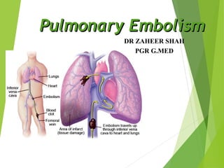
Pulmonary Emboli occurs when fragments of thrombus break loose and are carried through the right side of the heart into the pulmonary arterial tree.
  5. Cancers of primarily adenocarcinoma and CNS tumors most often cause thrombosis. We need to make special mention about patients with a prior history of DVT or PE. Studies have revealed these patients have between a 5 to 30 times increased risk of a new DVT in response to a triggering event compared to those who have not had prior episodes.
  6. All the above symptoms are a manifestation of cardiopulmonary stress caused by the cloth in the lung. These produce symptoms perceived by the patient and the signs observed by you! There are three common clinical presentations that you should be aware of: 1. Patient’s with pulmonary infarction may have pleuritic chest pain and can be hard to distinguish between that patient with infection pneumonitis 2. Submassive embolism are the hardest of all. By definition, they have an angiographically defined blockage of flow to an area served by less than two lobar arteries. These patients have acute or unexplained dyspnea with exertion or at rest. So, they can be easily confused with infection, asthma, CHF and the like. 3. Finally, Massive PE, or a clot which obstructs two lobar arteries, so-called “Saddle Embolus”. These patients have acute cor pulmonaly often with syncope. You might think there having an MI or look septic!
  7. These are the common symptoms that are associated with PE As we mentioned in the previous slide, dyspnea and chest pain are not always preset. The explanation is that with a small V/Q mismatch, the adaptive physiology of the pulmonary vasculature and bronchi produce intermittent shortness of breath. Because of this, we are easily distracted and looking for a cardiogenic cause of the dyspnea. What about pleuritic chest pain, still not a home run! In fact, up to 25% of patients ultimately diagnosed with a PE, never had any chest pain! This is what makes the diagnosis so difficult!
  8. Lets look a t a couple of these: Tachycardia! Myth #2 We are all taught this is a key component of the diagnosis. Right? In fact, actually not having tachycardia is more commonly seen in patients who are found to have a PE! What about fever? If a patient has a fever, it must not be a PE, right? Not true. Although not common, Among patients with PE and no other source of fever, fever was present in one study in 43 of 311 patients (14%).
  9. For example This could represent the likelihood that a specific patient , say a middle-aged man, with a specific past history, say hypertension and tobacco smoking who presents with a specific symptom complex: Chest pain, Dsypnea, or Diaphoresis has a specific diagnosis. Final Statement: Because PE can present with or with any of the “classic signs and symptoms” and even the risk factors which contribute to PE are varied in frequency, we are left with a intuition at best!
  10. As you can see there are a variety of test that we use to arrive at a diagnosis. Some better than others! So, lets talk about these individually.
  11. Granted that most are abnormal, but certainly not diagnostic. It is true that in the original PIOPED study it was recommended but the main value is to exclude diagnoses the mimic PE and to aid in the interpretation of the V/Q scan
  12. Here we see the dilated vessels and oligemia of westermark’s sign And below Hampton’s Hump
  13. A brief mention about the classic S1-Q3-T3, its appearance on the EKG may suggest PE, but study after study has shown it has no predictive value what so ever! But you got to know it because question writers for the boards love it!
  14. Actually, it is opposite of what you might think!
  15. As with most dogma, we are taken back by what we thought was truth. About 15% of patients with proven pulmonary embolism have a pO2 of 85mmHg or higher! In one study, researches drew from there data various combinations of the PaO2 of 80mm Hg or more, the PaCO2 of 35mmHg or higher, and the A-a gradient of 20 mmHg or less. They found that PE could not be excluded in more than 30% of patients with no prior cardiopulmonary disease. Moreover, PE could not be excluded in more than 14% of patients with prior cardiopulmonary disease. Conclusion, Blood gas levels are poor discriminate value to permit the exclusion of PE.
  16. Diagnosing of early right ventricular strain is important because it is a strong predictor of subsequent death Important to recommend echocardiogram with your admitting internist if a pattern of right heart strain is suggested by EKG. Studies have documented that lives are saved with early fibrinolysis is considered in these patients.
  17. Well, what is it? Basically, the assay is enzyme-linked monoclonal antibody test used to identify the protein, D-Dimer. D-Dimer itself is a unique degradation product that is produced by a plasmin mediated breakdown of cross-linked fibrin Good test with respect to its negative predictive value. The drawbacks are some of the false positives that we commonly see in the ER.
  18. Two types, Qualitative RBC agglutination assay, low sensitivity and specificity and not good enough to comfortably rule out PE. Quantitative, which measure the accurately the amount using a spectrophotometer. Our lab uses the 2nd generation VIDAS d-dimer with a negative predictive value of 99.3%!
  19. Essentially, a patient is injected with a radioisotope that travels through the pulmonary microcirculation and are detected by a gamma camera over the patient A normal Perfusion study will have evenly distributed blood flow. Then a patient inhales a radioactive gas and it is viewed as it washes over the bronchopulmonary tree. A mismatch, areas of blocked perfusion and normal ventilation is interpreted by the radiologist and given reading as normal (never), high probability, or non-diagnostic/low-probability The reason the low probability or non diagnostic scan category is so suspect is because in the PIOPED study this group had terrible inter-reader reliability. So, just beware.
  20. The entire lung can be scanned while the patient holds there breath. Advantages: CT most useful benefit is in providing evidence for an alternative diagnosis or excluding it entirely. Disadvantages: The clinical significance for subsegmental PE are not well known, but may be a marker for a larger PE Given that the majority of V/Q studies are non-diagnostic, I prefer the CT as the initial test of choice in place of V/Q scan.
  21. Right now there is no better test on the horizon of the immediate present to virtually rule out or in PE.
  22. . So what are our goals???
  23. Heparin is the most frequently used drug in the treatment of PE. Because heparin works by activating antithrombin III, this genetic mutation makes heparin ineffective.
  24. FDA approved dose for Unfractionated heparin is 80units/kg IV bolus, then 18 units/kg/hr infusion to maintain the INR at 2.5-3 Lovenox is dosed at 1mg/kg every 12 hours or 1.5 mg/kg per day. LMWH in pregnancy only preferred for convenience sake.
  25. This is because the anticoagulants protein C and S have short half-lives compared with the procoagulant vitamin K-dependent proteins. So, this gives rise to the clinical importance that heparin must be continued for at least five days after beginning Coumadin The incidence of progressive thrombosis and embolization is 40% when starting warfarin directly, compared to only 8% when beginning after a patient has been anticoagulated with heparin. Treatment is usually for 6 months, but may continue lifelong
  26. For critically ill patients, a very rapid infusion of 100mg over 10 minutes is preferred. Alternative is Retavase, you can give it as two IV doses of 10 units, each over two minutes.
  27. This is a procedure where a suction tip catheter is placed in contact with the thrombus under fluoroscopy and sucked out while catheter is withdrawn