Short case...Tuberous sclerosis
•
1 gostou•470 visualizações

Short case...Tuberous sclerosis

Recomendados
Mais conteúdo relacionado
Destaque
Destaque (20)
Semelhante a Short case...Tuberous sclerosis
Semelhante a Short case...Tuberous sclerosis (20)
Mais de Professor Yasser Metwally
Mais de Professor Yasser Metwally (20)
Último
Último (20)
Short case...Tuberous sclerosis
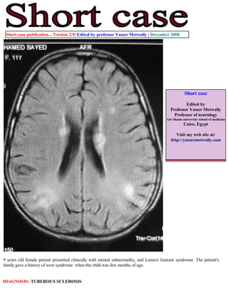
- 1. Short case publication... Version 2.9| Edited by professor Yasser Metwally | December 2008 Short case Edited by Professor Yasser Metwally Professor of neurology Ain Shams university school of medicine Cairo, Egypt Visit my web site at: http://yassermetwally.com 9 years old female patient presented clinically with mental subnormality, and Lennox Gastaut syndrome. The patient's family gave a history of west syndrome when the child was few months of age. DIAGNOSIS: TUBEROUS SCLEROSIS
- 2. Figure 1. Precontrast MRI T1 images showings bilateral subependymal nodules affecting the frontal horns and body of the lateral ventricles. A Cortical tuber can be seen in the occipito-parietal junction, it is hypointense, wedge shaped and involve gray matter and contiguous white matter. The wedge-shaped white matter lesions have their apex near the ventricle and their base at the cortex or at the cortical tuber. Two or more adjacent gyri are affected and appeared lissencephalic, notice gyral broadening and thickening. At histologic examination the laminar architecture of affected cortex is completely disorganized. Some scattered focal hypointense white matter changes are seen. Figure 2. MRI FLAIR images showing bilateral subependymal nodules affecting the frontal horns and body of the lateral ventricles. Bilateral wedge shaped, hyperintense cortical tubers can be seen in the occipito-parietal junction and the frontal area. The tubers involve gray matter and contiguous white matter. Two or more adjacent gyri are affected and appeared lissencephalic, notice gyral broadening and thickening. At histologic examination the laminar architecture of affected cortex is completely disorganized. Some scattered focal hyperintense white matter changes are seen. The wedge-shaped white matter lesions have their apex near the ventricle and their base at the cortex or at the cortical tuber.
- 3. Figure 3. MRI FLAIR images showing bilateral subependymal nodules affecting the frontal horns and body of the lateral ventricles. Bilateral wedge shaped, hyperintense cortical tubers can be seen in the occipito-parietal junction and the frontal area. The tubers involve gray matter and contiguous white matter. Two or more adjacent gyri are affected and appeared lissencephalic, notice gyral broadening and thickening. At histologic examination the laminar architecture of affected cortex is completely disorganized. Some scattered focal hyperintense white matter changes are seen. The wedge- shaped white matter lesions have their apex near the ventricle and their base at the cortex or at the cortical tuber. Figure 4. MRI FLAIR images showing bilateral cortical tubers in the temporal lobes. The tubers are wedge shaped, hyperintense and involve the cortical gray matter and the adjacent white matter. The cortex is broadened, thickened, lissencephalic and pachygyric. Notice the periventricular, periaqueductal white matter changes. The wedge-shaped white matter lesions have their apex near the ventricle and their base at the cortex or at the cortical tuber.
- 4. Figure 5. MRI T2 images showing bilateral cortical tubers in the temporal lobes. The tubers are wedge shaped, hyperintense and involve the cortical gray matter and the adjacent white matter. The cortex is broadened, thickened, lissencephalic and pachygyric. Notice the periventricular, periaqueductal white matter changes. The wedge-shaped white matter lesions have their apex near the ventricle and their base at the cortex or at the cortical tuber. Figure 6. MRI T2 (A) and FLAIR (B) images showing bilateral cortical tubers in the temporal lobes. The tubers are wedge -shaped, hyperintense and involve the cortical gray matter and the adjacent white matter. The cortex is broadened, thickened, lissencephalic and pachygyric. Notice the scattered focal and diffuse white matter changes. The wedge-shaped white matter lesions have their apex near the ventricle and their base at the cortex or at the cortical tuber.
- 5. References 1. Metwally, MYM: Textbook of neurimaging, A CD-ROM publication, (Metwally, MYM editor) WEB-CD agency for electronic publishing, version 9.4a October 2008 Addendum A new version of short case is uploaded in my web site every week (every Saturday and remains available till Friday.) To download the current version follow the link quot;http://pdf.yassermetwally.com/short.pdfquot;. You can download the long case version of this short case during the same week from: http://pdf.yassermetwally.com/case.pdf or visit web site: http://pdf.yassermetwally.com To download the software version of the publication (crow.exe) follow the link: http://neurology.yassermetwally.com/crow.zip At the end of each year, all the publications are compiled on a single CD-ROM, please contact the author to know more details. Screen resolution is better set at 1024*768 pixel screen area for optimum display For an archive of the previously reported cases go to www.yassermetwally.net, then under pages in the right panel, scroll down and click on the text entry quot;downloadable short cases in PDF formatquot; Also to view a list of the previously published case records follow the following link (http://wordpress.com/tag/ case-record/) or click on it if it appears as a link in your PDF reader