Short case...Cortical dysplasia
•
2 gostaram•384 visualizações

Short case...Cortical dysplasia http://yassermetwally.com http://yassermetwally.net

Recomendados
Recomendados
Mais conteúdo relacionado
Destaque
Destaque (20)
Semelhante a Short case...Cortical dysplasia
Semelhante a Short case...Cortical dysplasia (20)
Mais de Professor Yasser Metwally
Mais de Professor Yasser Metwally (20)
Último
Último (20)
Short case...Cortical dysplasia
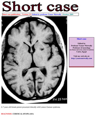
- 1. Short case publication... Version 3.3| Edited by professor Yasser Metwally | October 2009 Short case Edited by Professor Yasser Metwally Professor of neurology Ain Shams university school of medicine Cairo, Egypt Visit my web site at: http://yassermetwally.com A 7 years old female patient presented clinically with Lennox Gastaut syndrome. DIAGNOSIS: CORTICAL DYSPLASIA
- 2. Figure 1. Cortical dysplasia. Precontrast MRI T1 images showing lissencephaly, microgyria, pachygyria, hypointense cystic white matter changes specially affecting the right head of caudate nucleus and the globus pallidus on the right side. The lissencephalic changes are most marked in the bifrontal regions. Notice the subependymal nodular heterotopia specially involving the frontal horns bilaterally. There is also reduction of the brain volume and moderate degree of central atrophy. Figure 2. Cortical dysplasia. MRI FLAIR images showing lissencephaly, microgyria, pachygyria, hyperintense cystic white matter changes specially affecting the right head of caudate nucleus and the globus pallidus on the right side. The lissencephalic changes are most marked in the bifrontal regions. Notice the subependymal nodular heterotopia specially involving the frontal horns bilaterally, nodules are also seen subependymally in the left body of the lateral ventricles (C). There is also reduction of the brain volume and moderate degree of central atrophy.
- 3. Figure 3. Cortical dysplasia. MRI T2 images showing lissencephaly, microgyria, pachygyria, hyperintense cystic white matter changes specially affecting the right head of caudate nucleus and the globus pallidus on the right side. The lissencephalic changes are most marked in the bifrontal regions. Notice the subependymal nodular heterotopia specially involving the frontal horns bilaterally. There is also reduction of the brain volume and moderate degree of central atrophy. Figure 4. Cortical dysplasia. MRI FLAIR images showing lissencephaly, microgyria, pachygyria, hyperintense cystic white matter changes specially affecting the right head of caudate nucleus and the globus pallidus on the right side. The lissencephalic changes are most marked in the bifrontal regions. Notice the subependymal nodular heterotopia specially involving the frontal horns bilaterally, nodules are also seen subependymally in the left body of the lateral ventricles (C). There is also reduction of the brain volume and moderate degree of central atrophy. The hippocampi are atrophic and hyperintense bilaterally (possible mesial temporal sclerosis).
- 4. References 1. Metwally, MYM: Textbook of neurimaging, A CD-ROM publication, (Metwally, MYM editor) WEB-CD agency for electronic publishing, version 10.4a October 2009 Addendum A new version of short case is uploaded in my web site every week (every Saturday and remains available till Friday.) To download the current version follow the link "http://pdf.yassermetwally.com/short.pdf". You can download the long case version of this short case during the same week from: http://pdf.yassermetwally.com/case.pdf or visit web site: http://pdf.yassermetwally.com To download the software version of the publication (crow.exe) follow the link: http://neurology.yassermetwally.com/crow.zip At the end of each year, all the publications are compiled on a single CD-ROM, please contact the author to know more details. Screen resolution is better set at 1024*768 pixel screen area for optimum display For an archive of the previously reported cases go to www.yassermetwally.net, then under pages in the right panel, scroll down and click on the text entry "downloadable short cases in PDF format" Also to view a list of the previously published case records follow the following link (http://wordpress.com/tag/case- record/) or click on it if it appears as a link in your PDF reader