Diffuse Brain Stem Glioma in 11-Year-Old Boy
•
4 gostaram•1,496 visualizações

- An 11-year-old male presented with right facial nerve palsy and bilateral eye movement limitations. MRI showed a diffuse intrinsic brain stem glioma involving the pons. - Diffuse intrinsic brain stem gliomas are diffuse astrocytomas (grades II-IV) that insinuate throughout the brain stem. Radiologically they appear as diffuse enlargement and T1 hypointensity/T2 hyperintensity. - Treatment options include radiotherapy but not surgery due to diffuse infiltration. Chemotherapy has shown little benefit. Prognosis is poor with most children dying within a year despite therapy.

Recomendados
Mais conteúdo relacionado
Mais procurados
Mais procurados (20)
Destaque
Semelhante a Diffuse Brain Stem Glioma in 11-Year-Old Boy
Semelhante a Diffuse Brain Stem Glioma in 11-Year-Old Boy (20)
Mais de Professor Yasser Metwally
Mais de Professor Yasser Metwally (20)
Último
Último (20)
Diffuse Brain Stem Glioma in 11-Year-Old Boy
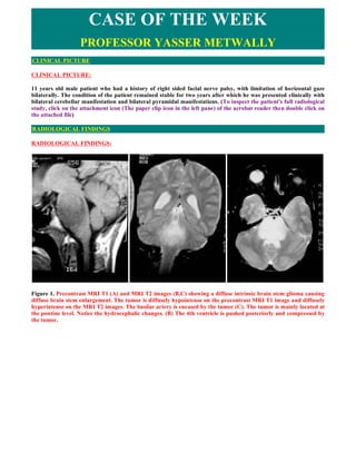
- 1. CASE OF THE WEEK PROFESSOR YASSER METWALLY CLINICAL PICTURE CLINICAL PICTURE: 11 years old male patient who had a history of right sided facial nerve palsy, with limitation of horizontal gaze bilaterally. The condition of the patient remained stable for two years after which he was presented clinically with bilateral cerebellar manifestation and bilateral pyramidal manifestations. (To inspect the patient's full radiological study, click on the attachment icon (The paper clip icon in the left pane) of the acrobat reader then double click on the attached file) RADIOLOGICAL FINDINGS RADIOLOGICAL FINDINGS: Figure 1. Precontrast MRI T1 (A) and MRI T2 images (B,C) showing a diffuse intrinsic brain stem glioma causing diffuse brain stem enlargement. The tumor is diffusely hypointense on the precontrast MRI T1 image and diffusely hyperintense on the MRI T2 images. The basilar artery is encased by the tumor (C). The tumor is mainly located at the pontine level. Notice the hydrocephalic changes. (B) The 4th ventricle is pushed posteriorly and compressed by the tumor.
- 2. Figure 2. MRI T2 images, notice the hydrocephalic changes. Cystic tumor masses could be seen at the midbrain level indicating that the tumor has extended to the midbrain level. Two points must be addressed in this case report 1) the cause of the MRI signal intensity of pontine gliomas and 2) the pattern of spread of this tumor. Radiologically low grade gliomas are usually identified by diffuse enlargement of the brain stem, abnormal signal intensity on MR or abnormal attenuation on CT. The lesions typically have precontrast CT attenuation and MRI signal changes suggesting increased water content and lower than normal specific gravity (lower CT scan densities with MRI T1 hypointensities and diffuse MRI T2 hyperintensities). It is tempting to consider that these changes represent edema. The question then arises: Is this vasogenic edema or cytotoxic edema? Because the blood-brain barrier is intact in these tumors, vasogenic edema is unlikely. The cells are not dead or dying, so that cytotoxic edema is also unlikely. Perhaps the edema results from the increased number of astrocytic cells that spread apart the normal myelinated axons of the white matter. The presence of significant amount of normal appearing astrocytes results in total increase in the water content of the brain stem. These cells may merely have different physical and chemical properties than the normal tightly packed bundles of axons that traverse through the brain stem. As the blood brain barrier is intact in low grade brain stem astrocytomas (grade II astrocytomas according to the WHO), no significant enhancement occurs, either on MRI or CT scan. Enhancement is characteristic of the more aggressive anaplastic astrocytomas (grade III) or glioblastoma multiforme. (6) Diffuse intrinsic brain stem gliomas are actually diffuse astrocytomas Grade II,III,IV which have the following characteristics Common pathological characteristics of diffuse astrocytomas Diffuse astrocytomas are tumors predominantly composed of astrocytes. Unless otherwise indicated, the term usually applies to diffusely infiltrating neoplasms (WHO grades II through IV). Diffuse astrocytoma is unusual in the first decade of life and most commonly presents in older children or young adults up to the age of 40 to 45. All diffuse astrocytomas, particularly the diffusely infiltrating variety, have a tendency toward progression to more malignant forms. Diffuse astrocytomas have a peculiar tendency to change its grade over time into the next higher grade of malignancy and the condition is age dependant. A change in the grade of diffuse astrocytoma is more likely to occur in the older age group. Diffuse astrocytomas commonly start as grade II at a younger age group then gradually change its grade over time into the next higher grade until they ultimately dedifferentiate into glioblastomas (secondary glioblastoma multiforme), on the other hand, glioblastoma multiforme in older patients are usually primary- that is, they occur as glioblastoma multiforme from their inception, without progression from a lower- grade tumor.
- 3. Diffuse astrocytomas appear to form a continuum of both biological and histological aggression. They vary from lesions with almost normal cytology (grade I and grade II astrocytomas) through intermediate stages (grade III, anaplastic astrocytomas) and up to the most aggressive of all human brain tumours (grade IV astrocytomas or glioblastoma multiforme). Diffuse astrocytoma often spreads widely through the brain but without destruction and also without interruption of normal function. Microscopically, tumor cells infiltrate between myelinated fibers in a nondestructive manner (perineuronal satellitosis). The local spread of diffuse astrocytomas (forming gliomatosis cerebri and butterfly gliomas) does not mean that the tumour grade is grade IV (glioblastoma multiforme), local spread can occur in grade II and grade III and in the author experience gliomatosis cerebri and butterfly gliomas are much more commonly seen in grade II astrocytomas and has not been encountered in grade III (anaplastic astrocytomas) and grade IV (glioblastoma multiforme). It takes a long time for a diffuse astrocytoma to cross the corpus callosum to the opposite hemisphere to form a butterfly glioma. Patients harbouring glioblastomas have a much shorter life span for their tumours to form butterfly gliomas, however cases were reported for glioblastomas forming butterfly tumours. These glioma cells migrate through the normal parenchyma, collect just below the pial margin (subpial spread), surround neurons and vessels (perineuronal and perivascular satellitosis), and migrate through the white matter tracks (intrafacicular spread). This invasive behavior of the individual cells may correspond to the neoplastic cell's reacquisition of primitive migratory behavior during central nervous system development. The ultimate result of this behavior is the spread of individual tumor cells diffusely over long distances and into regions of brain essential for survival of the patient. The extreme example of this behavior is a condition referred to as gliomatosis cerebri, in which the entire brain is diffusely infiltrated by neoplastic cells with minimal or no central focal area of tumor per se. Furthermore, 25% of patients with GBM have multiple or multicentric GBMs at autopsy. Although GBMs can be visualized on MRI scans as mass lesions that enhance with contrast, the neoplastic cells extend far beyond the area of enhancement. In practice considerable histological heterogeneity in astrocytic tumours is found ( i.e., low grade areas with Rosenthal fibers and calcification can be intermixed with with frankly malignant ones). The differences in histologic features, potential for invasiveness, and extent of progression likely reflect genetic differences acquired during astrocytoma growth. Grade IV astrocytomas (glioblastoma multiforme) differ from diffuse astrocytoma grade II and grade III (anaplastic astrocytomas) in the presence of gross necrosis, and microscopically in the presence of vascular endothelial hyperplasia and tumour hemorrhage. DIAGNOSIS: DIAGNOSIS: DIFFUSE INTRINSIC BRAIN STEM GLIOMA DISCUSSION DISCUSSION: Brain stem tumors are perhaps the most dreaded cancers in pediatric oncology, owing to their historically poor prognosis, yet they remain an area of intense research. Brain stem tumors account for about 10 to 15% of childhood brain tumors. Peak incidence for these tumors occurs around age 6 to 9 years. The term brain stem glioma is often used interchangeably with brain stem tumor. More precisely, glioma encompasses tumor pathology types such as ganglioglioma, pilocytic astrocytoma, diffuse astrocytoma, anaplastic astrocytoma, and glioblastoma multiforme. Rarely, other tumor pathologies such as atypical teratoid/rhabdoid tumor (ATRT), primitive neuroectodermal tumor (PNET)/embryonal tumor, and hemangioblastoma occur at the brain stem. These entities are quite different from brain stem gliomas, and the following comments do not apply. Classification Brain stem gliomas have been grouped in the past according to their pathology and location within the brain stem. Terms found in the medical literature include diffuse intrinsic gliomas, midbrain tumors, tectal gliomas, pencil gliomas, dorsal exophytic brain stem tumors, cervicomedullary tumors, focal gliomas, and cystic tumors. A simpler way to classify these tumors is by two categories: diffuse intrinsic pontine glioma and focal brain stem glioma.
- 4. Symptoms Children with DIPG present with ataxia (clumsiness or wobbliness), weakness of a leg and/or arm, double vision, and sometimes headaches, vomiting, tilting of the head, or facial weakness. Double vision (diplopia) is the most common presenting symptom for these tumors. Symptoms are usually present for 6 months or less at time of diagnosis. Patients with focal brain stem gliomas may display some of the same symptoms, although not the usual combination of ataxia, weakness, and double vision. Duration of symptoms is often greater than 6 months before the focal brain stem tumor is diagnosed. Diagnosis Throughout the United States, brain magnetic resonance imaging (MRI), with and without gadolinium contrast, remains the "gold standard" for diagnosis of brain stem gliomas. Biopsy is seldom performed outside specialized biomedical research protocols for DIPG, unless the diagnosis of this tumor is in doubt. Biopsy may be indicated for brain stem tumors that are focal or atypical, especially when the tumor is progressive or when surgical excision may be possible. Diffuse intrinsic pontine gliomas (DIPG) insinuate diffusely throughout the normal structures of the pons (the middle portion of the brain stem), sometimes spreading to the midbrain (the upper portion of the brain stem) or the medulla (the bottom portion of the brain stem). The term diffuse intrinsic glioma is synonymous. By pathology, these tumors are most often a diffuse (sometimes referred to as fibrillary) astrocytoma (World Health Organization [WHO] grade II) or its higher-grade counterparts, anaplastic astrocytoma (WHO III) and glioblastoma multiforme (WHO IV). Very rarely these tumors start in the medulla or midbrain. Focal brain stem gliomas--perhaps 20% or more of brain stem gliomas--include tumors that are more circumscribed, focal, or contained at the brain stem. These tumors may have cysts or grow out from the brain stem (i.e., exophytic). These tumors more often arise in the midbrain or medulla, rather than the pons. Pathology for these tumors is frequently pilocytic astrocytoma (WHO I) or ganglioglioma (WHO I), although rarely diffuse astrocytoma (WHO II). Treatment Since brain stem gliomas are relatively uncommon and require complex management, children with such tumors deserve evaluation in a comprehensive cancer center where the coordinated services of dedicated pediatric neurosurgeons, child neurologists, pediatric oncologists, radiation oncologists, neuropathologists, and neuroradiologists are available. In particular, for DIPG, because of its rarity and poor prognosis, children and their families should be encouraged to participate in clinical trials attempting to improve survival with innovative therapy. Neurosurgery Surgery to attempt tumor removal is usually not possible or advisable for DIPG. By their very nature, these tumors invade diffusely throughout the brain stem, growing between normal nerve cells. Aggressive surgery would cause severe damage to neural structures vital for arm and leg movement, eye movement, swallowing, breathing, and even consciousness. Surgery with less than total removal can be performed for many focal brain stem gliomas. Such surgery often results in quality long-term survival, without administering chemotherapy or radiotherapy immediately after surgery, even when a child has residual tumor. Surgery is particularly useful for tumors that grow out (exophytic) from the brain stem. Focal brain stem tumors that arise at the top back of the midbrain (tectal gliomas) should be managed conservatively, without surgical removal. Nevertheless, shunt placement or ventriculostomy for hydrocephalus (see below) is frequently necessary. These tumors have been described to be stable for many years or decades without any intervention other than shunting. Radiotherapy Conventional radiotherapy, limited to the involved area of tumor, is the mainstay of treatment for DIPG. A total radiation dosage ranging from 5400 to 6000 cGy, administered in daily fractions of 150 to 200 cGy over 6 weeks, is standard. Hyperfractionated (twice-daily) radiotherapy was used previously to deliver higher irradiation dosages, but such did not lead to improved survival. Radiosurgery (e.g., gamma knife, Cyberknife) has no role in the treatment of DIPG.
- 5. Chemotherapy and other drug therapies The role of chemotherapy in DIPG remains unclear. Studies to date with chemotherapy have shown little improvement in survival, although efforts (see below) through the Children's Oncology Group (COG), Pediatric Brain Tumor Consortium (PBTC), and others are underway to explore further the use of chemotherapy and other drugs. Drugs utilized to increase the effect of radiotherapy (radiosensitizers) have thus far shown no added benefit, but promising new agents are under investigation. Immunotherapy with beta-interferon and other drugs to modify biologic response have shown disappointing results. Intensive or high-dose chemotherapy with autologous bone marrow transplant or peripheral blood stem cell rescue has not demonstrated any effectiveness in brain stem gliomas and is not recommended. Future clinical trials may incorporate medicines to interfere with cellular pathways (signal transfer inhibitors) or other approaches that alter the tumor or its environment. For more information and a listing of the most up-to date trials, the reader is encouraged to check the websites of the National Institutes of Health clinical trials registry (http://clinicaltrials.gov/), the National Childhood Cancer Foundation/COG (http://www.curesearch.org/), and the PBTC (http://www.pbtc.org/). In focal brain stem gliomas, chemotherapy, such as carboplatin/vincristine, procarbazine/CCNU/vincristine, or temozolomide, may be useful in children whose tumors are progressive and not surgically accessible. In children younger than age 3 years, chemotherapy may be preferable to radiotherapy because of the effects of irradiation on the developing brain. Recurrent or Progressive Brain Stem Gliomas: Regrettably, DIPG has a high rate of recurrence or progression. At relapse, a variety of Phase I and Phase II drug trials are available through the national research consortiums COG and PBTC, as well as through individual pediatric institutions. Oral etoposide, temozolomide, and cyclophosphamide are drug options sometimes utilized outside a study. Prognosis DIPG often follows an inexorable course of progression, despite therapy. A large majority of children die within a year of diagnosis. Focal brain stem glioma, however, can carry an exceptional prognosis, with long-term survivals frequently reported. Other Management Issues: Shunts: Less than half of children with brain stem tumors will develop obstructive hydrocephalus, requiring a shunt or ventriculostomy, at some time during the course of their illness. Shunts are simple mechanical tubing devices that divert cerebrospinal fluid trapped in the brain's ventricles above the tumor to another location in the body, typically the abdomen (peritoneum), as in a ventriculoperitoneal shunt. A ventriculostomy is the surgical creation of an internal channel, often from the third ventricle to a lower portion of the brain, to allow cerebrospinal fluid to drain beyond the tumor. Steroids: Dexamethasone (brand name Decadron) is a steroid drug frequently administered to brain stem tumor patients for the swelling and "tightness" of their tumor at the base of their skull. Dexamethasone must be used sparingly! Dexamethasone should never be prescribed prophylactically or "just in case." That is, this steroid is an extremely effective medicine for symptomatic swelling associated with treatment of a brain stem glioma, particularly with radiotherapy. However, dexamethasone is not necessary unless a child has symptomatic swelling. Dexamethasone has a number of side effects which include mood changes, insomnia, weight gain, fluid retention, glucose instability, high blood pressure, and increased susceptibility to infection. SUMMARY SUMMARY Adult brainstem gliomas are different from the childhood subtypes. Overall, brainstem gliomas are less aggressive in adults than in children. However, survival merely reflects the course of the most frequent subtype of tumours. Diffuse intrinsic low-grade brainstem glioma
- 6. Interestingly, the most frequent type of of brainstem glioma in adults (representing 46% of the patients in this series) resembles the childhood diffuse gliomas of the pons in terms of clinical and radiological presentation but is radically different in course and survival. In both adults and children, the clinical picture is of a combination of cranial nerve and long tract signs. However, while the onset is rapid in children, the duration of symptoms is often long in adults. (6) In both children and adults, MRI at presentation reveals a diffuse infiltration of the pons, often increasing the size of the brainstem considerably. There is high signal on T2-weighted and low signal on T1-weighted images, which usually do not show contrast enhancement (100% in adults at diagnosis). It is worth noting that preferential location in the pons is less striking in adults than in children. When a biopsy is performed, which is far from routine practice in these diffuse intrinsic forms, a malignant glioma (grades III–IV) is found in many children, whereas a less aggressive histology is found the adults. (6) Malignant brainstem gliomas The other common tumour type identified in adult is clearly different from those discussed above. It occurs later than the diffuse, intrinsic, low-grade type and affects mainly older adults (most of them in their sixth decade). The clinical picture is characterized by the rapid onset of cranial nerve palsies and long tract signs leading to an early alteration in performance status. MRI reveals a brainstem mass that enhances after gadolinium infusion, often in a ring-like fashion. Contrast enhancement is a pejorative factor (particularly when the area of enhancement surrounded a low-signal area suggestive of necrosis) in contrast with children, in whom the prognostic value of contrast enhancement remains controversial. Pathologically, these tumours correspond to high-grade gliomas (grades III–IV) and median survival time is short (11.2 months) despite treatment with radiotherapy and chemotherapy. Thus, the clinical–radiological pattern, pathology and course closely resemble the common malignant supratentorial gliomas in adults and we suggest that this group be designated `malignant brainstem gliomas'. (6) Focal tectal gliomas Focal tectal gliomas represent the third type of adult brainstem glioma and constitute a small subgroup (8%) that also exists in children. The clinical picture is dominated by hydrocephalus. (6) Other types Other types of brainstem glioma can be observed in adults such as exophytic contrast-enhancing glioma arising from the floor of the fourth ventricle; this entity, which is associated with a good prognosis, is also well described in children (representing up to 10% of brainstem gliomas). A likely explanation for this discrepancy between the two age-groups is that most of the exophytic gliomas correspond to pilocytic astrocytoma, a very rare type of tumour in adults. (6) The brainstem is the second most frequent location of brain tumours after the optic pathways in patients with NF1. In contrast with children, in whom the course is usually very long, the tumour behaviour in adults with NF1 was much more aggressive, but larger series will be necessary to draw any conclusion on this point. (6) Complications Except for locoregional progression, two main complications are observed during the course of adult brainstem gliomas, namely hydrocephalus and leptomeningeal dissemination. Hydrocephalus is observed in 20% of cases. Whereas some pontine tumours may have an important mass effect on the fourth ventricle, hydrocephalus is always associated with mesencephalic involvement and blockage of the CSF at the level of the sylvian aqueduct. Leptomeningeal dissemination occurred in 13% of cases and is the cause of a quarter of the deaths. This complication has also been reported with a high frequency in children. Close proximity of the tumour and CSF pathways could explain such an increased trend for leptomeningeal dissemination, but this remains to be demonstrated. (6) The role of biopsy Finally, this classification may help in the selection of patients for biopsy. In children, MRI has become the reference for the diagnosis of brainstem glioma and is used for the current classification of these tumours. MRI has replaced biopsy in the diagnosis of paediatric diffuse brainstem gliomas, for which most authors agree that anticancer treatments can be administered without pathological confirmation if the clinical course is rapid. However, we believe that biopsy is not useful in the diagnosis of intrinsic, diffuse, low-grade brainstem gliomas in
- 7. adults when the clinical and radiological criteria described above are met. The issue is different in contrast- enhancing lesions because several reports have underlined the limits of MRI in differentiating tumours from infectious (e.g. tuberculomas) and inflammatory (sarcoidosis, Behciet's disease). (6) Addendum A new version of this PDF file (with a new case) is uploaded in my web site every week (every Saturday and remains available till Friday.) To download the current version follow the link "http://pdf.yassermetwally.com/case.pdf". You can also download the current version from my web site at "http://yassermetwally.com". To download the software version of the publication (crow.exe) follow the link: http://neurology.yassermetwally.com/crow.zip The case is also presented as a short case in PDF format, to download the short case follow the link: http://pdf.yassermetwally.com/short.pdf At the end of each year, all the publications are compiled on a single CD-ROM, please contact the author to know more details. Screen resolution is better set at 1024*768 pixel screen area for optimum display. Also to view a list of the previously published case records follow the following link (http://wordpress.com/tag/case-record/) or click on it if it appears as a link in your PDF reader To inspect the patient's full radiological study, click on the attachment icon (The paper clip icon in the left pane) of the acrobat reader then double click on the attached file. Click here to download the short case version of this case record in PDF format REFERENCES References 1. Fisher PG, Breiter SN, Carson BS, Wharam MD, Williams JA, Weingart JD, Foer DR, Goldthwaite PT, Burger PC. A clinicopathologic reappraisal of brainstem tumor classification: identification of pilocytic astrocytoma and fibrillary astrocytoma as distinct entities. Cancer 89:1569-1576, 2000. 2. Donaldson SS, Laningham F, Fisher PG. Advances toward an understanding of brain stem gliomas. J Clin Oncol 24:1266-1272, 2006. 3. Paul Graham Fisher, M.D., MHS, is Professor of Neurology and Pediatrics, and The Beirne Family Professor of Neuro-Oncology, at the Stanford University School of Medicine and Lucile Salter Packard Children's Hospital. Dr. Fisher voluntarily serves as a member of the Childhood Brain Tumor Foundation's Scientific/ Medical Advisory. 4. Michelle Monje, M.D., Ph.D., is Instructor of Neurology at Stanford University School of Medicine and Lucile Packard Children’s Hospital. Dr. Monje’s research focuses on the biology of brain stem gliomas and neural stem cell biology. 5. Metwally, MYM: Textbook of neuroimaging, A CD-ROM publication, (Metwally, MYM editor) WEB-CD agency for electronic publication, version 11.1a. January 2010 6. Metwally, MYM (2001): Brain stem glioma, A clinico-radiological study: A classification system with prognostic significance is suggested. Ain Shams medical journal, VOL. 51, NO. 10,11,12, pp 1085-1115 7. Metwally, MYM: Textbook of neuroimaging, A CD-ROM publication, (Metwally, MYM editor) WEB-CD agency for electronic publication, version 11.1a. January 2010 8. Metwally, MYM (2001): Brain stem glioma, A clinico-radiological study: A classification system with prognostic significance is suggested. Ain Shams medical journal, VOL. 51, NO. 10,11,12, pp 1085-1115 [Click to download in PDF format] 9. Case of the week...Brain stem glioma. [Click to download in PDF format] 10. Case of the week...Focal midbrain tumor. [Click to download in PDF format] 11. Case of the week...Tectal plate glioma. [Click to download in PDF format]