![2
INDEX
Expt. No. Aim Page No.
1 To introduce different tests used in qualitative analysis of
carbohydrates
2 To perform qualitative analysis of given unknown sample of
carbohydrates [Glucose/dextrose ]
3 To perform qualitative analysis of given unknown sample of
carbohydrates [Starch ]
4 To perform qualitative analysis of given unknown sample of
carbohydrates [Fructose ]
5 To perform qualitative analysis of given unknown sample of
carbohydrates [Lactose ]
6 To perform qualitative analysis of given unknown sample of
carbohydrates [Maltose ]
7 To perform qualitative analysis of given unknown sample of
carbohydrates [Sucrose ]
8 To introduce different test used in qualitative analysis of proteins
9
To perform Qualitative analysis of given unknown sample of protein
(albumin)
10
To perform Qualitative analysis of given unknown sample of protein
(CASEIN)
11 To perform Quantitative analysis of given unknown sample of protein
(Albumin)
12 To demonstrate estimation of quantity of creatinine in given
sample of urine by Alkaline picrate method (Jaffe's reaction)
13 To demonstrate estimation of quantity of glucose in given sample of
serum. (GOD/POD method ).
14 To detect abnormal constituents in given sample of urine by qualitative
tests.
15 To demonstrate Estimation of Cholesterol in serum by enzymatic
method](data:image/gif;base64,R0lGODlhAQABAIAAAAAAAP///yH5BAEAAAAALAAAAAABAAEAAAIBRAA7)
Recomendados
Mais conteúdo relacionado
Mais procurados
Mais procurados (20)
Semelhante a Biochemistry lab manual II semester
Semelhante a Biochemistry lab manual II semester (20)
Mais de Dipali Kulkarni
Mais de Dipali Kulkarni (20)
Último
Último (20)
Biochemistry lab manual II semester
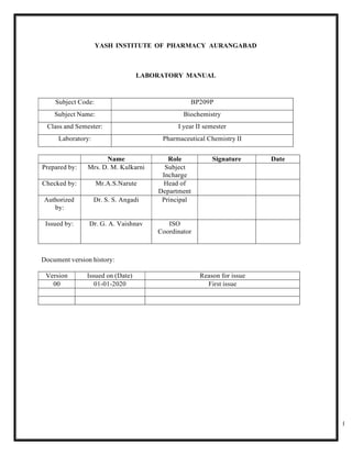
- 1. 1 YASH INSTITUTE OF PHARMACY AURANGABAD LABORATORY MANUAL Subject Code: BP209P Subject Name: Biochemistry Class and Semester: I year II semester Laboratory: Pharmaceutical Chemistry II Name Role Signature Date Prepared by: Mrs. D. M. Kulkarni Subject Incharge Checked by: Mr.A.S.Narute Head of Department Authorized by: Dr. S. S. Angadi Principal Issued by: Dr. G. A. Vaishnav ISO Coordinator Document version history: Version Issued on (Date) Reason for issue 00 01-01-2020 First issue
- 2. 2 INDEX Expt. No. Aim Page No. 1 To introduce different tests used in qualitative analysis of carbohydrates 2 To perform qualitative analysis of given unknown sample of carbohydrates [Glucose/dextrose ] 3 To perform qualitative analysis of given unknown sample of carbohydrates [Starch ] 4 To perform qualitative analysis of given unknown sample of carbohydrates [Fructose ] 5 To perform qualitative analysis of given unknown sample of carbohydrates [Lactose ] 6 To perform qualitative analysis of given unknown sample of carbohydrates [Maltose ] 7 To perform qualitative analysis of given unknown sample of carbohydrates [Sucrose ] 8 To introduce different test used in qualitative analysis of proteins 9 To perform Qualitative analysis of given unknown sample of protein (albumin) 10 To perform Qualitative analysis of given unknown sample of protein (CASEIN) 11 To perform Quantitative analysis of given unknown sample of protein (Albumin) 12 To demonstrate estimation of quantity of creatinine in given sample of urine by Alkaline picrate method (Jaffe's reaction) 13 To demonstrate estimation of quantity of glucose in given sample of serum. (GOD/POD method ). 14 To detect abnormal constituents in given sample of urine by qualitative tests. 15 To demonstrate Estimation of Cholesterol in serum by enzymatic method
- 3. 3 EXPERIMENT NO. 1 AIM – To introduce different tests used in qualitative analysis of carbohydrates REFERENCE: - 1. Kale S.R.,Kale R.R., “Practical Biochemistry And Clinical Biochemistry”, Nirali Prakashan, 24 th edition, 2015 page no 4 to 6 2. David T. Plummer “Introduction of Practical Biochemistry” 3rd Edition 3. Rajagopal and Ramakrishna “Practical Biochemistry for Medical students” 5 th edition . THEORY: - A carbohydrate is an organic compound with the general formula Cn(H2O)n, that is, consists only of carbon, hydrogen and oxygen, with the last two in the 2:1 atom ratio. Carbohydrates make up the bulk of organic substances on earth and perform numerous roles in living things. The carbohydrates (saccharides) are divided into four chemical groups: monosaccharides, disaccharides, oligosaccharides and polysaccharides. Polysaccharides serve for the storage of energy (e.g., starch in plants and glycogen in animals) and as structural components (e.g., cellulose in plants and chitin in arthropods). Structural polysaccharides are frequently found in combination with proteins (glycoproteins or mucoproteins) or lipids (lipopolysaccharides). The 5-carbon monosaccharide ribose is an important component of coenzymes (e.g., ATP, FAD and NAD) and the backbone of the genetic molecule known as RNA. The related deoxyribose is a component of DNA. Saccharides and their derivatives include many other important biomolecules that play key roles in the immune system, fertilization, preventing pathogenesis, blood clotting and development [1]. This experiment aims to introduce you with the identification of unknown carbohydrates. To gain maximum benefit, observations should be related, as far as possible, to the structure of the substances examined. Some important points: 1. Most of the tests and reactions described are not quantitative and volumes are approximate, despite these facts some tests do not work if quantities greatly in excess of those stated are used. 2. DO NOT place your pipettes in reagent bottles as this leads to contamination. 3. In most tests, it is important to apply a control test using water instead of the solution under examination. If you are in doubt about the result of a test, perform the reaction with a suitable known compound. 4. In this experiment, sugar samples are given in their solid state. To perform each procedure, you should prepare your own sugar solution by taking very small amounts of solid sugars.
- 4. 4 5. When you need to boil your sample in a test tube, prepare a hot water in a large beaker and put your test tube inside the beaker. DO NOT forget to put boiling chips in the beaker.
- 5. 5 TESTS ON CARBOHYDRATES: 1) Molisch’s Test: Molisch’s Test is a sensitive chemical test for all carbohydrates, and some compounds containing carbohydrates in a combined form, based on the dehydration of the carbohydrate by sulfuric acid to produce an aldehyde (either furfural or a derivative), which then condenses with the phenolic structure resulting in a red or purple-colored compound. PROCEDURE: - Apply this test two different carbohydrate solutions of your own choice, preferably to one monosaccharide and one polysaccharide. - Place 2 mL of a known carbohydrate solution in a test tube, add 1 drop of Molisch’s reagent (10% α-naphthol in ethanol). - Pour 1-2 mL of conc. H2SO4 down the side of the test tube, so that it forms a layer at the bottom of the tube. - Observe the color at the interface between two layers and compare your result with a control test. A brown color due to charring must be ignored and the test should be repeated with a more dilute sugar solution. 2) Carbohydrates as Reducing Sugars: A reducing sugar is any sugar that, in a solution, has an aldehyde or a ketone group. The enolization of sugars under alkaline conditions is an important consideration in reduction tests. The ability of a sugar to reduce alkaline test reagents depends on the availability of an aldehyde or ketone group for reduction reactions. A number of sugars especially disaccharides or polysaccharides have glycosidic linkages which involve bonding a carbohydrate (sugar) molecule to another one, and hence there is no reducing group on the sugar; like in the case of sucrose, glycogen, starch and dextrin. In the case of reducing sugars, the presence of alkali causes extensive enolization especially at high pH and temperature. This leads to a higher susceptibility to oxidation reactions than at neutral or acidic pH. These sugars, therefore, become potential agents capable of reducing Cu+2 to Cu+, Ag+ to Ag and so fort. Most commonly used tests for detection of reducing sugars are Fehling’s Test, Benedict’s Test and Barfoed’s Test. a) Fehling’s Test: Fehling’s Solution (deep blue colored) is used to determine the presence of reducing sugars and aldehydes. Perform this test with fructose, glucose, maltose and sucrose. Procedure: - To 1 mL of Fehling’s solution A (aqueous solution of CuSO4) add 1 mL of Fehling solution B (solution of potassium tartrate).
- 6. 6 - Add 2 mL of the sugar solution, mix well and boil. Try to see the red precipitate of cuprous oxide that forms at the end of the reaction. b) Barfoed’s Test: Barfoed’s reagent, cupric acetate in acetic acid, is slightly acidic and is balanced so that is can only be reduced by monosaccharides but not less powerful reducing sugars. Disaccharides may also react with this reagent, but the reaction is much slower when compared to monosaccharides. Perform this test with glucose, maltose and sucrose. Procedure: - To 1-2 mL of Barfoed’s reagent, add an equal volume of sugar solution. - Boil for 5 min. in a water bath and allow to stand. You will observe a brick-red cuprous oxide precipitate if reduction has taken place. c) Seliwanoff’s Test: Seliwanoff’s Test distinguishes between aldose and ketose sugars. Ketoses are distinguished from aldoses via their ketone/aldehyde functionality. If the sugar contains a ketone group, it is a ketose and if it contains an aldehyde group, it is an aldose. This test is based on the fact that, when heated, ketoses are more rapidly dehydrated than aldoses. Perform this test with glucose, fructose, maltose and sucrose. Procedure: - Heat 1 mL of sugar solution with 3 mL Seliwanoff’s reagent (0.5 g resorcinol per liter 10% HCl) in boiling water. In less than 30 seconds, a red color must appear for ketoses. Upon prolonged heating, glucose will also give an appreciable color. . d) Bial’s Test: Bial’s Test is to determine the presence of pentoses (5C sugars). The components of this reagent are resorcinol, HCl, and ferric chloride. In this test, the pentose is dehydrated to form furfural and the solution turns bluish and a precipitate may form. Perform this test with ribose and glucose. Procedure: - To 5 mL of Bial’s reagent, add 2-3 drops of sugar solution and boil. Upon boiling, note the green-blue color formed. 3) Action of Alkali on Sugars: Procedure:
- 7. 7 - Heat 1 mL glucose solution with 1 mL 40% NaOH for 1 min. - Cool and apply test for reducing sugars (e.g.; Fehling’s Test). - Apply a control test with glucose solution to observe the difference. 4) The Inversion of Sucrose: Sucrose is a disaccharide, which means that it is a molecule that is derived from two simple sugars (monosaccharides). In the case of sucrose, these simple sugars are glucose and fructose. Inverted sugar is a mixture of glucose and fructose. It is obtained by splitting sucrose into these two components. The splitting of sucrose is a hydrolysis reaction which can be induced simply by heating an aqueous solution of sucrose. Acid also accelerates the conversion of sucrose to invert. Procedure: - Add 5 mL of sucrose solution to two test tubes. - Add 5 drops of conc. HCl to one test tube. - Heat both tubes in boiling water bath for 10 min. - Cool and neutralize with diluted NaOH (use litmus paper). - Test both solutions for the presence of reducing sugar with Fehling’s Test. 5) Iodine Test: Iodine test is an indicator for the presence of starch. Iodine solution (iodine dissolved in an aqueous solution of potassium iodide) reacts with starch producing a blue-black color. Apply this test to all the polysaccharides provided. Procedure: - To 2-3 mL of polysaccharide solution, add 1-2 drops of iodine solution. - Observe the different colors obtained for each of the polysaccharide solutions. 6) Unknown Part: - Take an unknown solid from your assistants and please DO NOT forget to write your unknown number in your lab reports. - Carry out the carbohydrate tests in a reasonable sequence to determine your
- 8. 8 EXPERIMENT 2 AIM – To perform qualitative analysis of given unknown sample of carbohydrates [Glucose/dextrose ] REFERENCE: - 1. Kale S.R.,Kale R.R., “Practical Biochemistry And Clinical Biochemistry”, Nirali Prakashan, 24 th edition, 2015 page no 4 to 6 2. David T. Plummer “Introduction of Practical Biochemistry” 3rd Edition 3. Rajagopal and Ramakrishna “Practical Biochemistry for Medical students” 5 th edition . Chemicals Requirement Sr. Chemical Grade Qty/ group Qty/Batch Qty/ 3 batches 1 Molish reagent LR 2ml 40 ml 120ml 2 felhing A reagent LR 2ml 40 ml 120ml 3 felhing B reagent LR 2ml 40 ml 120ml 4 Barford reagent LR 2ml 40 ml 120ml 5 Tommers Reagent LR 2ml 40 ml 120ml 6 selwinof’s reagent LR 2ml 40 ml 120ml 7 Barford reagent LR 2ml 40 ml 120ml 8 sodium hydroxide LR 2ml 40 ml 120ml 9 potassium ferricyanide LR 2ml 40 ml 120ml 10 Dextrose LR 1gm 20 gm 60gm Glassware Requirement Sr. Glassware Qty/ group Qty/Batch 1 Test tubes 5 20 2 Test tubes holder 4 20 3 Test tubes stand 1 5 4 Beaker 2 10 5 Glass rod 5 20
- 9. 9 1 Microscope 1 4 Observation table: Sr.No. Test Observation Inference 1 Molish test Place 2 mL of a carbohydrate solution in a test tube, add 1 drop of Molisch’s reagent. Add 1-2 mL of conc. H2SO4 down the side of the test tube, so that it forms a layer at the bottom of the tube. Violet color ring is formed at the interface between two layers. Carbohydrates may be present. 2. Solubility Test Compound + 2 ml water Soluble Monosaccharide /disaccharide may be present 3 Fehling test To 1 mL of Fehling’s solution A add 1 mL of Fehling solution B Add 2 mL of the sugar solution, mix well and boil. Red, orange, violet precipitate of cuprous oxide Reducing sugar may be present 4 Benedicts test To 1-2 ml of benedicts reagent, add an equal volume of sugar solution. - Boil for 5 min. in a water bath and allow to stand. brick-red precipitate Reducing sugar may be present 5 Tommers test To 1-2 ml of Tommers reagent, add an equal volume of sugar solution. Boil for 5 min. in a water bath and allow to stand. Orange /yellow precipitate Reducing sugar may be present 6 Barfoed’s test To 1-2 mL of Barfoed’s reagent, add an equal volume of sugar solution. Boil for 5 min. in a water bath and allow to stand. brick-red cuprous oxide precipitate at the bottom of test tube Monosaccharide may be present 7 Selwinof’s test To 1-2 mL of Selwinof’s reagent, add an equal volume of sugar solution. Boil for 5 min. in a water bath and allow to stand No red precipitate Aldoses may be present
- 10. 10 8 Rapid ferfural test To 1-2 mL of sugar solution add 1 ml of 1% alpha naphthol solution +5 ml conc. HCl No deep purple colour or precipitate Aldoses may be present C.T. of Glucose /Dextrose 1 Test solution +NaOH solution Brown resinous formed Glucose present 2 3 ml of water + drop of methylene blue + NaOH solution+ sugar solution Blue colour decolorized Glucose present 3 3 ml of potassium ferricyanide + 1 ml NaOH solution+ sugar solution drop by drop keep solution boiling Yellow colour of ferricynide is discharged Glucose present 4 3 ml of sugar solution + 1 ml of NaOH solution + heat Red colour developed Glucose present 5 Osazone test 0.2 g of sugar sample + 0.4 g of phenyl hydrazine hydrochloride + 0.6 g of sodium acetate + 4 ml water+ heat on boiling water bath for 30 min , allow to cool and crystals developed observe them under microscope Greenish, brownish fan shaped crystals were observer Glucose confirmed Structure:
- 11. 11 Conclusion: All the naturally occurring carbohydrates are dextrorotatory in nature, hence glucose and dextrose are used simultaneously. Result : Qualitative tests of given unknown sample of carbohydrates was performed and it was found to be glucose / dextrose
- 12. 12 EXPERIMENT 3 AIM – To perform qualitative analysis of given unknown carbohydrate sample [Starch ] REFERENCE: - 1. Kale S.R.,Kale R.R., “Practical Biochemistry And Clinical Biochemistry”, Nirali Prakashan, 24 th edition, 2015 page no 4 to 6 2. David T. Plummer “Introduction of Practical Biochemistry” 3rd Edition 3. Rajagopal and Ramakrishna “Practical Biochemistry for Medical students” 5 th edition . REQUIREMENT: Chemicals required : Sr. Chemical Grade Qty/ group Qty/Batch Qty/ 3 batches 1 Molish reagent LR 2ml 40 ml 120ml 2 Sstarch solution LR 2ml 40 ml 120ml 3 Iodine Solution LR 2ml 40 ml 120ml Glassware required : Sr.no. Glassware Qty/ group Qty/Batch 1 Test tubes 5 20 2 Test tubes holder 4 20 3 Test tubes stand 1 5 4 Beaker 2 10 5 Glass rod 5 20 Observation table: Sr.No. Test Observation Inference 1 Molish test Place 2 mL of a carbohydrate solution in a test tube, add 1 drop of Molisch’s reagent. Add 1-2 mL of conc. H2SO4 down the side of the test tube, so that it forms a layer at the bottom of the tube. Violet color ring is formed at the interface between two layers. Carbohydrates may be present. 2. Iodine test Carbohydrate sample + 2 ml water + drop of iodine solution Blue colour obtained starch may be present
- 13. 13 3 C.T. for starch Carbohydrate sample + 2 ml water + drop of iodine solution+ heat Blue colour disappear And reappears on cooling Starch confirmed Structure: Resul : Qualitative tests of given unknown sample of carbohydrates was performed and it was found to be Starch
- 14. 14 EXPERIMENT 4 AIM – To perform qualitative analysis of given unknown sample of carbohydrates [fructose] REFERENCE: - Kale S.R.,Kale R.R., “practical biochemistry and clinical biochemistry”, Nirali prakashan 24 th edition 2015 page no 4 to 6 REQUIREMENT: Chemicals: Sr. Chemical Grade Qty/ group Qty/Batch Qty/ 3 batches 1 Molish reagent LR 2ml 40 ml 120ml 2 felhing A reagent LR 2ml 40 ml 120ml 3 felhing B reagent LR 2ml 40 ml 120ml 4 Barford reagent LR 2ml 40 ml 120ml 5 Tommers Reagent LR 2ml 40 ml 120ml 6 selwinof’s reagent LR 2ml 40 ml 120ml 7 Barford reagent LR 2ml 40 ml 120ml 8 sodium hydroxide LR 2ml 40 ml 120ml 9 potassium ferricyanide LR 2ml 40 ml 120ml 10 Fructose LR 1gm 20 gm 60gm Sr. Glassware Qty/ group Qty/Batch 1 Test tubes 5 20 2 Test tubes holder 4 20 3 Test tubes stand 1 5 4 Beaker 2 10 5 Glass rod 5 20
- 15. 15 Observation table: 1 Molish test Place 2 mL of a carbohydrate solution in a test tube, add 1 drop of Molisch’s reagent. Add 1-2 mL of conc. H2SO4 down the side of the test tube, so that it forms a layer at the bottom of the tube. Violet color ring is formed at the interface between two layers. Carbohydrates may be present. 2. Solubility Compound + 2 ml water soluble Monosaccharide /disaccharide may be present 3 Fehling test To 1 mL of Fehling’s solution A add 1 mL of Fehling solution B Add 2 mL of the sugar solution, mix well and boil. Red, orange, violet precipitate of cuprous oxide Reducing sugar may be present 4 Benedicts test To 1-2 ml of benedicts reagent, add an equalvolume of sugar solution. - Boil for 5 min. in a water bath and allow to stand. brick-red precipitate Reducing sugar may be present 5 Tommers test To 1-2 ml of Tommers reagent, add an equal volume of sugar solution. Boil for 5 min. in a waterbath and allow to stand. Orange /yellow precipitate Reducing sugar may be present 6 Barfoed’s test To 1-2 mL of Barfoed’s reagent, add an equal volume of sugar solution. Boil for 5 min. in a water bath and allow to stand. brick-red cuprous oxide precipitate at the bottom of test tube Monosaccharide may be present 7 Selwinof’s test To 1-2 mL of Selwinof’s reagent, add an equal volume of sugar solution. Boil for 5 min. in a water bath and allow to stand red precipitate Ketoses may be present 8 Rapid ferfural test To 1-2 mL of sugar solution add 1 ml of 1% alpha naphthol solution +5 ml conc. HCl deep purple colour or precipitate Ketoses may be present
- 16. 16 hydrochloride + 0.6 g of sodium acetate + 4 ml water+ heat on boiling water bath for 30 min , allow to cool and crystals developed observe them under microscope crystals were observer Structure: Result : Qualitative tests of given unknown sample of carbohydrates was performed and it was found to be fructose 9 Osazone test 0.2 g of sugar sample + 0.4 g of phenyl hydrazine Greenish, yellowish needle shaped fructose confirmed
- 17. 17 EXPERIMENT 5 AIM – To perform qualitative analysis of given unknown sample of carbohydrates [Maltose ] REFERENCE: - Kale S.R.,Kale R.R., “practical biochemistry and clinical biochemistry”, nirali prakashan 24 th edition 2015 page no 4 to 6 REQUIREMENT: Sr. Chemical Grade Qty/ group Qty/Batch Qty/ 3 batches 1 Molish reagent LR 2ml 40 ml 120ml 2 felhing A reagent LR 2ml 40 ml 120ml 3 felhing B reagent LR 2ml 40 ml 120ml 4 Barford reagent LR 2ml 40 ml 120ml 5 Tommers Reagent LR 2ml 40 ml 120ml 6 selwinof’s reagent LR 2ml 40 ml 120ml 7 Barford reagent LR 2ml 40 ml 120ml 8 sodium hydroxide LR 2ml 40 ml 120ml 9 potassium ferricyanide LR 2ml 40 ml 120ml 10 Lactose LR 1gm 20 gm 60gm Sr. Glassware Qty/ group Qty/Batch 1 Test tubes 5 20 2 Test tubes holder 4 20 3 Test tubes stand 1 5 4 Beaker 2 10 5 Glass rod 5 20
- 18. 18 Observation Table: Sr.N o. Test Observation Inference 1 Molish test Place 2 mL of a carbohydrate solution in a test tube, add 1 drop of Molisch’s reagent. Add 1-2 mL of conc. H2SO4 down the side of the test tube, so that it forms a layer at the bottom of the tube. Violet color ring is formed at the interface between two layers. Carbohydrates may be present. 2. Solubility Compound + 2 ml water soluble Monosaccharide /disaccharide may be present 3 Fehling test To 1 mL of Fehling’s solution A add 1 mL of Fehling solution B Add 2 mL of the sugar solution, mix well and boil. Red, orange, violet precipitate of cuprous oxide Reducing sugar may be present 4 Benedicts test To 1-2 ml of benedicts reagent, add an equal volume of sugar solution. - Boil for 5 min. in a water bath and allow to stand. brick-red precipitate Reducing sugar may be present 5 Tommers test To 1-2 ml of Tommers reagent, add an equal volume of sugar solution. Boil for 5 min. in a water bath and allow to stand. Orange /yellow precipitate Reducing sugar may be present 6 Barfoed’s test To 1-2 mL of Barfoed’s reagent, add an equal volume of sugar solution. Boil for 5 min. in a water bath and allow to stand. greenish precipitate Disaccharide may be present 7 Osazone test 0.2 g of sugar sample + 0.4 g of phenyl hydrazine hydrochloride + 0.6 g of sodium acetate + 4 ml water+ heat on boiling water bath for 30 min , allow to cool and crystals developed observe them under microscope Greenish, yellowish sunflower shaped crystals were observer Maltose confirmed
- 19. 19 Structure: Result: Qualitative analysis of given unknown sample of carbohydrates was performed and it was found to be Maltose
- 20. 20 EXPERIMENT 6 AIM – To perform qualitative analysis of given unknown sample of carbohydrates [Lactose ] REFERENCE: - Kale S.R.,Kale R.R., “practical biochemistry and clinical biochemistry”, Nirali prakashan 24 th edition 2015 page no 4 to 6 REQUIREMENT: Sr. Chemical Grade Qty/ group Qty/Batch Qty/ 3 batches 1 Molish reagent LR 2ml 40 ml 120ml 2 felhing A reagent LR 2ml 40 ml 120ml 3 felhing B reagent LR 2ml 40 ml 120ml 4 Barford reagent LR 2ml 40 ml 120ml 5 Tommers Reagent LR 2ml 40 ml 120ml 6 selwinof’s reagent LR 2ml 40 ml 120ml 7 Barford reagent LR 2ml 40 ml 120ml 8 sodium hydroxide LR 2ml 40 ml 120ml 9 potassium ferricyanide LR 2ml 40 ml 120ml 10 Maltose LR 1gm 20 gm 60gm Glassware: Sr. Glassware Qty/ group Qty/Batch 1 Test tubes 5 20 2 Test tubes holder 4 20 3 Test tubes stand 1 5 4 Beaker 2 10 5 Glass rod 5 20
- 21. 21 Observation Table: Sr.N o. Test Observation Inference 1 Molish test Place 2 mL of a carbohydrate solution in a test tube, add 1 drop of Molisch’s reagent. Add 1-2 mL of conc. H2SO4 down the side of the test tube, so that it forms a layer at the bottom of the tube. Violet color ring is formed at the interface between two layers. Carbohydrates may be present. 2. Solubility Compound + 2 ml water soluble Monosaccharide /disaccharide may be present 3 Fehling test To 1 mL of Fehling’s solution A add 1 mL of Fehling solution B Add 2 mL of the sugar solution, mix well and boil. Red, orange, violet precipitate of cuprous oxide Reducing sugar may be present 4 Benedicts test To 1-2 ml of benedicts reagent, add an equal volume of sugar solution. - Boil for 5 min. in a water bath and allow to stand. brick-red precipitate Reducing sugar may be present 5 Tommers test To 1-2 ml of Tommers reagent, add an equal volume of sugar solution. Boil for 5 min. in a water bath and allow to stand. Orange /yellow precipitate Reducing sugar may be present 6 Barfoed’s test To 1-2 mL of Barfoed’s reagent, add an equal volume of sugar solution. Boil for 5 min. in a water bath and allow to stand. Greenish cuprous oxide precipitate disaccharide may be present 7 Osazone test 0.2 g of sugar sample + 0.4 g of phenyl hydrazine hydrochloride + 0.6 g of sodium acetate + 4 ml water+ heat on boiling water bath for 30 min , allow to cool and crystals developed observe them under microscope Thin needle shaped crystals were observed all are in prickles lactose confirmed
- 22. 22 Structure: Result: Qualitative analysis of given unknown sample of carbohydrates was performed and it was found to be maltose
- 23. 23 EXPERIMENT 7 AIM – To perform qualitative analysis of given unknown sample of carbohydrates [Sucrose] REFERENCE: - Kale S.R.,Kale R.R., “practical biochemistry and clinical biochemistry”, nirali prakashan 24 th edition 2015 page no 4 to 6 REQUIREMENT: Chemicals: Sr. Chemical Grade Qty/ group Qty/Batch Qty/ 3 batches Sr. Glassware Qty/ group Qty/Batch 1 Test tubes 5 20 2 Test tubes holder 4 20 3 Test tubes stand 1 5 4 Beaker 2 10 5 Glass rod 5 20 1 Molish reagent LR 2ml 40 ml 120ml 2 felhing A reagent LR 2ml 40 ml 120ml 3 felhing B reagent LR 2ml 40 ml 120ml 4 Barford reagent LR 2ml 40 ml 120ml 5 Tommers Reagent LR 2ml 40 ml 120ml 6 selwinof’s reagent LR 2ml 40 ml 120ml 7 Barford reagent LR 2ml 40 ml 120ml 8 sodium hydroxide LR 2ml 40 ml 120ml 9 potassium ferricyanide LR 2ml 40 ml 120ml 10 Sucrose LR 1gm 20 gm 60gm
- 24. 24 Observation table: Sr.N o. Test Observation Inference 1 Molish test Place 2 mL of a carbohydrate solution in a test tube, add 1 drop of Molisch’s reagent. Add 1-2 mL of conc. H2SO4 down the side of the test tube, so that it forms a layer at the bottom of the tube. Violet color ring is formed at the interface between two layers. Carbohydrates may be present. 2. Solubility Compound + 2 ml water soluble Monosaccharide /disaccharide may be present 3 Fehling test To 1 mL of Fehling’s solution A add 1 mL of No Red, orange, violet precipitate of Nonreducing sugar may be present
- 25. 25 Fehling solution B Add 2 mL of the sugar solution, mix well and boil. cuprous oxide 4 Benedicts test To 1-2 ml of benedicts reagent, add an equal volume of sugar solution. - Boil for 5 min. in a water bath and allow to stand. No brick-red precipitate Nonreducing sugar may be present 5 Tommers test To 1-2 ml of Tommers reagent, add an equal volume of sugar solution. Boil for 5 min. in a water bath and allow to stand. No orange /yellow precipitate Nonreducing sugar may be present 6 Acid Hydrolysis Test/Inversion Test: To 5 mL of sugar solution to two test tubes. Add 5 drops of conc. HCl to one test tube. - Heat both tubes in boiling water bath for 10 min. - Cool and neutralize with diluted NaOH (use litmus paper). Test both solutions for the presence of reducing sugar with Fehling’s Test 7 Fehling test To 1 mL of Fehling’s solution A add 1 mL of Fehling solution B Add 2 mL of solution,from above test. mix well and boil. Red, orange, violet precipitate of cuprous oxide Sucrose confirmed 8 Selwinof’s test To 1-2 mL of Selwinof’s reagent, add an equal volume of sugar solution. Boil for 5 min. in a water bath and allow to stand Red precipitate Ketoses may be present (fructose) Sucrose confirmed 9 Osazone test 0.2 g of sugar sample + 0.4 g of phenyl hydrazine hydrochloride + 0.6 g of sodium acetate + 4 ml water+ heat on boiling water bath for 30 min , allow to cool and crystals developed observe them under microscope Greenish, yellowish needle shaped crystals were observer Sucrose confirmed Structure: Result: Qualitative analysis of given unknown sample of carbohydrates was performed and it was found to be sucrose
- 26. 26 Experiment no 8 Aim: To introduce different tests used in Qualitative analysis of proteins Theory: Amino acids are molecules containing an amine group, a carboxylic acid group and a side chain that varies between different amino acids. Amino acids of the general formula R- CH(NH2)COOH are amphoteric, behaving as amines in some reactions and as carboxylic acids in others. At a certain pH known as the isoelectric point an amino acid has no overall charge, since the number of protonated ammonium groups (positive charges) and deprotonated carboxylate groups (negative charges) are equal. Since the amino acids at their isoelectric points have both negative and positive charges, they are known as zwitterions. Amino acids are critical to life. They have particularly important functions like being the building blocks of proteins and being the intermediates in metabolism. Amino acids are generally classified by the properties of their side chain into four groups. The side chain can make an amino acid a weak acid or a weak base, and a hydrophile if the side chain is polar or a hydrophobe if it is nonpolar.
- 27. 27 Proteins (also known as polypeptides) are organic compounds made of amino acids arranged in a linear chain. The amino acids in a polymer are joined together by the peptide bonds between the carboxyl and the amino groups of adjacent amino acid residues. If large number of amino acid molecules combine, the product formed is called polypeptide. A polypeptide having molecular mass greater than 10000 is called a protein. Proteins differ from one another primarily in their sequence of amino acid. There are about more than 20 amino acids. Some amino acids are not made by the body and are supplied through diet. They are called essential amino acids. Like other biological macromolecules such as polysaccharides and nucleic acids, proteins are essential parts of organisms and participate in virtually every process within cells. Proteins are important in: 1. catalyzing biochemical reactions (enzymes) 2. structural and mechanical functions (actin and myosin) 3. cell signaling 4. immune responses 5. cell adhesion 6. cell cycle Biuret Test This test is given by all peptides having at least two peptide bonds. So, it is given by all proteins. Reagents: 10% NaOH: Take 10gm NaOH pellets and make it up to 100ml with DI water. 1% CuSO4: 1 gm of CuSO4 in 100 ml DI water. Principle:
- 28. 28 Cupric ions of copper sulphate solutions in alkaline medium form coordinate complex with at least two nitrogens of the peptide bonds to form purple colored complex. Thus color intensity is proportionate to the presence of number of peptide linkages. Minimum of 2 peptide bonds (3 amino acids) are required for binding of Cu2 + with peptide. single amino acids and dipeptides do not give positive test. The name of reaction is derived from organic compound biuret which is formed by condensation of 2 urea molecules at high temperature. Figure of Biuret Biurat is formed when solid urea powder is heated in a tube. The resultant Biurat is solid at room temperature and soluble in water. The test produces color proportionate to number of peptide bonds which can be correlated with amount of protein. Similar reagent is used for estimation of serum proteins quantitatively. Ninhydrin Test This test is given by all compounds having free α-Amino groups. ex: peptides, proteins, free α- Amino acid. Different Proline and hydroxyproline give yellow color in this test. Prepare reagent: 1 % Ninhydrine solution : 1 gm of Ninhydrine powder disolved in 100 ml DI water. Principle: Ninhydrine +α- Amino acid hydrindantin + aldehyde + CO2 + NH3 Hydrindantin + NH3 + Ninhydrine blue colored complex Ninhydrin oxidises an α-amino acid to an aldehyde liberating NH3 and CO2 and is itself reduced to hydrindantin. Hydrindantin then react with NH3 and another molecule of ninhydrine to form a purple colored complex. All amino acids that have a free amino group will give positive result (purple color) .While not free amino group-proline and hydroxy-proline (amino acids) will give a (yellow color).
- 29. 29 Note: Many substances other than amino acids, such as amines will yield a blue color with ninhydrin, particularly if reaction is carried out on filter paper. Xanthoproteic Test: This test is answered by aromatic amino acids. ( Tyrosine, Tryptophane) Reagent: Concentrated HNO3 40 % NAOH : 40 gm NAOH in 100 ml DI water. Principle Concentrated nitric acid causes nitration of activated benzene ring of tyrosine and tryptophan. (Benzene ring is considered activated when additonal groups are attached to it) The nitrated activated benzene is yellow in color. It turns to orange in alkaline medium.. Phenylalanine also contains benzene ring, but ring is not activated, so it does not undergo nitration. The reaction can be hastened by heating. The heat may be produced by dilution of concentrated HNO3 with OS or may require heating. Aldehyde Test Reagents 1:500 Formaldehyde Reagent: Take 1 ml of Formaldehyde solution (37-41 % W/V) and make upto 500 ml with DI water.Use only for 1 week. Old Formaldehyde may not give test. 1 % Sodium Nitrite solution : Take 1 gm sodium nitrite powder and make upto 100 ml with DI water. Use only for 1 week. Old Sodium nitrite may not give test.
- 30. 30 Sulphuric acid AR : Use sulphuric acid Bottle directly for use as reagent. Use for 1 week.Old Sulphuric acid may not give test Principle Indole ring is present in tryptophan. Formaldehyde react with indole ring to give violet colored complexes in presence of H2SO4. Addition of Sodium nitrite intensify and stabilize colour. Millon’s reagent Reagent: Millon's reagent: Dissolve 10 gm of mercuric sulphate(HgSO4) +100ml DI water + 7 ml Conc.H2SO4 1% sodium nitrite:1 gm in 100 ml DI water Principle Tyrosine has hydroxyphenyl(Phenol) group. The hydrophobic group is in the core of protein. The protein is denatured by mercuric sulphate in boiling water exposing hydroxyphenyl group. Sodium nitrite reacts with sulfuric acid to form nitrous acid. The exposed hydroxyphenyl groups react with nitrous acid. The compound formed chelates Hg2+ & give red colour precipitates. Sakaguchi’s Test This test is for Guanido group Which is the R-group of arginine. Reagent: 1%w/v α-Napthol: Dissolve 1 gm α-Napthol in 100 ml of methanol 10%w/v NaOH: Dissolve 10gm of NaOH & make it upto 100ml with DI water.
- 31. 31 Alkaline hypochloride : Make 100 ml 10 % NaOH & add 8 ml 5-6 % Analytical grade Sodium hypochloride. Principle In an alkaline medium, alpha-Napthol combines with guanidino group of arginine to form a complex, which is oxidized by bromine/chlorine. Sulphur Test (Lead acetate test): Reagent: 2% Lead acetate in 10% NaOH: add 20 gm lead acetate, 100 gm NaOH in 1 liter of water. There is no need to make exactly up to 1 liter. Above solution will be more than 1 liter in volume. Principle: When protein containing cysteine & cystine is boiled with strong alkali, organic sulphur(R-SH) is converted to sulphide (Na2S]. Addition of lead acetate to this solution causes precipitation of insoluble lead sulphide (PbS), which is black-gray in colour. Methionine does not give this test due to the presence of thioether linkage (H3C-S-CH2-R) which does not allow the release of sulphur in this reaction. Heat coagulation test: Reagent: 1% acetic acid: 1 ml acetic acid up to 100 ml with DI water. Principle Proteins have net zero charge at their iso-electric pH (pI). So, at pI, protein molecules have minimum repelling force. Thus proteins are easily precipitated at pI. When proteins are heated, weak bonds like hydrogen-bonds, salt bonds and van-der-wal forces are broken. Proteins are said to be denatured. Core hydrophobic regions of denatured Albumin can form intermolecular associations and cause precipitation.Thus, in order to precipitate proteins like albumin, two conditions are required. 1) Bring albumin to its pI(5.4) by adding few drops of 1% acetic acid. 2)Heat the solution Half & Full Saturation Test: Reagent: Saturated ammonium sulphate [(NH4)2SO4]: Add ammonium sulphate in 500 ml DI water till it stops dissolving. Ammonium sulphate [(NH4)2SO4] power Principle
- 32. 32 When ammonium sulphate is added to protein solution, water concentration decreases. This removes shell of water from outer surface of protein molecules, favoring formation of hydrogen bonds among protein molecules and causing their precipitation. While proteins like globulin, gelatin and casein are precipitated in half-saturated ammonium sulphate solutions, albumin is precipitated in full-saturated ammonium sulphate solution. Protein molecules contain both hydrophilic & hydrophobic aminoacids. In aqueous medium, hydrophobic amino acids form protected areas while hydrophilic amino acids form hydrogen bonds with surrounding water molecules (solvation layer).When proteins are present in salt solutions (e.g.ammonium sulfate), some of the water molecules in the solvation layer are attracted by salt ions. When salt concentration gradually increases, the number of water molecules in the solvation layer gradually decreases until protein molecules coagulate forming a precipitate; this is known as “salting out”. For example,albumin requires higher salt concentration for precipitation than casein or gelatin.Albumin particals are smaller in size & so have larger surface area,so they hold more water molecules around them.so a higher concentration of Ammonium sulphate is required.The salt concentration used is described as 'half saturation'(for casein,gelatin,globulin) or ' full saturation' (for albumin).
- 33. 33 Sr. no. Procedure Observation Inference 1 Biuret test: Main test: Protein solution treated with copper sulphate solution in presence of alkali (NaOH or KOH), Control test: with copper sulphate solution in presence of alkali (NaOH or KOH), More intense violet colour Obtained in main test solution than control peptide bond present 2 Ninhydrin test Take 2 ml of the sample in a test tube and add 3-4 drops of Ninhydrin solution and boil the contents. an intense blue coloured complex is formed protein present with free –NH2 group 3 Xanthoproteic test: Take about 2 ml of the sample in a test tube and add few drops of conc. HNO3 into it and heat the test tube. a yellow coloured substance is formed due to xanthoproteic acid which is formed by the nitration of certain amino acids with amino acid with aromatic group may present in protein such as tyrosine and tryptophan
- 34. 34 4 Millon’s test: Take 1-2 ml of the sample in a test tube and add 2 drops of Millon’s reagent. white coloured precipitate which then changes to brick red on boiling. egg albumin may be present 5 Millon’s test: Take 1-2 ml of the sample in a test tube and add 2 drops of Millon’s reagent. No change in colour or ppt formed Gelatin may be present 6 Hopkins-Cole Test: Place 2 to 3 ml of protein solution add an equal volume of Hopkins-Cole reagent in a test tube and mix. Add permit 5 to 6 ml of conc.H2SO4 to flow slowly down the side of the tube, thus forming a sharp layer of acid beneath the amino acid solution. reddish-violet color forms at junction of contact of the two fluids presence of the indoly group (tryptophan)may present in protein 7 Sakaguchi test Formation of a red color indicates presence of guanidine group. This is a very sensitive and specific test Mix 1 ml of NaOH with 3 ml of test solution and add 2 drops of alpha napthol. Mix thoroughly, add 4 to 5 drops of bromine water. 8 Nitroprusside test an intensely red but somewhat unstable colour observed . Protein may contains sulphahydryl groups in its structure Mix 0.5 ml of a fresh solution of sodium nitropruside with 2 ml of the test solution and add 0.5 ml of ammonium hydroxide 9 Folin test: Add 5ml of the alkaline solution to 1 ml of the test solution. Mix thoroughly and allow to stand at room temperature for 10 mins. Add 0.5 ml diluted Folin- Ciocalteau reagent rapidly with immediate mixing. indicates presence of indolyl or phenol group. Observe for development of color after 30 mins. Development of characteristic blue color Indolyl or phenol group containing amino acid may present iin protein. 10 Ehrlich Test: 0.5 mL of the amino acid solution to a test tube. Add 2 mL Ehrlich reagent Observe colour change Aromatic amines may be part of protein structure 11 Protein precipitation reaction: Add solid ammonium sulfate to about 5 mL of protein solution in a test tube the (salt should be added in quantities of approximately 1 g at a time) Agitate the solution gently after each addition to dissolve the ammonium sulfate proteins can aggregate and form precipitates from the solution Protein confirmed
- 35. 35 12 salts of Heavy Metals: Heavy metal salts usually contain Hg2+, Pb2+, Ag1+, Tl1+, Cd2+ and other metals with high atomic weights. Since salts are ionic, they disrupt salt bridges in proteins. The reaction of a heavy metal salt with a protein usually leads to an insoluble metal protein salt. - Treat 3 mL of the protein solution provided with a few drops of mercuric nitrate. A white precipitate formation should be observed. Protein confirmed 13 By Acid Reagents The precipitation of a protein in the presence of acid reagents is probably due to the formation of insoluble salts between the acid anions and the positively charged protein particles. These precipitants are only effective in acid solutions. Treat 3 mL of protein solution provided with a few drops of trichloroacetic acid solution. A white precipitate formation should be observed. Protein confirmed Result: Different tests used in Qualitative analysis of proteins were introduced.
- 36. 36 Experiment no 9 Aim: To perform Qualitative analysis of given unknown sample of protein (albumin) REFERENCE: - 1. Kale S.R.,Kale R.R., “Practical Biochemistry And Clinical Biochemistry”, Nirali Prakashan, 24 th edition, 2015 page no 4 to 6 2. David T. Plummer “Introduction of Practical Biochemistry” 3rd Edition 3. Rajagopal and Ramakrishna “Practical Biochemistry for Medical students” 5 th edition . Chemicals Requirement Sr. Chemical Grade Qty/ group Qty/Batch Qty/ 3 batches 1 Molish reagent LR 2ml 40 ml 120ml 2 Conc. HNO3 LR 2ml 40 ml 120ml 3 Conc.H2SO4 LR 2ml 40 ml 120ml 4 1% Ninhydrin solution LR 2ml 40 ml 120ml 5 NaOH solution 10%,20% 40% LR 2ml 40 ml 120ml 6 CuSO4 solution LR 2ml 40 ml 120ml 7 Formalin solution LR 2ml 40 ml 120ml 8 Millon’s reagent LR 2ml 40 ml 120ml 9 1% Alpha naphthol LR 2ml 40 ml 120ml 10 Sodium nitrite LR 1gm 20 gm 60gm 11 Chlorophennol red LR 2ml 40 ml 120ml 12 Ammonium Sulphate LR 10gm 20 ml 60ml 13 Albumin LR 1gm 20 gm 60gm Glassware Requirement Sr. Glassware Qty/ group Qty/Batch 1 Test tubes 5 20 2 Test tubes holder 4 20
- 37. 37 3 Test tubes stand 1 5 4 Beaker 2 10 5 Glass rod 5 20 Theory: Proteins (also known as polypeptides) are organic compounds made of amino acids arranged in a linear chain. The amino acids in a polymer are joined together by the peptide bonds between the carboxyl and the amino groups of adjacent amino acid residues. Like other biological macromolecules such as polysaccharides and nucleic acids, proteins are essential parts of organisms and participate in virtually every process within cells. Proteins are important in: Preparation of Protein solutions: Egg albumin solution (1:21): Mix 50 ml of egg(both white and yellow) in 1 liter of tap water. Use only for 24 hours Sr. no. METHOD OBSERVATION INFERENCE 1 BIURET TEST 10% NaOH (2 ml ) +1% CuSO4 (2 ml) divide above mixture in two parts of 2 mlpart 1: add 2 ml OS part 2: add 2 ml H2O Pink or Violet Colour develops in part 1. No such color develop in part 2 Two or more peptide linkages present. Protein present 2 XANTHO-PROTEIC TEST OS (0.5 ml) + HNO3con (1 ml) Mix it. (Solution turns yellow) + 40%NaOH (1 ml) in above mixture. Solution turns orange Note: Use Fresh(tightly packed) conc.HNO3otherwise test come negative. Yellow-Orange colour develops. Aromatic Amino Acids Tyrosine and Tryptophan present in protein. 3 NINHYDRIN TEST OS (1 ml) + 1% Ninhydrine (2 drops)Mix, Boil (1 min). Cool. Blue or Purple colour develops. Alpha Amino groups of proteins at N-terminal are responsible for positive test with proteins. 4 Aldehyde Test 1 ml Protein Solution + 1 drop of 1:500 formalin. Mix. Violet color is formed. Indole group present in protein. Tryptophan present in the protein.
- 38. 38 Slant the test tube and slowly add 1 ml of conc. H2SO4 . Mix. Add 1 drop of 1% sodium nitrite solution in Test tube. Mix. Use Fresh(tightly packed) conc.H2SO4 &1:500formaline otherwise test come negative. 5 MILLION’S TEST 0.5 ml protein sol. +50 ul sodium nitrate sol.n +100 ul Millon’s reagent. mix well &Heat Red coloured precipitate 0bserved. Hydroxyphenyl group present in protein. Tyrosine present in protein. 6 SAKAGUCHI TEST 1 ml Protein sol.n + 2 drops of alpha Napthol +1 ml Alkaline sodium Hypoochloride Carmine Red colour observed. Guanidino gro up present in protein. Arginine present in protein. 7 SULPHER TEST (Lead acetate test) 0.5 ml OS + 0.5 ml Lead acetate reagentBoil for 1 minute Black- Grey colour seen. Sulfhydryl group (-SH) present in protein. Cysteine & Cystine present in protein 8 HEAT COAGULATION TEST 5 ml Protein solution + 1- 2 drops of chlorophenol red. If faint pink colour appears, add 1% aceticacid drop wise till you get faint pink colour with yellowish tinge (pH 5.4 is obtained) If yellow colour appears, add 2% Na2CO3 solution drop wise till you get faint pink colour with yellowish tinge (pH 5.4 is obtained) White precipitates seen in upper part of solution, as compared to clear lower part of solution Albumin is precipitated when denatured at its pI~5.4 9 HALF SATURATION TEST 2 ml of the protein sol.n + 2 ml of saturated sol.n of (NH4)2 SO4 (Thus, saturated (NH4)2 SO4 is half diluted) No White precipitate formed. Casein, Gelatin and Globulin are precipitated at half saturation with (NH4)2 SO4
- 39. 39 10 FULL SATURATION TEST 5 ml. Of protein sol.n + a pinch ofAmmonium Sulphate powder, Shake Repeat above steps till some undissolved (NH4)2 SO4 remains at the bottom of the testtube. White precipitate formed Albumin precipitates at full saturation with (NH4)2 SO4 11 MOLISCH’S TEST 1ml OS + 2 drops of α-napthol solution,mix Add 2 ml. of conc. Sulphuric acidcarefullythrough the side of the test tube without shaking. Purple ring is formed at the junction of acid and solution. Proteins conta inCarbohydrates Result: Qualitative analysis of given protein sample was performed and it was found to be Albumin.
- 40. 40 Experiment no 10 Aim: To perform Qualitative analysis of given unknown sample of protein (Casein) REFERENCE: - 1. Kale S.R.,Kale R.R., “Practical Biochemistry And Clinical Biochemistry”, Nirali Prakashan, 24 th edition, 2015 page no 4 to 6 2. David T. Plummer “Introduction of Practical Biochemistry” 3rd Edition 3. Rajagopal and Ramakrishna “Practical Biochemistry for Medical students” 5 th edition . Chemicals Requirement Sr. Chemical Grade Qty/ group Qty/Batch Qty/ 3 batches 1 Molish reagent LR 2ml 40 ml 120ml 2 Conc. HNO3 LR 2ml 40 ml 120ml 3 Conc.H2SO4 LR 2ml 40 ml 120ml 4 1% Ninhydrin solution LR 2ml 40 ml 120ml 5 NaOH solution 10%,20% 40% LR 2ml 40 ml 120ml 6 CuSO4 solution LR 2ml 40 ml 120ml 7 Formalin solution LR 2ml 40 ml 120ml 8 Millon’s reagent LR 2ml 40 ml 120ml 9 1% Alpha naphthol LR 2ml 40 ml 120ml 10 Sodium nitrite LR 1gm 20 gm 60gm 11 Chlorophennol red LR 2ml 40 ml 120ml 12 Ammonium Sulphate LR 10gm 20 ml 60ml 13 Casein LR 1gm 20 gm 60gm Glassware Requirement
- 41. 41 Preparation of protein solution: Casein solution(0.5%) : Dissolve 5 gm of Casein powder in 20 ml of 40% NaOH & make upto 1 Liter with water Sr. no. METHOD OBSERVATION INFERENCE 1 BIURET TEST 10% NaOH (2 ml ) +1% CuSO4 (2 ml) divide above mixture in two parts of 2 ml part 1: add 2 ml OS part 2: add 2 ml H2O Pink or Violet Colour develops in part 1. No such color develop in part 2 Two or more peptide linkages present. Protein present 2 XANTHO-PROTEIC TEST OS (0.5 ml) + HNO3con (1 ml) Mix it. (Solution turns yellow) + 40%NaOH (1 ml) in above mixture. Solution turns orange Note: Use Fresh(tightly packed) conc.HNO3 otherwise test come negative. Yellow-Orange colour develops. Aromatic Amino Acids Tyrosine and Tryptophan present in protein. 3 NINHYDRIN TEST OS (1 ml) + 1% Ninhydrine (2 drops) Mix, Boil (1 min). Cool. Blue or Purple colour develops. Alpha Amino groups of proteins at N-terminal are responsible for positive test with proteins. Sr. Glassware Qty/ group Qty/Batch 1 Test tubes 5 20 2 Test tubes holder 4 20 3 Test tubes stand 1 5 4 Beaker 2 10 5 Glass rod 5 20
- 42. 42 4 Aldehyde Test 1 ml Protein Solution + 1 drop of 1:500 formalin. Mix. Slant the test tube and slowly add 1 ml of conc. H2SO4 . Mix. Add 1 drop of 1% sodium nitrite solution in Test tube. Mix. Use Fresh(tightly packed) Violet color is formed. Indole group present in protein. Tryptophan present in the protein.
- 43. 43 conc.H2SO4 &1:500formaline otherwise test come negative. 5 MILLION’S TEST 0.5 ml protein sol. +50 ul sodium nitrate sol.n +100 ul Millon’s reagent. mix well & Heat Red coloured precipitate 0bserved. Hydroxyphenyl group present in protein. Tyrosine present in protein. 6 SAKAGUCHI TEST 1 ml Protein sol.n + 2 drops of alpha Napthol + 1 ml Alkaline sodium Hypoochloride Carmine Red colour observed. Guanidino group present in protein. Arginine present in protein. 7 SULPHER TEST (Lead acetate test) 0.5 ml OS + 0.5 ml Lead acetate reagent Boil for 1 minute Black- Grey colour seen. Sulfhydryl group (- SH) present in protein. Cysteine & Cystine present in protein 8 HEAT COAGULATION TEST 5 ml Protein solution + 1- 2 drops of chlorophenol red. If faint pink colour appears, add 1% acetic acid drop wise till you get faint pink colour with yellowish tinge (pH 5.4 is obtained) If yellow colour appears, add 2% Na2CO3 solution drop wise till you get faint pink colour with yellowish tinge (pH 5.4 is obtained) White precipitates seen in upper part of solution, as compared to clear lower part of solution Albumin is precipitated when denatured at its pI~5.4 9 HALF SATURATION TEST 2 ml of the protein sol.n + 2 ml of saturated sol.n of (NH4)2 SO4 (Thus, saturated (NH4)2 SO4 is half diluted) No White precipitate formed. Casein, Gelatin and Globulin are precipitated at half saturation with (NH4)2 SO4 10 FULL SATURATION TEST 5 ml. Of protein sol.n + a pinch of Ammonium Sulphate powder, Shake Repeat above steps till some undissolved (NH4)2 SO4 remains at the bottom of the test tube. White precipitate formed Albumin precipitates at full saturation with (NH4)2 SO4 11 MOLISCH’S TEST 1ml OS + 2 drops of α-napthol solution, mix Add 2 ml. of conc. Sulphuric acid carefully through the side of the test tube without shaking. Purple ring is formed at the junction of acid and solution. Proteins contain Carbohydrates
- 44. 1 Result: Qualitative analysis of given protein sample was performed and it was found to becasein
- 45. 2 Experiment no 11 Aim: To perform Quantitative analysis of given unknown sample of protein (Albumin) REFERENCE: - 1. Kale S.R.,Kale R.R., “Practical Biochemistry And Clinical Biochemistry”, Nirali Prakashan,24 th edition, 2015 page no 4 to 6 2. David T. Plummer “Introduction of Practical Biochemistry” 3rd Edition 3. Rajagopal and Ramakrishna “Practical Biochemistry for Medical students” 5 th edition . Chemicals Requirement Theory: One commonly used method for determining the total protein in a sample is the Biuret method. The Biuret method is based on the complexation of Cu2+ to functional groups in the protein’s peptide bonds as shown in Figure 1.The formation of a Cu2+-protein complex requires two peptide bonds and produces a violet-colored chelate product which is measured by Colourimeter 540 nm. Over a given concentra tion range,the measured absorption at 540 nm is linear with respect to the concentration of total protein. This relationship allows a standard curve to be created that is used to calculate the concentration of an unknown sample. COLOURIMETER Colourimeter - A colourimeter is used to measure the concentration of substance in coloured sample by comparing the amount of light it absorbs, with that absorbed by a standard preparation that contains a known amount of substance being tested. In colourimetric determination a specific reagent or reagents are used which react with substance under determination to form a coloured complex. The concentration of coloured complex is directly proportional to the concentration of substance under determination. The depth of coloured complex is measured on photometer or spectrophotometer. Most colourimetric analytical tests are based on Beer-
- 46. 3 Lambert's law which states that the absorbance of a solution at the appropriate wavelength is directly proportional to its concentration and the light path through the solution. This law can be applied for measuring the concentration of substance in an unknown (test) solution by using formula. Concentration of test C Absorbance of test A, - x Concentration of standard C Absorbance of standard As Formerly the amount of light absorbed by a coloured solution i.e. absorbance was referred as optical density (O.D.) Absorbance A = 2-log%T Absorbance is zero when there is 100% transmission. In colourimetric tests, the light path is kept constant by using optically matched cuvettes usually of 10mm light path distance or tubes of known light path distance. Basic components of a photometer and spectrophotometer are a) A light source b) Monochromator / filter c) Cuvette d ) Photodetector and e) Galvanometer. The instrument which uses a light filter for wavelength at which photometric measurements are made is called photometer, while the instrument which uses a prism or diffraction grating with a slit to get monochromatic light is called spectrophotometer. The colourimeter is supplied with colour filters which ranges between wavelength 400-700 nm. Wavelength 400-419 420-449 450-479 480-504 505-534 Filter Deep violet Violet Blue Blue green Green Wavelength 535-564 565-589 590-639 640-689 690-700 Filter Yellow green Yellow Orange Red Deep red Basic steps involved in use of colourimeter / spectrophotometer are 1. Put on the main switch 2. Select a proper filter / wavelength 3. Adjust 0% without cuvette 4. Place a cuvette filled with blank / distilled water and adjust 100%T. 5. Replace the blank / distilled water with test and standard solution and record their absorbance (optical density). Reagent blank is used to correct any absorbtion of light by reagents. It contains reagents and chemicals used for development of colour but do not contain the substance being tested. Procedure: The Biuret reagent was prepared by adding 3 g of CuSO4•5H2O and 9 g of sodium potassium citrate to 500 mL of 0.2 N NaOH solution, followed by the addition of 5 g of KI. The resultingsolution was then brought to a total volume of 1 L with 0.2 N NaOH. Protein standards and the sample were prepared with saline solution (8.5 g/L) according to Table
- 47. 4 1. 3.0 mL of Biuret reagent was added to each standard and sample, the solution was mixed welland incubated at room temperature for 30 minutes. Result: quantitative analysis of protein is performed quickly and easily using the Biuret reagent methodand colourimeter
- 48. 5 Experiment no 12 AIM: To demonstrate estimation of quantity of creatinine in given sample of urine by Alkaline picrate method (Jaffe's reaction) REFERENCE: - 1. Kale S.R.,Kale R.R., “Practical Biochemistry And Clinical Biochemistry”, Nirali Prakashan,24 th edition, 2015 page no 4 to 6 2. David T. Plummer “Introduction of Practical Biochemistry” 3rd Edition 3. Rajagopal and Ramakrishna “Practical Biochemistry for Medical students” 5 th edition APPARATUS: Glassware Conical flask, pipette, test tubes, volumetric flask, cuvettes, photo electric colorimeter / spectrophotometer. Chemicals 24 hours urine sample, picric acid (0.9%w/v), Sodium hydroxide (3%w/v), Distilled water Creatinine stock (1mg/ml) 100mg creatinine in 100ml of 0.1N HCI, Creatinine working standard (0.01mg/ml) Dilute 1.0 ml of stock creatinine solution to 100ml with distilled water in volumetric flask. To estimate quantity of creatitine in given sample of urine Theory : Urine It is chief excretory fluid eliminated through kidney. Waste products are eliminated through urine. Normal Constituents of urine Urine Contains organic constituents urea, uric acid, creatinine and inorganic constituents chlorides, phosphates, sulphates, bicarbonates, ammonia and calcium etc. 1: Creatinine Creatinine is end product of creatine metabolism. It is an anhydride of creatine. Proposition 2 : Creatine Creatine is present in muscles, brain and blood in free form as well as in the form of creatine phosphate. Creatine phosphate gives supply of readily available energy to muscles. Concept structure: Creatine phosphate + ADP ATP + Creatine (in muscles) During severe exercise creatine phosphate stored in muscles is converted to ATP Proposition 3: Excretion of Creatinine Creatinine is formed in body from spontaneous
- 49. 6 breakdown of creatine phosphate. It is a nonthreshold substance. It is filtered by glomerulli. It's excretion is not related with food protein. It is remarkably constant. It's variation in excretion indicates metabolic disorder. It's excretion in urine increases in fever, starvation, on carbohydrate free diet and in diabetes mellitus. Proposition 4 : Alkaline Picrate Method Creatinine reacts with picric acid in alkaline medium to form reddish yellow coloured complex creatinine picrate. The intensity of colour developed is directly proportional to the amount of creatinine present in urine and is compared with that of standard creatinine solution similarly treated against reagent blank at green filter / 520nm using colourimeter or spectrophotometer respectively. Estimation of quantity of creatinine in urine is useful in calculation of creatinine clearance. Concept Structure: Creatinine in urine colourless Picric acid in alkaline media Creatinine plcrate Reddish yellow coloured complex 1. Ability to add proper amount of urine, creatinine working standard, picric acid, sodium hydroxide solution and distilled water in blank, standard and test solution respectively. 2. Ability to operate colourimeter. 3. Ability to measure % Transmittance and Absorbance. PROCEDURE: 1. Prepare creatinine working standard by diluting 1.0ml of stock creatinine solution to 100ml with distilled water in volumetric flask. So the concentration of creatinine will be 0.01mg/ml. Dilute 1.0ml of given urine sample to 100ml in volumetric flask. Prepare blank standard, test solution as mentioned in following table Standard Test Blank 5.0 ml 5.0 ml Distilled water Creatinine working standard (0.01mg/ml) Diluted urine Picric acid (0.9%w/v) | Sodium hydroxide (3%w/v) 1.0ml 1.Oml 1.Oml 1.Oml 5.0ml 1.Oml 1.Oml WONO
- 50. 7 Allow it to stand for 15 minutes. Set photoelectric colorimeter at green filter / spectrophotometer at 520 nm Adjust 0% T (transmission) Adjust 100% T (transmission) with distilled water. Measure Absorbance or calculate Absorbance for blank standard and test OBSERVATION : Absorbance of blank Ag = Absorbance of standard As= Absorbance of Test A. = CALCULATIONS : A Concentration of creatinine in test solution C A.-A Where Cs = 0.05mg of creatinine A Concentration of creatinine in test solution C, 98 x 0.05 - Ag-Ą, Now as 1.0ml of urine is diluted to 100ml and 5.0 ml is taken in test solution concentration of creatinine in 0.05ml undiluted urine = C, Hence concentration of creatinine in 100ml urine C,x100 0.05 A -AB x 0.05 x100 As -Ą, 0.05 Ą, -A As-A - X 100 Hence mg of creatinine in 100ml sample of urine x100 mg/100ml - ...... - ........ xmg/100ml ............ mg/100ml G of creatinine in 1000 ml sample of urine – – - 1000 X 10 g/lit x 10 g/lit 1000
- 51. 8 Yg/lit | .......... g/lit Volume of 24 hours urine sample in liters x Y hence amount of creatinine in 24 hours urine sample X....... 1 ....... g/lit RESULT: Amount of creatinine in given sample of urine .. Normal range is 1.5 to 3.0 g/24 hrs. ........ g / 24 hrs QUESTIONS: Name the method used for estimation of creatinine in urine? Which colour developing agent is used for estimation of creatinine in urine by colorimeter? What is colour of creatinine picrate? State the wavelength and filter at which estimation of creatinine in urine is done by colorimeter If patient's urine report 3.5g creatinine in 24 hours, Name the diseases he is suffering from? State the condition in which creatinine excretion is increased in urine. Name the standard used for estimation of creatinine in urine by colorimeter How creatinine is formed in body? State the factors by which creatinine excretion in urine is affected. What is normal value of excretion of creatinine in urine?
- 52. 9 Experiment no 13 AIM : To demonstrate estimation of quantity of glucose in given sample of serum. (GOD / POD method ). REFERENCE: - 1. Kale S.R.,Kale R.R., “Practical Biochemistry And Clinical Biochemistry”, Nirali Prakashan,24 th edition, 2015 page no 4 to 6 2. David T. Plummer “Introduction of Practical Biochemistry” 3rd Edition 3. Rajagopal and Ramakrishna “Practical Biochemistry for Medical students” 5 th edition APPARATUS: Glassware : Test tubes, Beakers, Test tube stand, Glass rod, Photoelectric Colorimeter, Centrifuge, Incubator etc. Chemicals: 1. (Buffer Enzyme) This reagent is prepared by mixing following constituents in a 100 ml of phosphate buffer (M/10, pH 7). Glucose Oxidase : 650 unit 2. Peroxidase : 500 unit 3. 4 amino phenozone : 20 mg 4. Sodium azide : 30 mg 2. Phenol reagent : 100 mg/100 ml 3. Glucose Standard : 100 mg/100 ml Theory: Diabetes Mellitus Increase in sugar (glucose) level in blood than normal level is known as diabetes mellitus. Proposition 2: Hypoglycemia Decrease in blood glucose level than normal level is known as hypoglycemia. Preposition 3: GOD / POD method Glucose oxidase (GOD ) oxidises glucose to gluconic acid and hydrogen peroxide. Glucose + 0, +H,0 Glucose oxidase Gluconic Acid + H, O, In presene of enzyme peroxidase. hydrogen peroxide reacts with phenol and 4-amino antipyrine (4 AAP)to form red coloured Quinoneimine dye absorbance of coloured dye is measured at 510 nm and directly proportional to glucose concentration in sample. H,O, + Phenol + 4 Amino antipyrine_Peroxidase Q uinoneimine dye + H2O PROCEDURE: Preparation of glucose reagent. Mix 2 parts of buffer enzyme reagent and one part of phenol reagent to gate glucose reagent. 2. Preparation of test, standard and blank solutions as follows. Blank 3.0 ml Standard 3.0 ml Sr. No.Reagent
- 53. 10 Glucose reagent Serum / Plasma Glucose standard 4. Distilled water Test 3.0 ml 0.02 ml 0.2 ml 0.02 ml Mix well incubate at a 370c for 15 minutes. The final colour is stable for one hour. Set the spectrophotometer at 530 nm or photoelectric colourimeter on green filter. Adjust 100 % Transmission and 0 absorbance with a distilled water. Read absorbance for standard test and blank OBSERVATION TABLE: 1. Absorbance (optical density) of Blank : 2. Absorbance ( optical density) of Standard: 3. Absorbance ( optical density) of Test : CALCULATION : A Ag A X C, = As — Ag When Cs = 0.02 mg C = quantity of glucose in 0.02 ml serum. Hence quantity of glucose in a 100 ml serum = C X 100 - mg/100 ml 0.02 A-Ą X 0.02 - x 100 mg/100 ml As — A 0.02 A - AB - x 100 mg/100 ml Ą A .. - x 100 mg/100 ml RESULT : Given sample of serum/plasma contains Glucose ----- ----------------- mg/100 ml Normal values : 70 - 105 mg/100 ml
- 54. 11 Experiment No. 14 AIM: To detect abnormal constituents in given sample of urine by qualitative tests. REFERENCE: - 1. Kale S.R.,Kale R.R., “Practical Biochemistry And Clinical Biochemistry”, Nirali Prakashan,24 th edition, 2015 page no 4 to 6 2. David T. Plummer “Introduction of Practical Biochemistry” 3rd Edition 3. Rajagopal and Ramakrishna “Practical Biochemistry for Medical students” 5 th edition APPARATUS: Glassware Test tubes, Beakers, Test tube stand, Gas, Burner, Glass rod etc. Chemicals Concentrated HNO3, 0.1 N.Chlorophenol red solution, Ammonium sulphate powder 1 N Sulphosalicylic acid solution, Fehling A solution, Glacial Acetic acid solution, Benzidine powder Sulphur powder Strong Ammonia., Solution, Fehling B solution, 0.1 N. HCl solution, Hybrogen Peroxide Solution Theory: Pathological urine The urine which contains essential of body like glucose, proteins, ketone bodies, bile salt, bile pigment, blood , etc. Abnormal constituent Sugar (Glucose) Protein Ketone bodies Bile salt Bile pigment Blood Significante disease Glycosuria Protinuria Ketonuria Jaundice Hepatitis Haematuria PROCEDURE : Table for performing test on urine sample with reagents. 1. Use urine sample as a original solution 2. Perform following tests on given urine sample and interpret as mentioned in following table. Physical properties Table for performing test on urine sample with reagents.
- 55. 12 Test Inference Volume Normal Observation (a) 1000 ml-1500 ml per day (b) More than 1500 ml per day (c) Less than 1000 ml/ day (1) Polyuria may be due to more water intake, less perspiration, high protein diet diuretic substance like alcohol, coffee, tea, diseased state like diabetes inspidius may be due to 1. Hard physical work 2. Fever 3. Dehydration 4. Vomitting, diarrhoea 5. Acute nephrittis. Anuria due to acute renal insufficiency. (a) Normal due to presence of urochrome pigment. (d) No urine (a) Pale yellow Colour (observe within 60 minutes) (b) Marked yellow (b) Normal due to decreased urine output. (c) Light yellow (Slightly (c) Seen after heavy meals. yellow than pale yellow) (d) Yellowish green (d) Abnormal indicates condition like jaundice. brown (e) Reddish (e) Abnormal due to haematuria. (1) Dirty bluish (f) Abnormal like in cholera and typhus. (g) Milky (g) Abnormal like in pyuria or chyluria (Pus and fat respectively) (h) Brown colour (h) Abnormalprecipitates of urate, phosphates etc. (a) Peculier, aromatic (a) Normal. (b) Unpleasant aromatic (b) Due to various drug metabolites and microbial decomposition. (a) 6 - 7.5 (a) Normal (b) Acidic (below 6) (b) Normally seen after high meat diet. Abnormally seen in acidosis. (c) Alkaline (above 8) (c) Abnormally seen in alkalosis. Odour :PH ABNORMAL CONSTITUENTS ANALYSIS OF URINE FOR ABNORMAL CONSTITUENTS White ppt appears Albumin present Test for proteins (Albumin and globulin) (i) Sulphosalicylic acid test: 3 ml clear urine + sulphosalicyclic acid drop by drop. (ii) Hellers nitric acid ring test : 3 ml conc. HNO, + add from side of test tube dropwise urine Albumin confirmed
- 56. 13 White ring at the junction of two fluids. Test Observation Turbidity or precipitates Inference Albumin confirmed (iii) Heat co-agulation test : 3ml urine + 2 drops of chlorophenol red, adjust the pH faint pink colour by adding 1 % Na, co, boil for two minutes. Add 5 drops acetic acid. Turbidity remains Albumin confirmed Test for sugar (Glucose) (i) Benedicts test: 5ml urine + 5ml benedicts reagent boil for two minutes and cool (i) Green ppt. (ii) Yellow ppt. (iii) Red ppt. Glucose present -1% Glucose present -2% Glucose present more than 2% Glucose confirmed. Red/Yellow ppt appears (ii) Fehlings test: 2 ml fehlings A + 2 ml fehlings B boil for few minutes, add 2-3 ml urine boil again. Test for Ketones : (Rothera's test) 5 ml urine + solid (NH), SO, to saturate it completely + 2 drops of sodium nitropruside solution + 2 ml strong ammonia solution from sides of test tube wait for 10 minutes. Permanganate colour develops Ketones like acetone present. Test for bile salts 5 ml urine in beaker sprinkle sublimed sulphur powder on surface. Bile salts present. (i) Powder sinks to bottom (ii) Powder floats on the surface Bile salts present. Bile pigments present. Test for bile pigments (a) Gmelins test (modified) 10 ml urine + 2-3 drops of dilute. HCI filter it through paper, allow it to dry put a drop of conc. HNO, at the apex of paper. (b) Nitric acid test : 3 ml conc. Nitric acid, add urine slowly from side of test tube.
- 57. 14 Coloration on paper in following order green, blue, violet, red and yellowish red seen. Fine play of colours. Bile pigments present. Test for blood (Benzidine test) Pinch of benzidine powder + 1 ml of glacial acetic acid shake for 1 minute + 2 ml urine + few drops of H,O, Blood present. Green/blue colour due to iron benzidine formation.
- 58. 15 Experiment No. 15 AIM: To demonstrate Estimation of Cholesterol in serum by enzymatic method REFERENCE: - 1. Kale S.R.,Kale R.R., “Practical Biochemistry And Clinical Biochemistry”, Nirali Prakashan,24 th edition, 2015 page no 4 to 6 2. David T. Plummer “Introduction of Practical Biochemistry” 3rd Edition 3. Rajagopal and Ramakrishna “Practical Biochemistry for Medical students” 5 th edition Theory: Cholesterol is a steroid with a secondary hydroxyl group in the C position and found in blood, bile and brain tissue. It serves as a precursor to bile acids, steroids and vitamin D. It is synthesized in many types of tissues, but particularly in the liver and intestinal walls. Approximately 75% of cholesterol is newly synthesized and a 25% originates from dietary intake. Measurements of serum cholesterol levels are important in the diagnosis and classifications of hyper-cholesteremias. Elevated cholesterol levels may occur with hypothyroidism, nephritic syndrome, diabetes and various liver diseases. There is a correlation between elevated serum cholesterol level and incidence of coronary artery disease. Normal cholesterol levels are affected by stress, diet, age, gender, hormonal balance and pregnancy. Depressed levels are associated with hyperthyroidism and severe liver diseases. Principle: Cholesterol analysis was first reported by Liebermann in 1885 followed by Burchard in 1889. In Leibermann Burchard reaction, cholesterol forms a blue green dye from polymeric unsaturated carbohydrates in an acetic acid/ acetic anhydride/ concentrated sulfuric acid medium. The Abell and Kendall method is specific for cholesterol but is technically complex and requires corrosive agents. In 1974, Allain and Roeschlau were able to combine cholesterol esterase and cholesterol oxidase into a single enzymatic regent for the determination of total cholesterol. Vitro cholesterol reagent combines the use of three enzymes with the peroxidase/phenol/4-aap system
- 59. 16 of Trinder11 for the measurements of total cholesterol in human serum. The series of reaction involved in the assay system are as follows: 1. Cholesterol esters are enzymatically hydrolysed by cholesterol esterase (CE) to cholesterol and free fatty acids. 2. Free cholesterol is then oxidized by cholesterol oxidase (CHOD) to cholest-4-ene-3-one and H2O2. 3. In presence of peroxidise (POD), the formed hydrogen peroxide affects the oxidative coupling of phenol and 4-aminoantipyrine(4-AAP) to form a red coloured quinoneimine dye. Cholesterol esters + H2O → cholesterol + fatty acids Cholesterol+O2→ cholest4-ene-3 one +H2O2 2 H2O2+4-AAP+Phenol → quinoneimine dye+4 H2O The intensity of the colour produced is directly proportional to cholesterol concentration. It is determined by measuring the increase in absorbance at 500-550nm. Reagents: R1: Cholesterol standard 200 mg/dl R2: Pipes buffer, pH-6.9, 90 mmol/l Phenol 26 mmol/l Cholesterol oxidase 500 µ/l Cholesterol esterase 500 µ/l Peroxidase 1250 µ/l 4-Aminoantipyirine 0.4 mmol/l Procedure: 1. Prepare the test tubes as shown below: Blank Standard Sample R2 (Reagent) 1.0 ml 1.0 ml 1.0 ml dH2O 10 µl R1 (Standard) - 10.0 µl - Serum - - 10.0µl Absorbance
- 60. 17 2. Mix and incubate for 5 minutes at 37ºC or 10 minutes at 20-25ºC. Measure the absorbance of specimen and standard against reagent blank at 545. The colour is stable for 60 minutes. Calculation: Calculate the total Cholesterol concentration by using the following formulae Total Cholesterol concentration = (Absorbance of specimen/Absorbance of standard) x [Standard] (Unit conversion: mg/dl x 0.0259 = ___ mmol/L) Expected value: Risk classification Total Cholesterol Desirable <200 mg/dl (5.2 mmol/l) Borderline high 200-239 mg/dl (5.2-6.2 mmol/l) High >240 mg/dl (6.2 mmol/l)