
Recommended
More Related Content
What's hot
What's hot (20)
Viewers also liked
Viewers also liked (19)
Similar to ABO incompatibility's role in neonatal jaundice
Similar to ABO incompatibility's role in neonatal jaundice (20)
Recently uploaded
Recently uploaded (20)
ABO incompatibility's role in neonatal jaundice
- 1. The International Journal Of Engineering And Science (IJES) ||Volume||2 ||Issue|| 11||Pages|| 17-23||2013|| ISSN(e): 2319 – 1813 ISSN(p): 2319 – 1805 ABO Incompatibility And Its Role In Neonatal Jaundice In Zaria, Kaduna State Of Nigeria 1, 1, Ella, E. E., 2,Garba, S. A. , 3,Ogala, W. N Centre for Biotechnology Research and Training, Ahmadu Bello University, Zaria 2, Dept. of Microbiology, F. U. T. Minna 3, Dept of Pediatrics, Ahmadu Bello University Teaching Hospital Zaria -----------------------------------------------------------ABSTRACT----------------------------------------------The blood group of mother and child assay with ABO incompatible was 5 (10%). The values Percentage Reticulocyte count, Percentage Neutrophils and Basophils were elevated in the ABO compatible group as compared with the compatible group. The severity of the jaundice as evident by the mean bilirubin levels observed for the infants with ABO incompatibility was 20.84 ± 8.1 mg/100ml. This was higher than the mean value of 16.35 ± 6.5 mg/100ml observed for the group ABO compatible group. The bilirubin levels was high in the samples of babies with blood group A or B born to mothers with blood group O with 27.1 mg/100ml for the baby with blood group B. The difference was however not statistically significant (P > 0.05). All (100%) of the cases with ABO incompatibility were Coomb’s positive while only 6 (16.2%) of the ABO compatible cases were Coomb’s positive. The level of maternal antibodies in the neonatal circulation was as high as 512 in both cases with over 50% of ABO incompatible group having titres of 256 and above. All (100%) of the ABO incompatible cases were hemolytic in the presence serum compliments and 4 (80%) were hemolytic even in the absence of compliments. However, in the ABO compatible cases, 56.8% were hemolytic in the presence of serum compliments while only 19% were hemolytic in the absence of compliments. ------------------------------------------------------------------------------------------------------------------------------- --------Date of Submission: 25October 2013 Date of Acceptance: 20 November 2013 --------------------------------------------------------------------------------------------------------------------------------------- I. INTRODUCTION Despite the advances made in the diagnosis and treatment of Rh hemolytic diseases of the newborn, the role of the A and B factors, as a cause of a similar disease has not received sufficient treatment.1 Zuelzer and Cohen pointed out the importance of ABO incompatibility as a contributing factor to neonatal jaundice 2 and Robinson, et al. associated hyperbilirubinemia to ABO incompatibility. 3 The factors that hindered the general appreciation of A-B hemolytic disease according to Rosenfield could generally be that whereas Rh disease is readily recognizable with the aid of the direct anti-globulin test, A-B disease may not be recognized easily.1 An unusual antibody is not present in the serum of the mother since anti-A and anti- B are normally present, the usual form of the disease has little or no anemia and a variable degree of jaundice and less than 10 percent of all severe cases of hemolytic disease of the newborn are due to the A-B factor.1,4 Alongside the discovery, of the ABO system in 1901, Karl Lansteiner also reported the presence of antibodies directed against the A and B antigens in the sera of his colleagues. 5, 6 Thus, individuals belonging to Blood group A has antibodies against B antigen in their sera while those with group B blood has antibodies against the A antigen in their sera. Group O individuals have both anti-A and anti-B antibodies in their sera while those with AB cells possess neither of the antibodies. These antibodies are referred to as iso-antibodies 5, 7, 8 Cases of hemolytic diseases do result from pregnancies involving a group A, B, and AB infants in which the mother is O group. It is said to be an important diagnosis in cases of neonatal jaundice and anemia, with very severe cases requiring exchange blood transfusion.9 This often occurs where the infants inherit a blood group from the father that is not compatible with that of the mother, leading to the maternal response to the fetal cells.6, 10 Determination of hemolytic anemia is usually presumed in cases of an Rh negative woman immediately after birth once the baby’s blood is known to be positive. 10 This is not routinely carried out in cases involving the ABO incompatible births. Newborns incompatible with their mothers in the ABO system presents a potentially higher risk of severe hyperbilirubinemia especially in the presence of a positive direct coomb’s test.11 The objective of this study therefore is to asses the extent to which ABO incompatibility is implicated in neonatal jaundice in Zaria www.theijes.com The IJES Page 17
- 2. ABO Incompatibility And Its Role In Neonatal… II. MATERIALS AND METHODS The Study design: The project spans a period of four months from the month of March to July. Within this period, a total of 50 newborn admitted into the Hospital and diagnosed, as having jaundice were included in the study. Subject Study and inclusion criteria: - The period under study, only neonates (ages 0-28) admitted into the hospital and clinically diagnosed, as having jaundice was included. This excluded every other admission in respect of illnesses other than jaundice as well as children above 28 days old. Sample Collection A total of three milliliter (mls) of blood was obtained aseptically from neonates and another three milliliters (mls) were obtained from the mother into separate tubes. All the samples were collected by venous puncture using a sterile disposable 5 mls syringe and needle. The blood was transferred into a 5 mls sterile Kahn tubes respectively containing 0.01 gm of Ethylene diamine tetra – acetic acid - E.D.T.A. (BDH) and well stoppered. The sample were transported to the laboratory in a cool pack and subjected to the following analysis. Blood Group Determination: Blood grouping was carried out by the slide agglutination method,12 using monoclonal antibody reagents obtained from Biotec laboratories (U.K.). Packed Cell Volume (P.C.V.): This was done using whole blood for the neonate only. The heparinised blood was allowed to flow into a capillary tube by capillary action. One end of the capillary tube was then sealed using the flame of the Bunsen burner. It was placed in the haematocrit centrifuge (Hawksley microhaematocrit centrifuge Patent No 891481) and centrifuged at 5000rpm for 10 minutes. At the end of centrifugation the haematocrit was read using the PCV reader (Hawksley micro-haematocrit reader 850179). The haematocrit reading is equal to the P.C.V. White Blood Count: The total white blood count was determined for the neonate blood using the haemocytometer (Neubauer improved chamber S.748) In the process 0.05ml of heparinised blood was pipetted into the white blood cell pipette and it was filled with the white cell diluting fluid (3.0 percent Acetic acid containing 0.01gm of gentian violet in distilled water) to the 1ml mark to make a dilution of 1:20. It was allowed to stand at room temperature for 10 minutes after which a Pasteur’s pipette was used to fill the haemocytometer chamber with a cover slip in place by capillary action. The preparation was placed on the binocular microscope (Olympus model BH2) stage and the cells counted using the medium power objective (x40), and the total white blood count determined per cubic ml of blood. Differential Leucocyte Count: A thin blood film was prepared using the neonate blood on a clean grease free microscope slide and stained with Wright’s stain. The stained film was air dried and observed using the high power objective (x100) of a binocular microscope (Olympus model BH2) and the various cells and their percentages determined i.e. number of each cell type per 100 white cells counted. Reticulocyte Count: This was also performed for the neonate’s blood. Three drops of the whole blood was placed in a Kahn tube and three drops of 10 percent New Methylene blue stain was added (New Methylene blue in citrate-saline). The mixture was incubated at 37˚c for 30 minutes in an incubator (Gallenkamp model 150). After staining a thin film was prepared from it and was allowed to air dry .It was then viewed using the high power objective (x100) of a binocular microscope. Reticulocytes were counted and percentage occurrence calculated. This is the total number of reticulocytes (nucleated red cells) per 1000cells counted. Coomb’s Test: Both direct and indirect Coomb’s tests were carried out on the neonate’s red blood cells, using Human Antiglobulin reagent (Biotec Laboratories U.K.). In the direct test, the neonate’s washed cells were mixed and centrifuged at 1500rpm for 15 seconds. The tubes was gently shaken and observed for clumping of cells under a binocular microscope (x40) (Olympus BH2). While in the indirect test, the washed neonate cells were first sensitized in a tube with the mother’s serum and excess serum washed by addition of saline and centrifuging repeatedly for three times. Human Antiglobulin reagent (Biotec Lab. U.K.) was then added to the sensitized cells and the cells centrifuged at 1500rpm for 15 seconds. After which it was placed on a microscope slide and observed for clumping of cells under the medium power objective (x40) of a binocular microscope. www.theijes.com The IJES Page 18
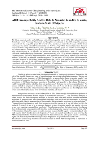
- 3. ABO Incompatibility And Its Role In Neonatal… Bilirubin Determination: This was determined by the method of Malloy and Evelyn.12 The method is based on the formation of a purple compound Azobilirubin, when bilirubin reacts with the diazotized sulphanilic acid introduced by Vander Bergh. Into 2.7mls of distilled water was added 0.1ml of serum in two different tubes. 0.7mls of a mixed diazo reagent was added to the test while the blank was mixed with 0.7mls of diazo A reagent only. A standard was prepared using 0.2mls of standard bilirubin as control. 2.6mls of distilled water was added to a third tube containing 0.2mls of the standard bilirubin in 3.5mls of methanol to which was also added 0.7mls of mixed diazo reagent. All three tubes were incubated at room temperature for 5 minutes. The absorbance was read at 540nm using a spectrophotometer (Cecil 1000 Series). The bilirubin level was calculated using the formula; Key. T=Test Bilirubin = T-B × 171µmol/L S=Standard S-B B=Blank Titration of Maternal Sera: The maternal sera were titrated in a two-fold dilution to a dilution of 1:2048 using sterile saline. To each dilution was added 2mls of a 5 suspension of washed neonatal red cells. All the wells were incubated for one hour at 37°C and were observed for agglutination or settling of cells. Hemolysin Determination: Using a sterile pipette, 0.2mls of maternal sera was transferred into two serological tubes separately. One was placed in a water bath (GallenKamp 20H) held at 56°C for 30 minutes to inactivate complement. The other was used unheated. Into both tubes was added a 0.2mls of 5 suspension of the neonate’s red cell and both were incubated at 37 ˚C for one hour. They were then centrifuged to determine presence of cells or occurrence of hemolysis of the cells by the action of the maternal sera. Saline Agglutination: This was performed using the slide agglutination method used for blood grouping using 5 of the neonate’s red cell. A drop of the cell suspension was mixed with one drop of undiluted serum of the mother. The method was repeated using saline diluted serum. All were mixed individually and observed for agglutination. Serum from a different mother was used to serve as a positive control. Statistical Analysis: The results were analyzed statistically using the student’s t test and the chi’s squares to determine the significance of the results obtained and used to interpret the data obtained from all the analysis. RESULTS The blood group of mother and child assay showed that the number that was incompatible by this reaction was 11(22%) and in this ABO incompatible were 5 (10%) as presented in Fig. 1. the other six (12%) were rhesus incompatible and excluded in the result. 45 39 40 Nouber of cases 35 30 25 20 15 10 5 6 5 0 ABO incompatible Rhesus incompatible ABO and Rhesus compatible Compatibility status Fig. 1: Determination of Blood group compatibility of mother and child The hematological indices indicated reduced levels of Packed cell volume (PCV), Full blood count (FBC), Percentage Lymphocyte, Percentage Monocyte and Percentage Eosinophils in the ABO incompatible as www.theijes.com The IJES Page 19
- 4. ABO Incompatibility And Its Role In Neonatal… compared with the values from the compatible groups. The values Percentage Reticulocyte count, Percentage Neutrophils and Basophils were elevated in the ABO compatible group as compared with the compatible group (Table 1). The severity of the jaundice as evident by the mean bilirubin levels observed for the infants with ABO incompatibility was 20.84 ± 8.1 mg/100ml. This was higher than the mean value of 16.35 ± 6.5 mg/100ml observed for the group ABO compatible group. The bilirubin levels was high in the samples of babies with blood group A or B born to mothers with blood group O with 27.1 mg/100ml for the baby with blood group B. The difference was however not statistically significant (P > 0.05) (Table 2). All (100%) of the cases with ABO incompatibility were coomb’s positive while only 6 (16.2% of the ABO compatible cases were coomb’s positive (Table 3). The level of maternal antibodies in the neonatal circulation was as high as 512 in both cases with over 50% of ABO incompatible group having titres of 256 and above (Table 4). All (100%) of the ABO incompatible cases were hemolytic in the presence serum compliments and 4 (80%) were hemolytic even in the absence of compliments. However, in the ABO compatible cases, 56.8% were hemolytic in the presence of serum compliments while only 19% were hemolytic in the absence of compliments (Table 5). Table 1: Hematological parameters of ABO compatible and incompatible infants Parameter Reticulocyte (%) PCV (%) FBC (cumm) Lymphocytes (%) Neutrophils (%) Monocytes (%) Eosinophils (%) Basophils (%) Bloodgroup Compatible ABO incompatible Compatible ABO incompatible Compatible ABO incompatible Compatible ABO incompatible Compatible ABO incompatible Compatible ABO incompatible Compatible ABO incompatible Compatible ABO incompatible N 39 5 32 5 38 5 39 5 39 5 39 5 39 5 39 5 Mean / SD 2.98 ± 1.27 3.4 ± 0.84 45.78 ± 5.19 42.4 ± 4.6 10250.53 ± 3089.9 7850 ± 3449.3 43.5 ± 9.2 37.6 ± 7.2 32.95 ± 9.6 41 ± 4.9 12.3 ± 3.9 10.8 ± 2.0 10.5 ± 2.8 9.6 ± 2.7 0.41 ± 0.82 1.2 ± 0.83 Table 2: Determination of bilirubin levels in neonates ABO incompatible ABO Compatible Blood group Baby Mother B+ O+ A+ O+ OAO+ B+ A+ O+ Mean Mean Bilirubin mg/100ml 27.1 16.8 26.2 8.4 25.7 20.84 ± 8.1 16.35 ± 6.5 Table 3: Determination of maternal antibody in neonate’s circulation by coomb’s reaction Case Bloodgroup ABO Compatible O+/O+ A+/A+ B+/B+ A+/AB+ Total ABO Incompatible Total www.theijes.com B+/O+ A+/O+ O-/AO+/B+ Coomb’s reaction Positive Negative 5 25 0 3 0 2 1 1 6 (16.2%) 31 (83.8%) 1 0 2 0 1 0 1 0 5 (100%) 0 The IJES Total 30 3 2 2 37 1 2 1 1 5 Page 20
- 5. ABO Incompatibility And Its Role In Neonatal… Table 4: Determination of maternal antibodies in the neonate’s circulation Titer 512 256 128 64 32 16 8 4 2 Total ABO Compatible 1 2 _ _ 2 5 11 10 6 37 ABO Incompatible 1 2 _ _ _ 1 1 _ _ 5 Total 2 4 _ _ 2 6 12 10 6 42 Table 5: Hemolysin reaction in the presence and absence of compliments Blood Group B M Unheated Serum + Total - B+ O+ A+ O+ OAO+ B+ TOTAL 1 2 1 1 5 (100%) O+ O+ A+ A+ B+ B+ A+ AB+ Total 17 2 0 2 21(56.8%) Heated Serum + ABO incompatible group 1 1 2 2 1 1 1 0 5 4 (80%) ABO compatible group 13 30 9 1 3 1 2 2 0 0 2 0 16(43.2%) 37 10(27%) 0 0 0 0 0 Total 0 0 0 1 1 (20%) 1 2 1 1 5 21 2 2 2 27 (73%) 30 3 2 2 37 Table 6: Determination of Saline Agglutination reaction 1(-) 2(+) 3(++) 4(+++) Total 13 22 2 37 Compatible 2 2 1 5 Incompatible 13 24 4 1 42 Total Key +++ Highly Positive ++ Mildly Positive + Weakly Positive DISCUSSION The study showed that ABO incompatible was responsible for 10% of the cases studied. The values obtained from the hematological findings showed a slight variation from the normal values. The mean reticulocyte count for the compatible group was slightly lower than the values of the incompatible group. However the statistics showed no significance in the values (P > 0.05). Moreover, both figures were within the range indicated by Gupta.13 The PCV values were significantly lower (P < 0.05) in the incompatible group as compared to the compatible group. This reveals a mild degree of anemia in the incompatible group. Anemia is a known symptom associated with ABO incompatibility.10 The value of the compatible group was within the range stated by Gupta.13 Our findings agree with the report of Koenig.14Likewise, the bilirubin levels differ significantly in the two groups (P < 0.05), the level in the incompatible group being higher than that in the compatible group. This is indicative of the severity of the jaundice characterized by massive destruction of red blood cells and the accumulation of bilirubin in the baby’s circulation. These agree with other findings that jaundice resulting from incompatibility usually present higher levels of serum bilirubin above20mg/100ml.15, 16 Physiologic jaundice is therefore likely in the case of most of the compatible group though some of them presented a positive coomb’s result. These antibodies so detected could be cross-reacting antibodies or other maternal immunoglobulin, which usually occur in the fetal circulation. This finding is supported by the work of Beard and Nathanielsz.17 www.theijes.com The IJES Page 21
- 6. ABO Incompatibility And Its Role In Neonatal… A survey of the white cell counts analysis did not reveal any unusual trend. The count for both compatible and incompatible were low as compared to the normal value given by Gupta.13 When the values were subjected to chi-square 2 analyses at P = 0.05, no significant difference was indicated in the compatible group and the normal accepted value while the incompatible group showed a remarked difference. The differential leukocyte counts obtained from both groups revealed a similar pattern. The range of the counts was found to be within the range indicated by Lanzkousky (1980)18 as presented in Table 5. These figures also agree with the findings of Read, et al that blood counts in cases of hepatic and obstructive jaundice are usually normal unless there are cases of blood loss.19 The count was fairly low with the incompatible cases, which is indicative of mild anemia associated with hemolytic jaundice. Similarly the white cell counts (W.C.C.) for the compatible group fell within the range of that indicated as normal values for neonates in the first week of life. These values are useful indices in detecting cases of bacterial infections, which are known to result in neutrophilia (Baker, et al., 2001). The incompatible cases were all Coomb’s positive (a case of 100% positivity). The findings of Rosenfield on the ABO incompatible cases, showed that 88.4% cases of the total samples they analyzed as Coomb’s negative.1 Risemberg et al on the other hand reported that Coomb’s reaction of infants with a bilirubin level at which hyperbilirubinemia is indicated was 60% positive and 40% negative in ABO incompatible cases.4 The present study showed that all the cases of ABO incompatibility were Coomb’s positive thus agreeing with the use of Coomb’s test for the establishment of cases of hemolytic disease due to incompatibility.11 The high level of cases with a positive Coomb’s result could be due to incompatibility in other blood group antigens erstwhile not given attention. The existences of incompatibility in these rare antigens and the attending reactions have been documented.5, 6, 7 Findings from these reports revealed that incompatibility in these blood group antigens occurring between mother and child result in maternal antibodies which cross the placenta barrier into fetal circulation.7 The findings from this research brings to the fore the need to consider the role of ABO incompatibility, a contributing factor to neonatal jaundice in the population studied. As other factors are considered in the diagnosis of neonatal jaundice, the role of ABO incompatibility should be considered as well as it contributes considerably to the incidence and severity of neonatal jaundice. III. CONCLUSION It can be seen from the studies that ABO incompatibility is responsible for 10% of cases of neonatal jaundice with a mean bilirubin level of up to of 20.77mg/100ml. it is therefore a factor to be considered in the diagnosis of neonatal jaundice and should be considered as such. IV. ACKNOWLEDGEMENTS The contribution of the hospital staff in the collection of samples is dully acknowledged as well as the ethical board for the permit to undertake the work. REFERENCES [1] [2] [3] [4] [5] [6] [7] [8] [9] [10] [11] [12] [13] Rosenfield, R.E. (1955). A-B hemolytic disease of the newborn. Analysis of 1480 cord blood specimen with special reference to the direct antiglobulin test and to the group O mother. Blood 10: 17.28. Zueler, W.W. and Cohen,I. (1957). ABO Hemolytic disease and hetero-specific pregnancy. Pediatric Clinics of North America 4: 405-428. Robinson, G.C., Dunn, H.H., and Wong, L.C. (1960). Clinical and laboratory findings in hetero-specific pregnancy with a note on the incidence of ABO hemolytic disease. Acta Paediatrica 49(120): 53-62. Risemberg, H.M., Mazzi, E., Macdonald, M.G., Peratta, M. and Heldrich, F. (1977). Correlation of cord bilirubin levels with hyperbilirubinemia in ABO incompatibility. Archives of Disease of Childhood 52: 219-222. Zmijewski, C. M. and Fletcher, J. L. (1972). Immunohamatology (2eds). Appleton –Century – Croft. New York. Pp 334. Bryant, N. J. (1976). An introduction to Immunohematology. (eds) W. B. Saunders Company. U. S. A. Pp 253 Bloodbook, (2001). http://www.bloodboook.com/type-sys.html. Pp.1-6 Hull, J.W. (2003). ABO incompatibility. http://www.drhull.com/EncyMaster/A/ABO-incompatibility.html. Pg1. Brimblecombe, F., and Barltrop, D. (1978). Children in Health and Disease. (eds) Williams Clones, Bailliere,Tindall Publishers, London. Pp 91-145. Merck manual. (2003). Hematologic disorders. http://www.merck.com/pubs/mmanual/section19/chapter260/260h.htm Pp 1-7. Orzalesi, M., Gloria, F., Lucarelli, P. and Bottini, E. (1973). ABO system incompatibility: relationship between direct coomb’s test positivity and neonatal jaundice. Pediatrics 51: 288-289. Baker, F.J., Silverton, R.E. and Pallister, C.J. (2001). Introduction to Medical Laboratory Technology. (7eds). Bounty Press, Ltd. London. Pp. 348-419. Gupta, S. (1978). A Text Book Of Pediatrics. (eds) Vikas Publishing House. (PVT): New Delhi. Pp 375-408. www.theijes.com The IJES Page 22
- 7. ABO Incompatibility And Its Role In Neonatal… [14] [15] [16] [17] [18] [19] Koenig, J.M. (2000). Evaluation and treatment of erythroblastosis fetalis in the neonate. In: Christensen R, ed. Hematologic Problems of the Neonate. Philadelphia, Pa: WB Saunders: Pp. 185-207 Hansen, T.W.R. (2002). Jaundice neonatal. http:// www.emedicine.com/ped/topic/061.htm. Pg 1-21. Beeby, P. (2003). Jaundice.http://www.cs.nsw.gov.au/rpa/neonatal/htm/newprot/jaund2.htm pp1-9. Beard, R.W., and Nathanielsz, P.W. (1976). Fetal physiology and Medicine. W.B. Saunders Co. London. Pp17-42. Lanzkousky, P. (1980). Pediatric Hematology – Oncology. McGraw-Hill Inc. U.S.A. Pp 224-249. Read, A.E., Barritt, D.W., and Hewer, R.L. (1979). Modern Medicine. A textbook for Students. (2eds). The Pitman Press. Great Britain. Pp 61-64 www.theijes.com The IJES Page 23