Hydronephrosis: Causes, Symptoms and Treatment
•
19 likes•9,829 views

definition of hydronephrosis, causes and types of hydronephrosis pathophysiology of hydronephrosis clinical manifestation and diagnostic test for hydronephrosis management

Recommended
More Related Content
What's hot
What's hot (20)
Similar to Hydronephrosis: Causes, Symptoms and Treatment
Similar to Hydronephrosis: Causes, Symptoms and Treatment (20)
More from NEHA BHARTI
More from NEHA BHARTI (15)
Recently uploaded
Recently uploaded (20)
Hydronephrosis: Causes, Symptoms and Treatment
- 1. PRESENTED BY- NEHA BHARTI NURSING TUTOR M.Sc (N) MEDICAL SURGICAL NURSING SMVDCoN, KAKRYAL
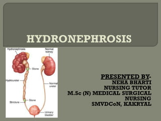
- 2. Hydronephrosis is swelling of one or both kidneys.
- 3. Kidney swelling happens when urine can't drain from a kidney and builds up in the kidney as a result. This can occur from a blockage in the tubes that drain urine from the kidneys (ureters) or from an anatomical defect that doesn't allow urine to drain properly.
- 4. Hydronephrosis can happen at any age. Hydronephrosis in children may be diagnosed during infancy or sometimes during a prenatal ultrasound before the baby is born.
- 5. Normally, urine goes from the kidney to the tube that drains the kidney (ureter), to the bladder and then out of the body. But, sometimes urine backs up or remains inside the kidney or in the ureter. That's when hydronephrosis can develop. Some common causes of hydronephrosis include:
- 6. PARTIAL BLOCKAGE IN THE URINARYTRACT Urinary tract blockages often form where the kidney meets the ureter, at a point called the uretero- pelvic junction. Less commonly, blockages may occur where the ureter meets the bladder at what's called the ureterovesical junction. VESICOURETERAL REFLUX Vesicoureteral reflux happens when urine flows backward through the ureter from the bladder up into the kidney. Normally, urine flows only one way in the ureter. Urine flowing the wrong way makes it difficult for the kidney to empty properly and causes the kidney to swell
- 7. Less-common causes of hydronephrosis include kidney stones, a tumor in the abdomen or pelvis.
- 8. Hydronephrosis may be Unilateral involving just one kidney or Bilateral involving both kidneys.
- 9. UNILATERAL HYDRONEPHROSIS Unilateral hydronephrosis usually results from a blockage in one of ureters, or where the renal pelvis (the wide, urine-collecting system of each kidney) joins the narrow ureter. BILATERAL HYDRONEPHROSIS Both kidneys will be affected by hydronephrosis if there is a blockage in or near the bladder.
- 10. Normally, the kidney filters waste products from blood and disposes of it in the urine. The urine drains into individual calyces (single -calyx) that form the renal pelvis. This empties into the ureter, a tube that connects the kidney to the bladder. The urethra is the tube that empties the bladder.
- 11. The main function of kidneys is to filter the blood and eliminate the waste materials from the body. In this process,they also absorb the useful materials such as glucose. The waste materials eliminate in the form of urine that goes out through a tube called urethra.
- 12. DUETO ANY CAUSE OR FACTOR UPJ OBSTRUCTIONWILL OCCUR AND JUNCTION IS BLOCK URINE BECOMES BACKED UP CAUSES A BUILD-UP OF URINE INSIDE THE KIDNEY, PUTTING PRESSURE ON THE KIDNEY AND CAUSESTHE KIDNEY PLUMBING TO DILATE IT MAY ALSO CAUSE URINARY INFECTION, KIDNEY STONES OR PAIN. THIS LEADSTO INFLAMMATION AND SWELLING OF THE KIDNEY.
- 13. There may or may not be direct symptoms of hydronephrosis depending upon the underlying cause. 1. ACUTE HYDRONEPHROSIS: When the blockage happens quickly, patient may experience a severe pain (renal colic)in back or side, between ribs and hip. The pain will be on the side of the affected kidney or on both sides if both kidneys are affected.
- 14. Other symptoms can include: Swelling in abdomen . Fever Urinary tract infection Pain during urination Blood seen in the urine
- 15. 2. CHRONIC HYDRONEPHROSIS: Chronic hydronephrosis develops over time and there may be no specific symptoms. If hydronephrosisis‘ caused by a slow-developing blockage, patient may have the same symptoms as acute hydronephrosis, no symptoms at all, or a dull, aching pain in side that comes and goes.
- 16. These are very nonspecific and may include weakness, malaise, nausea and vomiting. If electrolyte abnormalities occur because the kidneys are unable to regulate sodium, potassium, and calcium,there may be heart rhythm disturbances and muscle spasms.
- 17. Tests for diagnosing hydronephrosis may include: A blood test to evaluate kidney function A urine test to check for signs of infection or urinary stones that could cause a blockage An ultrasound imaging exam, during which doctor can view the kidneys, bladder and other urinary structures to identify potential problems A voiding cysto- urethrogram, an X-ray exam that uses a special dye to outline the kidneys, ureters, bladder and urethra, capturing images before and during urination
- 18. Depending on the results from initial testing,doctor may recommend additional imaging exams,such as a Computerized tomography (CT) scan or Magnetic resonance imaging (MRI).
- 19. The goal of treatment for hydronephrosisis to restart the free flow of urine from the kidney and decrease the swelling and pressure that builds up and decreases kidney function. The initial care for the patient is aimed at minimizing pain and preventing urinary tract infections. Otherwise, surgical intervention may be required.
- 20. The timing of the procedure depends upon the underlying cause of hydronephrosis and hydro- ureter and the associated medical conditions that may be present. For example, patients with a kidney stone may be allowed 1-2 weeks to pass the stone with only supportive pain control if urine flow is not completely blocked by the stone.
- 21. If, however, the patient develops an infection or if they only have one kidney, surgical intervention may be done emergently to remove the stone.
- 22. Shock wave lithotripsy (SWL or extra corporeal shock wave lithotripsy) is the most common treatment for kidney stones. Shockwaves from outside the body are targeted at a kidney stone causing the stone to fragment into tiny pieces that are able to be passed out of the urinary tract in the urine.