TỔNG ÔN TẬP THI VÀO LỚP 10 MÔN TIẾNG ANH NĂM HỌC 2023 - 2024 CÓ ĐÁP ÁN (NGỮ Â...
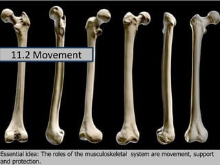
11.2 Movement
1. Essential idea: The roles of the musculoskeletal system are movement, support
and protection.
11.2 Movement
2. Understandings
Statement Guidance
11.2 U.1 Bones and exoskeletons provide anchorage
for muscles and act as levers.
11.2 U.2 Synovial joints allow certain movements but
not others.
11.2 U.3 Movement of the body requires musclesto
work in antagonistic pairs.
11.2 U.4 Skeletal muscle fibres are multinucleateand
contain specialized endoplasmic reticulum.
11.2 U.5 Muscle fibres contain many myofibrils.
11.2 U.6 Each myofibril is made up of contractile
sarcomeres.
The contraction of the skeletal muscleis
11.1 U.7 achieved by the sliding of actin and myosin
filaments.
11.2 U.8 ATP hydrolysis and cross bridge formationare
necessary for the filaments to slide.
11.2 U.9 Calcium ions and the proteins tropomyosin
and troponin control muscle contractions.
3. Applications and Skills
Statement Guidance
11.2 A.1 Antagonistic pairs of muscles in an insectleg.
Elbow diagram should include cartilage, 11.2
11.2 S.1 Annotation of a diagram of the human elbow. synovial fluid, joint capsule, named bonesand
named antagonistic muscles.
Drawing labelled diagrams of the structure ofa
11.2 S.2 Drawing labelled diagrams of the structure of a sarcomere should include Z lines, actin
sarcomere. filaments, myosin filaments with heads, and
the resultant light and dark bands.
Analysis of electron micrographs to find the Measurement of the length of sarcomeres will
11.2 S.3 state of contraction of muscle fibres. require calibration of the eyepiece scale ofthe
microscope.
4. Bones:
serve as a structural framework for tendons and ligaments to
attach, provide support for soft tissue. In addition your bones
protect internal organs from injury.
11.2 U.1 Bones and exoskeletons provide anchorage for muscles and act as levers
5. 11.2 U.1 Bones and exoskeletons provide anchorage for muscles and act as levers
Exoskeleton is the stiff covering on the outside of some creatures. There are often
flexible joints with underlying muscles that allow for a range of movement of the
exoskeleton. An exoskeleton is actually the opposite of how we are put
together (our protection and attachments are on the inside and
exoskeletons are on the outside).
6. 11.2 U.1 Bones and exoskeletons provide anchorage for muscles and act as levers
Exoskeletons: surround and protect the body surface of
animals. It provides assistance in movement since they are the
anchorage for muscles and act as levers. As with bone these
levers change the size and direction of forces generated by the
muscles.
7. 11.2 A.1 Antagonistic pairs of muscles in an insect leg.
Grasshoppers (Acrididae)
These insects have a skeleton on the outside of the body called an exoskeleton. The muscles are
inside the hard shell. The back leg is much longer than the others to aid jumping. Long legs
increase the distance over which the jumper can push on the ground.
8. • Grasshopper legs are third-class levers. The two main muscles inside are
the extensor tibiae muscle which contracts to extends the leg, and the
flexor tibiae muscle which contracts to flex the leg. These muscles pull on
tendons which are attached to the tibia on either side of the joint pivot.
• Skeletal muscles, such as the extensor and flexor that occur in pairs are
often antagonistic: when one contracts the other relaxes to produce
controlled movement in opposite directions.
11.2 A.1 Antagonistic pairs of muscles in an insect leg.
9. Levers can be used so that a small force can move a much bigger force. This is called
mechanical advantage. In our bodies bones act as lever, joints act as a pivot, and muscles provide the forces
to move loads
Three Types:
First-class levers act like a seesaw. An example of a first class lever is when a human nods their
head
Second-class levers have a resistance in the middle, like a load in a wheel-barrow. An example is
calf raise ball of foot is fulcrum, the body’s mass is the resistance and the effort is applied by calf
muscle.
Third-class levers have the effort from the muscle in the middle of the lever. The majority of the
human body's musculoskeletal levers are third class. An example of the arm, the effort force is
provided by the contraction of the biceps, the fulcrum is the elbow joint and the resistance would be
provided by whatever weight is being lifted.
11.2 U.1 Bones and exoskeletons provide anchorage for muscles and act as levers
10. 11.2 U.2 Synovial joints allow certain movements but not others
A synovial joint, are made up of
bones with a joint capsule that
surrounds the bones articulating
surfaces with synovial fluid.
Synovial fluid reduce the friction
between the articular cartilage of
synovial joints during movement.
11. 11.2 U.2 Synovial joints allow certain movements but not others
Synovial joints are subdivided based on the shape. There are six types of synovial joints are
pivot, hinge, condyloid, saddle, plane, and ball-and socket-joints.
As an example the ball and socket joint
below, has the greatest range of motion.
These joints consist of a rounded head of
one bone (the ball) fits into the concave
articulation (the socket). The hip joint and
the shoulder joint are the only ball-and-
socket joints of the body.
12. 11.2 U.2 Synovial joints allow certain movements but not others
These are examples of the ball and socket joint
that make up the hip. The shoulder is also a ball
and socket joint. Ball and socket joints have the
greatest range of motion. The increase in range
is due to the shape of the joint, it allows for
moment in all axes and planes.
13. Hip MotionKnee Motion
11.2 U.2 Synovial joints allow certain movements but not others
Knee movement: is limited due to joint shape and moves in one plane with flexion
and extension.
Hip movement: has a much greater range of movement. This is due to the ball and
socket joint shape. The difference in shape means the joint moves in more than one
plane, flexion, extension, rotation, abduction and adduction.
15. Structure Function
Biceps
Triceps
Humerus
Radius / Ulna
Cartilage
Synovial fluid
Joint capsule
Tendons
Ligaments
11.2 S.1 Annotation of a diagram of the human elbow.
Can you annotate the structures? Remember structure dictates function
16. Can you annotate the structures? Remember structure dictates function
Structure Function
Biceps Bends the arm (flexor)
Triceps Straightens the arm (extensor)
Humerus Anchors the muscle (muscle origin)
Radius / Ulna Acts as forearm levers (muscle insertion) – radius for the biceps, ulna for the triceps
Cartilage Smooth surface to allow easy movement, absorbs shock and distributes load
Synovial fluid Provides lubrication, reduces friction in the joint.
Joint capsule Seals the joint, contains the synovial fluid.
Tendons non-elastic tissue connecting muscle to bone
Ligaments non-elastic tissue connecting bone to bone
11.2 S.1 Annotation of a diagram of the human elbow.
17. 11.2 U.3 Movement of the body requires muscles to work in antagonistic pairs.
https://youtu.be/SOMFX_83sqk
http://purchon.com/flash/elbow.swf
The triceps and
biceps are working in
opposite directions
and hence are
examples of
antagonistic muscles.
18. Striated Muscle Cell Organization:
• Each muscle fiber, (1 muscle cell) has many myofibrils (protein bundles)
• Each muscle fiber has many flattened or sausage shaped nuclei pushed
against the plasma membrane
• Each muscle fiber has: plasma membrane= Sarcolemma
• Each muscle fiber has: cytoploasm = Sarcoplasm
• Each muscle fiber has: many Mitochondria
11.2 U.4 Skeletal muscle fibers are multinucleate and contain specialized endoplasmic reticulum 11.2
U.5 Muscle fibers contain many myofibrils.
19. Structure of a Skeletal Muscle Fiber
11.2 U.4 Skeletal muscle fibers are multinucleate and contain specialized endoplasmic reticulum 11.2
U.5 Muscle fibers contain many myofibrils.
20. 11.2 U.4 Skeletal muscle fibers are multinucleate and contain specialized endoplasmic reticulum 11.2
U.5 Muscle fibers contain many myofibrils.
21. A single skeletal muscle cell is multinucleated, with nuclei positioned along the
edges
Muscle fiber cells are held together by the plasma membrane
Many mitochondria are present due to the high demand for ATP
Muscle cell contain sarcoplasmic reticulum, a specialized type pf endoplasmic
reticulum, that stores calcium ions and pumps them out into the sarcoplasm when
the muscle fibers is stimulated
Myofibrils are the basic rod-like contractile units with a muscle cells. Myofibrils are
grouped together inside muscle cells, which are known as muscle fibers.
11.2 U.4 Skeletal muscle fibres are multinucleate and contain specialized endoplasmic reticulum 11.2
U.5 Muscle fibers contain many myofibrils.
22. 11.2 U.6 Each myofibril is made up of contractile sarcomeres.
Sarcomere: are repeating units of muscle fiber. They are the site
where contractions occur. This causes muscles fibers to shorten pulling z discs
closer to each other.
23. 11.2 U.7 The contraction of the skeletal muscle is achieved by the sliding of actin
and myosin filaments.
Sliding filament theory: Actin and Myosin Cross-Bridge Formation
•Actin is the binding sites for the myosin heads. The head is covered by a blocking
complex (troponin and tropomyosin)
•Calcium ions bind to troponin (found on the Actin filament) and reconfigure the
complex, exposing the binding sites for the myosin heads
•The Myosin heads (after the release of a phosphate from ATP) then form a
cross-bridge with the actin filaments
ATP
24. 11.2 U.8 ATP hydrolysis and cross bridge formation are necessary for the
filaments to slide. 11.2 U.9 Calcium ions and the proteins tropomyosin and
troponin control muscle contractions.
Muscle Contraction Part 1
Skeletal Muscle Contraction (Part I)
Step Outline of each step of the contraction
1 Thick filaments (Myosin) anchored on the M line and thin filaments (Actin) anchored on the Z line. A
Contraction begins when ATP is hydrolyzed into ADP causing Myosin to extend.
2 When an action potential is reached in a striated muscle cell, the sarcoplasmic reticulum releases
calcium ions into the myofibrils. Ca+2 binds on actin
3 Actin is made of two protein fibers Troponin and Tropomyosin that respond to the presence of Ca+2.
Troponin which binds to Ca+2, which moves Tropomyosin which moves to create binding sites
25. 11.2 U.8 ATP hydrolysis and cross bridge formation are necessary for the
filaments to slide. 11.2 U.9 Calcium ions and the proteins tropomyosin and
troponin control muscle contractions.
Muscle Contraction Part 2
Skeletal Muscle Contraction (Part 2)
Step Outline of each step of the contraction
4 The myosin head now can attach to Actin at the cross bridge, triggering a power stroke. The energy
stored in the myofilament causes pulling of the filament towards the M line, shortening the Sarcomere
and releasing ADP
5 Myosin and Actin remain attached until ATP attaches once again breaking the bond at the cross bridge.
This free Myosin making it possible for another contraction or to relax.
6 The ATP release a phosphate (P) to became ADP and the energy is stored in the Myosin heads
26. 1.2 S.2 Drawing labelled diagrams of the structure of a sarcomere.
27. 11.2 S.3 Analysis of electron micrographs to find the state of contraction of
muscle fibers.
28. 11.2 S.3 Analysis of electron micrographs to find the state of contraction of
muscle fibers.
29. 11.2 S.3 Analysis of electron micrographs to find the state of contraction of
muscle fibers.
30. 11.2 S.3 Analysis of electron micrographs to find the state of contraction of
muscle fibers.
Electron micrograph of human skeletal muscle
1μm
Analyze the micrograph and use it
to answer the following:
1. Deduce whether the myofibrils are
contracted or relaxed
2. Calculate the magnification of the
electron micrograph
3. Measuring an individual sarcomere
accurately is difficult due to their
small size (Usual size is 2 um).
Commonly scientists use the
formula below:
= total length of n sacromeres
n
a. Measure the total length of
five sarcomere from z-line to
z-line
b. Calculate the mean length of a
sarcomere
mean
sarcomere
length
(μm)
31. The light emissions are detected and recorded using specially adapted microscopes and cameras.
• Monitoring calcium fluxes in real time could help to understand the functioning of the central nervous
system and its interactions with muscles. In jellyfish, the chemiluminescent calcium binding aequorin protein
is associated with the green fluorescent protein and a green bioluminescent signal is emitted upon
Ca2+ stimulation
• A number of researchers have used fluorescent dyes to visualize and measure the movement of myosin
and actin.
• Aequorin and the fluorescent dyes used in research only emit for a few short nano-seconds making them
ideal to measure the rapid movements found in muscle cells.
Nature of science: Developments in scientific research follow improvements in apparatus -
fluorescentcalcium ions have been used to study the cyclic interactions in muscle contraction.(1.8)
32. Nature of science: Developments in scientific research follow improvements in apparatus - fluorescentcalcium ions
have been used to study the cyclic interactions in muscle contraction.(1.8)
These studies were first done by Ashley and
Ridgway (1968) were the first to study the role that
Calcium ions (Ca+2) plays in the coupling of nerve
impulses and muscle contraction. Their work was
made possibly by the use of aequorin, a Ca+2
binding bioluminescent protein. Upon Ca+2
binding aequorin emits light. The timing of light
emission peaks between the arrival of an electrical
impulse at the muscle fiber and the contraction of
the muscle fibers. This is consistent with theory of
release of Ca+2 from the sarcoplasmic reticulum.