Chronic Diarrhea and Malabsorption: Causes and Clinical Presentation
1. By: Yugeasan A/L M Arumugam
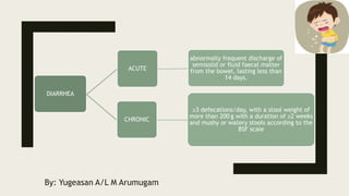
DIARRHEA
ACUTE
abnormally frequent discharge of
semisolid or fluid faecal matter
from the bowel, lasting less than
14 days.
CHRONIC
≥3 defecations/day, with a stool weight of
more than 200 g with a duration of ≥2 weeks
and mushy or watery stools according to the
BSF scale
3. Pathophysiology of diarrhea in general
1) Osmotic diarrhea:
Common cause: ingestion of non-absorbable substance, generalized malabsorption, specific
absorptive defect
2) Secretory diarrhea:
Common cause: enterotoxin (cholera toxin, E.coli thermolabile and thermosensitive toxins),
hormone (VIP), bile salt and fatty acid in illeal resection and laxatives
3) Inflammatory diarrhea (mucosal destruction):
Damage to intestinal mucosa due to infection and inflammation
4) Abnormal motility:
Common cause: diabetic, post-vagotomy, hyperthyroidism
7. Etiology:
■ Short-lived, usually <10 days
■ Requires no investigation or treatment
■ usually results from gastrointestinal infection
Common causes:
■ Food poisoning
■ Traveler's diarrhea
10. Food poisoning
■ Definition: any disease of an infective or toxic nature caused by or thought to
be caused by the consumption of food and water
■ Some food poisoning cause gastroenteritis expect food poisoning due to
botulism
■ flaws in the processing, storage and distribution of food products allow
massive amplification of infection, resulting in extensive contamination
11. Traveler’s diarrhea
■ passage of three or more unformed stools per day in a resident of an
industrialized country travelling in a developing nation
■ Infection is food- or water-borne
■ younger travellers are most often affected
■ Usually no treatment required but quinolone antibiotics speeds up recovery
12. Common clinical features:
■ Fever
■ Abdominal pain
■ Vomiting
■ If severe and left untreated can lead to dehydration
Clinical features according to cause:
■ Inflammatory causes: blood-stained and loose stool, increased frequency
■ Infective causes: blood-stained and loose stool, increased frequency
■ Steatorrhea: pale, offensive stool that floats and associated with loos of
weight and appetite
■ Organic causes: nocturnal, increased frequency and urgency
■ Cholera infection: Rice water stool
13. Investigation:
■ Investigation only carried out if diarrhea more than 5-7 days
Investigation Test for or expected results and
conditions
Stool sample culture and sensitivity
testing
Stool sample should be sent for
investigation immediately
Observe presence of ova, cyst or
parasite under microscope
Clostriduim. difficile assay Specimen: stool
Advantage: Safe, easy and ready to
use,
accurate, easy to interpret
To rule out hospital acquired
diarrhoea
Sigmoidoscopy and Rectal biopsy with aid
of colonoscopy
In patients where the diarrhea persist
even after taking medications, in
homosexual male and in
immunocompromised patients
14. Immature E. histolytica in a concentrated wet mount stained
with iodine. This early cyst has only one nucleus and a glycogen
mass is visible (brown stain). From CDC’s Division of Parasitic
Diseases
Giardia intestinalis on small
intestinal mucosa. (Courtesy of Dr A
Phillips, Department of Electron
Microscopy,
Royal Free Hospital, London.)
15.
16. Management:
■ Usually resolves spontaneously
■ If severe, oral fluid and electrolyte replacement therapy should be started
■ It is believed that prescribing antidiarrhea drugs can impair clearance of
causal organism or toxins
■ Antibiotics can be prescribed for infective gastroenteritis
19. Etiology:
■ Always needs to be investigated
■ 3 or more loose stool per day for more than 2 weeks or 4 weeks
■ impact on quality of life and overall health mildest: discomfort, severe: disabling
and life threatening
Common causes:
■ irritable bowel syndrome (IBS)
■ Inflammatory bowel disease (Crohn disease and ulcerative colitis)
■ Malabsorption syndromes in which food cannot be digested and absorbed
■ Chronic infections
■ Endocrine disorder (e.g. hyperthyroidism and long term uncontrolled diabetes)
22. Why is there chronic diarrhea in IBD?
■ Inflammation. When intestine is inflamed, it absorbs much less sodium and
water. It also may leak more fluids, resulting in loose, watery stool
■ Difficulty absorbing fat, starches, sugars, and bile acids
■ Bacterial infection
■ Fistulas
■ Malabsorption after surgery
24. Hyperthyroidism and chronic
diarrhea:
■ Intestinal hypermotility in thyrotoxicosis reduces small bowel transit time
■ Increased appetite and excessive fat-rich food intake may contribute to
excessive faecal fat due to fat malabsorption
■ hypersecretory state within the intestinal mucosa due to hypersecretion of
bile
■ mean anal resting and squeeze pressures are lowered
32. ■ Monastyrsky, K. (2016, April 26). How to evaluate stools with Bristol stool
chart. Retrieved December 18, 2017, from
https://www.gutsense.org/constipation/normal_stools.html
■ Treatment. (n.d.). Retrieved December 18, 2017, from
https://crohnsdisease.com/treatment/
■ Ahmed Emad Sami, Stagiaire at TUCOM Follow. (2017, August 17). Chronic
diarrhea & malnutrition. Retrieved December 18, 2017, from
https://www.slideshare.net/ahmedemad88/chronic-diarrhea-malnutrition
■ P., & M. (2017). Kumar and clalrk's Clinical Medicine NINTH EDITION (9th ed.).
Edinburgh London New York Oxford Philadelphia St Louis Sydney Toronto:
Elsevier .
■ B., N., & S. (Eds.). (2014). Davidson’s Principles and Practice of Medicine 22nd
Edition (22nd ed.). Edinburgh London New York Oxford Philadelphia St Louis
34. DEFINITION:
Defective absorption of fats, fat- and water-soluble vitamins, proteins,
carbohydrates, electrolytes and minerals and water.
- Presents most commonly as chronic diarrhea.
35. Pathophysiology:
- results from abnormalities of the three processes which are essential to normal digestion:
1. Intraluminal maldigestion occurs when deficiency of bile or pancreatic enzymes results in inadequate
solubilisation and hydrolysis of nutrients. Fat and protein malabsorption results. This may also occur with
small bowel bacterial overgrowth.
2. Mucosal malabsorption results from small bowel resection or conditions which damage the small intestinal
epithelium, thereby diminishing the surface area for absorption and depleting brush border enzyme activity.
3. ‘Post-mucosal’ lymphatic obstruction prevents the uptake and transport of absorbed lipids into lymphatic
vessels. Increased pressure in these vessels results in leakage into the intestinal lumen, leading to protein-
losing enteropathy.
36. Effects:
Carbohydrate malabsorption / carbohydrate intolerance:
- Example: lactase intolerance
- Undigested disaccharides an osmotic load that attracts water and electrolytes into the bowel watery
diarrhea
- Bacterial fermentation of carbohydrates in the colon produces gases excessive flatus, bloating and
distention, and abdominal pain.
Protein malabsorption
- Example: celiac disease ( gluten-sensitive enteropathy)
- Symptoms: - edema
- Weight loss
- chronic diarrhea
- abdominal distention
37. Fat malabsorption
- example: bacteria overgrowth
- Unabsorbed fats trap fat-soluble vitamins (A, D, E, K) and possibly some minerals, causing deficiency.
- Fat in stool Steatorrhea (foul-smelling, pale, bulky, and greasy stools) hallmark of malabsorption.
Vitamins and minerals malabsorption:
- Vitamin A / vitamin B12 peripheral neuropathy
- Vitamin B12, pyridoxine, folate megaloblastic anemia and mucositis
- Vitamin K bleeding
- Vitamin D, calcium, magnesium osteopenia, tetany
40. Coeliac disease (gluten-sensitive enteropathy)
inflammation of the mucosa of the upper small bowel that improves when gluten is withdrawn from the diet
and relapses when gluten is reintroduced.
Dermatitis herpetiformis
an uncommon, blistering, subepidermal eruption of the skin associated with a gluten-sensitive enteropathy .
Rarely, gross malabsorption occurs, but usually the jejunal morphological abnormalities are not as severe as in
coeliac disease.
The inheritance and immunological abnormalities are the same as for coeliac disease.
The skin condition responds to dapsone but a gluten-free diet improves both the enteropathy and the skin
lesion, and is recommended for long-term benefit.
Non-coeliac gluten intolerance
sensitive to dietary wheat and gluten containing foods but do not have coeliac disease, in so far as their
coeliac serology is negative and duodenal biopsies are normal.
range of symptoms, including diarrhoea, bloating and abdominal pain, which improve on avoidance of gluten.
The mechanism is not yet clear.
41. Tropical sprue
This condition presents with chronic diarrhoea and malabsorption, and occurs in residents of or visitors to
tropical areas where the disease is endemic: most of Asia, some Caribbean islands, Puerto Rico and parts of
South America.
Epidemics occur, lasting up to 2 years; in some areas, repeated epidemics are seen at varying intervals of up
to 10 years.
Exact causative factor is unknown, but an intestinal microbial infection is believed to be the initiating insult.
The infection results in enterocyte injury, intestinal stasis, and possible bacteria overgrowth.
Villous destruction and demonstrable nutrient malabsorption happens.
Usual organisms : Klebsiella, E coli and Enterobacter species.
Bacterial overgrowth
clinical manifestations that occur when the normally low number of bacteria that inhabit the stomach,
duodenum, jejunum, and proximal ileum significantly increases or becomes overtaken by other pathogens.
Normally found in association with a structural abnormalities of the small intestine (stricture or
diverticulum), although it can occur occasionally in the elderly without such an abnormality or disorders that
cause decreased gastric acidity, reduced peristaltic activity, and mucosal damage or atrophy.
Normally, colony counts of gram-positive bacteria and fungi in the duodenum and jejunum are less than 1X10⁵
organisms/mL.
Malabsorption of bile acids, fats, carbohydrates, proteins, and vitamins results in direct damage to the lining
of the luminal surface by bacteria or by transformation of nutrients into toxic metabolites, leading to many
of the symptoms of diarrhea and weight loss associated with bacterial overgrowth syndrome.
42. Small intestinal resection
usually well tolerated
massive resection leaving less than 1 m of small bowel in continuity is followed by the short bowel syndrome.
effects of resection (depends on the amount and location of the resection and the presence or absence of
the colon)
Effects of jejunum resection:
- may lead to gastric hypersecretion with high gastrin levels
Effects of ileal resection:
• Bile-salt-induced diarrhea Bile salts and fatty acids enter the colon and cause malabsorption of water
and electrolytes
• Steatorrhoea and gallstone formation Increased bile salt synthesis can compensate for loss of
approximately one-third of the bile salts in the faeces. Greater loss than this results in decreased micelle
formation and steatorrhoea, and lithogenic bile and gallstone formation.
• Oxaluria and oxalate stones Bile salts in the colon cause increased oxalate absorption with oxaluria,
leading to urinary stone formation.
• B12 deficiency Low serum B12, macrocytosis and other effects of B12 deficiency are seen.
43. Whipple's disease
rare infectious bacterial disease caused by Tropheryma whipplei.
presents with: - arthritis and arthralgia
- progressive weight loss
- diarrhoea with abdominal pain
- systemic symptoms of fever and weight loss
- Peripheral lymphadenopathy
- many neurological conditions.
features of chronic inflammation and malabsorption.
Endoscopy typically shows pale, shaggy duodenal mucosa with eroded, red, friable patches
Radiation enteritis
Radiation of >40 Gy will damage the intestine.
chronic effects: muscle fibre atrophy, ulcerative changes due to ischaemia, and obstruction due to radiation-
induced
fibrotic strictures.
Chronic radiation enteritis is diagnosed if symptoms persist for ≥3 months
Abdominal pain due to obstruction is the main symptom.
Malabsorption bacterial overgrowth in dilated segments and mucosal damage increased bowel
frequency.
44. Parasite infestation
• Giardia intestinalis diarrhoea and malabsorption with steatorrhoea with minor changes in the jejunal
mucosa
• Cryptosporidiosis malabsorption
• HIV infection prone to parasitic infestation malabsorption
Other causes
- Drugs bind bile salts (e.g. colestyramine) and some antibiotics (e.g. neomycin) produce steatorrhoea.
- Thyrotoxicosis diarrhoea, rarely with steatorrhoea, owing to increased gastric emptying and increased
motility.
- Zollinger–Ellison syndrome
- Lymphoma that has infiltrated the small bowel mucosa malabsorption.
- Diabetes mellitus diarrhoea, malabsorption and steatorrhoea, and bacterial overgrowth from autonomic
neuropathy that leads to small bowel stasis.
45. References:
- P. K., & M. C. (2017). Kumar and clark's clinical medicine (9th ed.).
- S. D. (2014). Davidson's Principles and Practice of Medicine (22nd ed.) (B. R. Walker, N. R. Colledge, S. H.
Ralston, & I. D. Penman, Eds.).
- Ruvolo-Wilkes, V. (2017, August 14). Symptoms of Protein Absorption Disorder. Retrieved December 18,
2017, from https://www.livestrong.com/article/456784-symptoms-of-protein-absorption-disorder/
- https://www.healthline.com/health/malabsorption
- http://www.msdmanuals.com/professional/gastrointestinal-disorders/malabsorption-syndromes/overview-
of-malabsorption#v893564
- https://emedicine.medscape.com/article/197483-overview#a4
- https://emedicine.medscape.com/article/212861-overview#a5
- https://emedicine.medscape.com/article/182986-overview#a5
46. Coeliac Disease
• Coeliac disease is also known as gluten-sensitive enteropathy
• an inflammatory disorder of the small bowel occurring in genetically
susceptible individuals, which results from intolerance to wheat gluten and
similar proteins found in rye, barley and, to a lesser extent, oats.
47. • Coeliac disease is commoner in Europeans.
• It is thought to be rare in Central Africa and East Asia.
• The prevalence in the UK is approximately 1%, although 50% of these
people are asymptomatic. These include both undiagnosed ‘silent’ cases of
the disease and cases of ‘latent’ coeliac disease – genetically susceptible
people who may later develop clinical coeliac disease.
• It is more common in females and can occur at any age.
• There is an increased incidence within families, and it is associated with
HLA-B8 and DR3.
• The precise mechanism of mucosal damage is unclear but immunological
responses to gluten play a key role
49. Clinical Features
■ Symptoms may be non-specific (e.g. lethargy and malaise).
■ There is usually a history of diarrhoea or steatorrhoea, with abdominal discomfort,
and there may be weight loss.
■ Other features include mouth ulcers, anaemia and less commonly tetany,
osteomalacia, neuropathies, myopathies and hyposplenism.
■ Nonetheless, the disease is mainly picked up by blood testing in patients with
anaemia or ‘IBS’.
■ There is an increased incidence of auto immunedisease (e.g. thyroid disease and
insulin-dependent diabetes).
■ Coeliac disease may be complicated by GI lymphoma and gastric or oesophageal
carcinoma.
50. Investigation of Coeliac Disease
■ Anti-tissue transglutaminase (TTG):positive in about 98% of patients with
celiac disease who are on a gluten-containing diet.
■ Duodenal biopsy: villous atrophy with chronic inflammatory cells in the lamina
propria.
■ FBC: may show anaemia (folate or iron deficiency – vitamin B12 deficiency is
rare as the stomach and terminal ileum are not involved).
■ Blood film may show Howell–Jolly bodies or other signs of hyposplenism.
■ Serum albumin: hypoalbuminaemia
54. Fecal
Fat Test
Microscopic
(Sudan III staining)
Quantitative
(72 hr stool
collection)
• best screening test for fat
malabsorption
• The presence of more than
100 globules greater than 6
µm in diameter per high-
powered field (×430)
indicates a definite increase
in fecal fat excretion.
• done over a period of three
days, during which the
patient consumes ≥ 100 g
fat/day.
• this test is available routinely
in only a few centers.
• Fecal fat > 7 g/day is
abnormal.
57. Small-bowel x-rays (eg, small-bowel follow-
through, enteroclysis, CT enterography)
• can detect anatomic conditions that predispose to bacterial
overgrowth.
• these include jejunal diverticula, fistulas, surgically created blind
loops and anastomoses, ulcerations, and strictures.
• Abdominal flat plate x-rays may show pancreatic calcifications
indicative of chronic pancreatitis.
• Barium contrast studies of the small bowel are neither sensitive
nor specific but may show findings suggestive of mucosal
disease (eg, dilated small-bowel loops, thinned or thickened
mucosal folds, coarse fragmentation of the barium column).
• CT, magnetic resonance cholangiopancreatography (MRCP), and
ERCP can establish the diagnosis of chronic pancreatitis.
58. • Fecal Calprotein
• Calprotectin is a stool (fecal) test that is used to detect inflammation
in the intestines.
• An elevated calprotectin level is a person's stool indicates that
inflammation is likely present in the intestines but does not indicate
either its location or cause. In general, the degree of elevation is
associated with the severity of the inflammation.
• not diagnostic but may be used to distinguish between IBD and non-
inflammatory disorders and to monitor the severity of IBD.
• Lactulose/glucose H2 breath test
• measures the exhaled hydrogen produced by the bacterial degradation
of carbohydrates.
• In patients with disaccharidase deficiencies, enteric bacteria degrade
nonabsorbed carbohydrates in the colon, increasing exhaled hydrogen.
• The lactose-hydrogen breath test is useful only to confirm lactase
deficiency and is not used as an initial diagnostic test in the
evaluation of malabsorption.
59. • Fecal Elastase
• Fecal elastase-1 estimation is a sensitive test to
assess to evaluate both children and adults for
pancreatic insufficiency.
• Elastase-1 is a stable endoprotease unaffected by
exogenous pancreatic enzymes.
• Pancreatic insufficiency is the inability of the
pancreas to produce and/or transport enough
digestive enzymes to break down food in the intestine
and aid in the absorption of nutrients.
• It typically occurs as a result of ongoing and
worsening pancreatic damage.
• A decreased amount of stool elastase may mean that
the person tested has pancreatic insufficiency.
60. • Se-HCAT scan
• The Se-HCAT test is most widely used and involves ingestion of this
synthetic analogue of the natural conjugated bile acid taurocholic
acid.
• The test involves two scans one week apart, and these are carried out
as outpatient appointments.
• During the first appointment, SeHCAT is administered orally and,once
localised in the body (after approximately one to three hours), the
radionuclide tracer atom is detected in a whole body baseline scan
using a standard gamma camera.
• During the second appointment, the patient is scanned to produce a
second
• count and the retained activity is expressed as a percentage of the
original value.
• A retention value of less than 10% is considered abnormal and
indicative of BAM.
• This can also be used to assess the functional integrity of the terminal
ileum in cases where localised disease is suspected.
• Serum 7α-hydroxycholestenone
• Elevated values are found in patients with bile acid
malabsorption and may be useful in the diagnosis of this
condition as high values are associated with low SeHCAT
61. Two basic principles underlie the management of patients with malabsorption,
as follows: (1) the correction of nutritional deficiencies, and (2) when possible,
the treatment of causative diseases.
Nutritional support
• Supplementing various minerals, such as calcium, magnesium, iron, and
vitamins, which may be deficient in malabsorption, is important.
• Caloric and protein replacement also is essential.
• Medium-chain triglycerides can be used as fat substitutes because they do
not require micelle formation for absorption and their route of transport is
portal rather than lymphatic.
• In severe intestinal disease, such as massive resection and extensive
regional enteritis, parenteral nutrition may become necessary.
Treatment of causative diseases
• A gluten-free diet helps treat celiac disease.
• Similarly, a lactose-free diet helps correct lactose intolerance;
supplementing the first bite of milk-containing food products with Lactaid
also helps.
• Protease and lipase supplements are the therapy for pancreatic
insufficiency.
• Antibiotics are the therapy for bacterial overgrowth.
• Corticosteroids, anti-inflammatory agents, such as mesalamine, and other
therapies are used to treat regional enteritis.
62. Reference
■ Colledge, N. R., Walker, B. R., Ralston, S., & Davidson, S. (2010).
Davidson's principles and practice of medicine. Edinburgh:
Churchill Livingstone/Elsevier.
■ Kumar, P. J., & Clark, M. L. (2009). Kumar & Clark's clinical
medicine (7th ed.). Edinburgh: Saunders/Elsevier.
■ Thomas, P. D., Forbes, A., Green, J., Howdle, P., Long, R.,
Playford, R., . . . Brydon, G. (2003, July 01). Guidelines for the
investigation of chronic diarrhoea, 2nd edition. Retrieved
December 18, 2017, from
http://gut.bmj.com/content/52/suppl_5/v1
■ Overview of Malabsorption - Gastrointestinal Disorders. (n.d.).
Retrieved December 18, 2017, from
http://www.merckmanuals.com/professional/gastrointestinal-
disorders/malabsorption-syndromes/overview-of-
malabsorption#v1605228