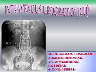
An IVU (Intravenous Urography) is an x-ray of your urinary tract (consisting of kidneys, ureters and bladder) following an injection of a clear dye called contrast into a vein in your arm.
The pictures produced are called intravenous urograms (IVU) or intravenous pyelograms (IVP).
A series of x-rays are taken of the abdomen at various time intervals. This usually takes up to an hour, but occasionally it may be necessary to take additional delayed images, which may continue for several hours.
Routine IVP[edit]
This procedure is most common for patients who have unexplained microscopic or macroscopic hematuria. It is used to ascertain the presence of a tumour or similar anatomy-altering disorders. The sequence of images is roughly as follows:
plain or Control KUB image;
immediate X-ray of just the renal area;
5 minute X-ray of just the renal area.
15 minute X-ray of just the renal area.
At this point, compression may or may not be applied (this is contraindicated in cases of obstruction).
In pyelography, compression involves pressing on the lower abdominal area, which results in distension of the upper urinary tract.[1]
If compression is applied: a 10 minutes post-injection X-ray of the renal area is taken, followed by a KUB on release of the compression.
If compression is not given: a standard KUB is taken to show the ureters emptying. This may sometimes be done with the patient lying in a prone position.
A post-micturition X-ray is taken afterwards. This is usually a coned bladder view.
Image Assessment[edit]
3. What is Intravenous Urography (IVU)
It commonly used investigation for evaluation of
urinary system.
Radiographic study of the renal parenchyma,
pelvicalyceal system, ureters and the urinary bladder.
After intravenous injection of contrast media.
4. In suspected obstructive uropathies like stone etc.
Congenital anomalies involving kidney.
Investigation of hypertension.
Suspected abdominal mass lesion arising from
kidney.
In Blunt injuries of abdomen with Hematuria.
Function of kidney.
Bladder pathology –diverticulum fistula.
5. Iodine sensitivity or previous reaction to
contrast media.
Intractable cardial or renal failure.
Multiple myeloma.
Renal insufficiency.
Thyrotoxicosis.
6. The kidneys are a pair of fist-sized organs located at the
bottom of the rib cage.
The organs of Urinary system.
-Kidneys
-Ureters
-Bladder and Urethra.
7.
8. Ask the patient history diabetics renal disease or allergy
to drugs
Fasting for 4 hours.
explain the exam to patient in layman language.
Check the patient creatine level. (RFT. Normal range: 0.6-1.2mg)
Bowel Preparation
Low residue diet.
Bowel wash is given till bowel is clear of feacal
matter on previous night.
Laxative Dulcolax is given 2-4 tablet at bedtime for
two day prior to exam .
9. Patient is placed in supine position with pelvis at
cathode side of the tube.
A scout film is taken including KUB and ureter region
on large film.
Contrast media injected intravenously into a
prominent vein in the arm.
Test injection of 1 ml contrast look for reaction and
observed 1 min.
Exposures are generally in the 65-75 kV range, mA of 600-1000.
(Higher kV ranges reduce contrast of the renal parenchyma)
10. Correct positioning .
supine full AP abdomen
include lower border of
symphysis pubis and
diphrams.
A KUB to allow the
determination adequate
bowel preparation.
Plain KUB
Plain x ray KUB/Scout film
14’’x 17’’
11. Contrast media is usually given as a IV bolus injection
within 30-60 sec.
IONIC -Low osmolar contrast material LOCM.
-Urografin
NON IONIC -High osmolar contrast material HOCM.
-Ultravist and Omnipaque.
Adult dose
1-2 ml for each 1 kg.
Pediatric dose
-1 ml for each 1 kg.
Mode of Injection
12. 1 minute film show nephrogram.
Radiograph is often omitted as the renal outline are
usually adequately visualize on 5 minute.
(after the 1 minute film compression band is applied)
1 Minute film10’’x 12’’
13. 5 minute film show nephrogram renal pelvis
upper part of ureter.
compression band applied on patient abdomen.
(note if calyces and pelvis are not visualize adequately
obstruction exist and band should not applied)
5 Minute film. 14’’x14’’
5MIN FILM
14. If compression is applied
10 minute film
center on kidney to
demonstrate distended
collecting system and
proximal ureter.
supine position.
All film taken in
expiratory phase.
10 Minute film. 14’’x 14’’
10MIN FILM
15. Visualize the ureter in
prone position as they
fill better.
Supine full length AP
Compression is
released when
satisfactory
Demonstration of
pelvicalyceal system
has been achived.
15 Minute film. 14’’x 17’’
15MIN FILM
16. Its give complete overview
of urinary track.
Bladder distention can be
evaluated.
Taken immediately after
voiding.
its use to assess for
-Residual urine
-Bladder mucosal lesions
- Diverticula
- Bladder tumor
- Outlet obstruction.
Post Void film. 8’’x 10’’
35 Minute film. 14’’x 17’’
35MIN FILM
17. Oblique film:
better visualize the calyaceal system.
filling defect that may overlap in routine AP View
Prone film:
better imaging the ureter.
Upright film:
Layering of contrast media is in severely
hydronephrotic system.
Post void film:
Filling defect in bladder post wall.
Diverticula.