

Recomendados
Mais conteúdo relacionado
Mais procurados
Mais procurados (20)
Destaque
Destaque (15)
Semelhante a Entoptic phenomena
Semelhante a Entoptic phenomena (20)
Último
Último (20)
Entoptic phenomena
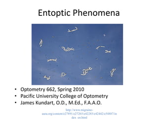
- 1. Entoptic Phenomena • Optometry 662, Spring 2010 • Pacific University College of Optometry • James Kundart, O.D., M.Ed., F.A.A.O. http://www.migraine- aura.org/content/e27891/e27265/e42285/e42442/e54887/in dex_en.html
- 2. Focus Questions 1. Why do we see the Purkinje Tree under the slit lamp, but not in the sunshine? 2. Why are “flying corpuscles” better described as “flying spots?” 3. Which entoptic phenomena can be used by an observant patient to monitor glaucoma? Which can be used to monitor diabetic retinopathy? 3. What part of the retinal causes the polarization responsible for the Hadinger Brush effect? 4. What is Maxwell’s spot? When is it seen?
- 3. What Are Entoptic Images? • “Visual perceptions that are produced or influenced by native structures in your own eyes” are entoptic phenomena • For example, you have all seen your retinal vessels when sitting for slit lamp exams. This is called the Purkinje tree. • Why don’t you see it all the time? • Why should you care? • We will answer these question today
- 4. Why Haven’t I Heard About This Before? • It’s not just because you are second years! Hart and Westheimer (in Adler) say: – “Because of their subjective nature, entoptic phenomena require a savvy, articulate patient to observe and describe them.” • They also can’t be photographed, so I can only show you drawings • We will cover these phenomena from anterior to posterior eye structures • Because entoptic phenomena improve your understanding of the physiology of vision and visual perception, and can sometimes be used to monitor ocular disease
- 5. Some Entoptic Phenomena 1. Corneal Mosaic 2. Physiologic Halos 3. Vitreous Floaters 4. Retinal Phosphenes 5. Purkinje Tree 6. Flying Spots 7. Blue Retinal Arcs 8. Haidinger’s Brushes
- 6. 1) Corneal Mosaic • You have seen what sodium fluorescein looks like on the tear film of your classmates, and the corneal epithelium by now • Did you know you can see your own corneal mosaic without a slit lamp? • If a small (0.1 mm) pinhole is placed at the spectacle plane (17 mm in front of the eyes) and backlit, you can see your own tear film, and irregularities in the cornea • The resulting image is limited in size by the pupil, not the pinhole
- 7. Imagining the Corneal Mosaic Adler, figure 16-4 Corneal Guttata http://www.flickr.com/photos/12212056 @N04/2035013811/
- 8. What Can Be Seen with the Pinhole Technique? • According to Adler, folds in corneal epithelium appear as horizontal bands • Excessive oil or mucus in the tear film look like bright blobs surrounded by a dark ring which “swim” up and down on blink • Shallow, linear channels made by ridges in Bowman’s membrane can be seen with sodium fluorescein, as are sometimes caused by contact lens wear
- 9. Endothelial Dystrophy and the Corneal Mosaic http://www.opt.pacificu.edu/ce/catalog/10603-AS/Cornea.html http://www.flickr.com/photos/jrmod/221356083/
- 10. Tattooing the Cornea • What is the treatment for symptomatic partial corneal scarring where transplants (penetrating keratoplasty or DSEK) are not available? • With a translucent scar, patient symptoms would decrease when the scar was made opaque by surgical tattoo of the cornea • This is because a true opacity reduces the light that reaches the retina, but does not reduce overall contrast like a translucent defect does • “Special pigments can be embedded in the cornea to hide corneal scars and to block light from entering the eye through iris defects” http://www.michigancornea.com/tech_Iris.htm
- 11. 2) Physiologic Halos • You may have learned about pathologic halos, such as those from a steamy, edematous cornea, from contact lens overwear or ocular hypertension • Physiologic halos are different -- they are colored rings from chromatic aberration caused by the corneal mosaic -- but they still come from the cornea • They are dimmer, and their size varies with wavelength (color) of the light • All colors of the rainbow are present -- which are smallest, and which biggest? _______________ (Remember: blue bends best!)
- 12. Which of These Will Cause a Pathologic Halo? http://www.atlasophthalmology.com/atlasimg/Img0086_51_low.jpg http://www.flickr.com/photos/jrmod/339707643/ Nuclear sclerotic cataract vs. corneal opacity The cornea causes haloes, and the lens…
- 13. Lenticular Diffraction Spikes • Instead of haloes, the surface of the crystalline lens causes diffraction of pinpoint light, such as starlight • Everyone knows that you don’t need a cataract to see starbursts around lights this way, so… • They must originate from a healthy lens, too • Physiologic sutre lines are the likely culprit • The same pinhole technique used for the corneal mosaic can be used to image lens opacities, which otherwise simply dampen light http://www.astronomie.de/astropraxis/starhopper/canis-major/sirius.jpg
- 14. Why Patients Don’t See Their Cataracts Directly Adler, figure 16-4Chttp://www.flickr.com/photos/whvick/132165203/
- 15. Early Cataract as It Appears Through a Pinhole Adler, 9th edition, figure 15-4http://www.flickr.com/photos/mak506/283085523/ Please resist temptation to use the abbreviation “cats” for cataracts
- 16. 3) Vitreous Floaters • We are all familiar with the muscae volitantes (flitting flies) that patients believe are in their tear film, but you know are actually in the vitreous • Some of these are remnants of the hyaloid artery that feeds the fetal lens. Others may be due to retinal tears or hemorrhage, like so-called tobacco dust floaters • When they settle to the inferior vitreous due to gravity, we don’t see them • Remember, the vitreous never circulates or gets replenished, so floaters are forever. Learn to love them.
- 17. Why Patients Do See Vitreous Floaters Adler, figure 16-4B
- 18. Which Floaters Are Harmless? • We quickly become accustomed to reassuring patients that floaters are normal, but sometimes they are not • Familiar, countable floaters can be normal might as well become your friends • Recent-onset, innumerable floaters often are due to retinal tear or detachment • Likewise, large, new spider-shaped floaters can be a retinal hemorrhage, so ask your patients to describe what they see • Will patients with active uveitis see their cells and flare? What about asteroid hyalosis? Why not? ___________
- 19. Are These Floaters Symptomatic? Some Treat Asteroid Hyalosis with Laser http://dro.hs.columbia.edu/asthyalosis.htm
- 20. When Do You Expect To See Floaters? • When the lighting is bright and there is stationary background, floaters are most visible • For example, you might see your floaters against the blue sky or snow on the SOA ski trip • As we have seen, they also have to be close to the retina to cause a penumbra (shadow) • Holding a pen tip so that it casts a shadow on a paper gives you an idea, as the shadow fades the further away you hold the pen from the paper
- 21. 4) Retinal Phosphenes • We are all familiar with the bright glow you see when rubbing your closed eyes • This is known to occur due to increase in vitreal pressure on, and deformation of, the retina • This causes the photoreceptors to fire, and for you to perceive light, especially if you are in a dark room • Why don’t you feel pain? Hint: where are pain sensors in the eye? How high does your IOP have to be to feel painful? • These are different than other entopic phenomena as they require a nonlight stimulus -- rubbing or quick eye or head movements (flick phosphenes) • Infants with low vision are thought to rub their eyes incessantly in order to trigger phosphenes and stimulate the optic nerve (this helps diagnosis)
- 22. Phosphenes and the B&L Proview Eye Pressure Monitor Adler, figure 16-6 http://coopereyecare.com/glaucoma.htm
- 23. Moore’s Lightning Streaks • These are entoptic phenomena that occur at the vitreal-retinal interface • They are most often seen in the temporal visual field and are vertically oriented • They were first described in 1935, but are very common in middle-aged patients • It is now thought that they are brought on by posterior vitreous detachment, or PVD • This is a universal condition that some patients never see because it happens in the periphery
- 24. Posterior Vitreous Detachment (PVD) vs. RD http://www.flickr.com/photos/nrgthedude/3359660841/
- 25. Why Do We Get PVD? • Since the vitreous never replenishes, it degenerates over a lifetime in all patients • Think of it like a bowl of jello left out of the refrigerator on a warm day. What happens? • The jello is the vitreous, and the bowl is the retina from which it can become detached • The jello becomes liquified and separates from the bowl the longer it sits out (or the older your patient is) • This liquefaction is called vitreous syneresis
- 26. 5) Purkinje Tree • The Purkinje tree is a good example of how the visual cortex separates self from non-self • It’s there, but we don’t see it most of the time, EXCEPT… • The retinal arteries (arterioles) and veins (venuoles) show up in stark relief when you sit in the slit lamp for your classmates. Have you seen it? • Why??? The slit lamp isn’t brighter than, say, the sun, and the sun doesn’t make it appear, right? • When you see the Purkinje Tree, one part of the biomicroscope is moving faster than the sun moves in the sky -- which part? light
- 27. Jan Evangelista Purkyně (1787-1869) A monk who raised flowers, like Mendel http://en.wikipedia.org/wiki/Jan_Evangelista_Purkyn%C4%9B
- 29. How the Purkinje Tree is Similar to PVD • Both are seen only when conditions change, like when the light moves, or when the vitreous becomes liquified • We see them because they are close to the retina, unlike corneal or lens defects • So, can O.D.’s use this phenomenon so that their diabetic patients can check themselves for retinal hemorrhages at home? • If you instruct the patient to move a penlight over their closed lids in a dark room, you can! • You can also use a blue light, as in the next example
- 30. 6) Flying Spots (not in Adler) • When looking at a brightly-lit blue field with no background (moving or stationary), a series of fast-moving whitish spots are seen that move along curves and leave a trail, like a comet • Since blue light is the type absorbed by hemoglobin, these flying spots are thought to be white blood cells in the retinal vessels • Thus, the flying spots are sometimes called flying corpuscles (an old name for blood cells), or the blue field entoptic phenomenon
- 31. Which Cells Cause the Flying Spots Entoptic Phenomenon? http://www.citylightsnews.com/randy/glossary/glossary_tuvwxyz.htm Hint: it looks green here, but it isn’t really green
- 32. Other Facts About Entoptic Flying Spots • The spots move in pulsatile fashion that accelerates with increased heart rate, as after exercise • Their aren’t enough of them to be caused by red blood cells and they are the wrong color (whitish) • Applying pressure to the eye may make them as easy to see as the Purkinje tree • They can potentially be used to monitor for clinically significant macular edema (CSME), because there should be no blood vessels in the foveal avascular zone
- 33. No Spots Seen in the Foveal Avascular Zone (FAZ) Source: Adler, page 493, 9th edition
- 34. From Which Vascular Beds Do the Spots Arise? • There are two possibilities – One is the precapillary arterioles of the nerve fiber layer – The other is the capillary loops of the inner nuclear/outer plexiform layers • Marshall determined long ago that it can’t be the nerve fiber layer, by using different blue lights to illuminate the Purkinje tree in one eye, and spots in the other • So it’s the capillary loops that make the spots appear
- 35. 7) Blue Retinal Arcs • The nerve fiber layer itself can cause entoptic images, also found by Purkinje • Purkinje used “a glowing tinder taken from a fire,” (don’t try this at home) • Red, rectangular lights apparently the most effective stimuli, held parallel to NFL bundles • For example, when a red target was seen by nasal field, two arcs were seen, and would change apparent distance from each other with target movement • The arcs are dim gray in the dark, and bright blue in the light, like an afterimage
- 36. What Blue Retinal Arcs Look Like OS Adler, figure 16-8
- 37. How Does Retinal Anatomy Explain Blue Arcs? • The nerve fiber layer (NFL) radiate from the optic nerve in bundles toward and around the fovea • They respect the horizontal midline and do not cross it, making a seam of sorts • Recall that this midline is called the horizontal raphe • The perceptual phenomenon lateral inhibition likely plays a significant role • Much more on lateral inhibition later!
- 38. Nerve Fibers Bundles and the Horizontal Raphe OD See also Adler, figure 16-7 http://www.glaucomaworld.net/english/010/e010i12.html
- 39. 8) Haidinger’s Brush • Our last type of entoptic phenomenon, these come from the fovea when a spinning polarized target is used • They are best seen with magnification and a glass cobalt blue filter (same color as the slit lamp filter for fluorescein) • You can see a “brush” that spins like an airplane propeller and moves with your eye, looking like a Maltese cross
- 40. The Maltese Cross -- Can You See It On Your Laptop Screen? You need to use polaroid glasses to see it on a laptop http://world.std.com/~mmcirvin/haidinger.htm l http://www.bernell.com/product/4092/184
- 41. Why Do We See the Haidinger Brush? • Plane-oriented molecules of pigment in the fovea causes some plane-polarized light to be absorbed, especially if it’s blue • So when a spinning polarizer is put in front of a blue light, you see a propeller • Subtle macular edema and other retinal disease may cause the brush to disappear, even if the retina looks normal to your ophthalmoscope • It is used in vision therapy to track the location of the fovea in patients with amblyopia and eccentric fixation
- 42. Which Part of the Retina Sees the Brush? http://cheme.che.caltech.edu/groups/jak/research/eyes/
- 43. Maxwell’s Spot • Maxwell’s Spot is a close relative to Haidinger’s Brushes • Seen as a dark reddish circle surrounded by a clear ring and brighter blue halo when looking at a diffuse flickering blue light • The size is 2-3 degrees, oval horizontally, and may look grainy • Xanthophyll foveal pigment is responsible for Maxwell’s Spot • Which color is xanthophyll? Hint: which color is the macula lutea? ______________ http://www.nature.com/nature/journal/v175/n4450/abs/175306a0.html
- 44. Review Capsule 1. Why do we see the Purkinje Tree under the slit lamp, but not in the sunshine? 2. Why are “flying corpuscles” better described as “flying spots?” 3. Which entoptic phenomena can be used by an observant patient to monitor glaucoma? Which can be used to monitor diabetic retinopathy? 3. What part of the retinal causes the polarization responsible for the Hadinger Brush effect? 4. What is Maxwell’s spot? When is it seen?
- 45. References & Readings • Today’s material can be found in chapter 16 (in the 10th edition) of Adler’s Physiology of the Eye, the chapter with the same title as this lecture, by Hart and Westheimer. Thanks to them! • Next time, we will start Schwartz, chapters 1 and 2. Better buy now and keep up! • I recommend a used copy since a new edition is coming out by next year, and you may want to keep your copy until after your Boards