
Recomendados
Mais conteúdo relacionado
Mais procurados
Mais procurados (20)
Semelhante a Microtubules
Semelhante a Microtubules (20)
Último
Último (20)
Microtubules
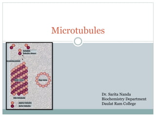
- 1. Microtubules Dr. Sarita Nanda Biochemistry Department Daulat Ram College
- 2. Characteristics They are 25nm in diameter They are dynamic structure which continually undergo assembly and disassembly They determine cell shape, cell locomotion, cell organelle movement, separation of chromosomes during mitosis. They are made up of monomers tubulin. The tubulins are alpha and beta. Gamma tubulin is present in centrosome
- 3. Characteristics Tubulin dimers polymerize to form microtubule which is made of 13 linear protofilaments. The dimers are arranged in head to tail manner in parallel fashion. They have the fast growing plus end and slow growing minus end. This gives polarity to microtubule.
- 4. Characteristic They undergo treadmilling in which fast growing end is adding GTP bound tubulin and minus end is continuously loosing GDP bound from the minus end. In dynamic instability individual microtubules shrink between cycles of growth and shrinkage. Dynamic instability is described by Tim Mitchison and Marc Kirschner in 1984.
- 5. Colchicine, colecimid, vincristine, vinblastine , taxol affect polymerisation and block cell division.
- 6. Treadmilling
- 7. Assembly of Microtubules They extend from centrosome (ist describes Theodor Boveri in 1888 During mitosis microtubules form spindle Plants don not have organised centrosome Centrosome is microtubule organising centre (MTOC) in which minus end is anchored. It serves as initiation site for MT assembly. The key protein of centrosome is gamma tubulin. Gamma tubulin forms a ring complex.
- 8. characteristic The centrosome of animal cell contains a pair of centrioles. Centrioles are cylindrical structures which contains nine triplets of microtubules. Centrioles are necessary to form basal body, cilia, flagella. The peritubular region initiates microtubular assembly. Centrioles have cartwheel like structure. Centrioles also include delta tubulin. Two centrioles are connected by protein called centrin.
- 9. Organisation of Microtubules within Cells They interact with microtubule associated protein(MAPs) MAPS stabilise MT by capping their ends MAPs can also destabilise MT by severing their ends Several MAPs are plus end tracking proteins. The several identified MAPs are MAP1,2, and TAU The neurons have dendrites and axons . The plus end of axons are away form the body and in dendrites plus and minus ends are oriented away from the body. Axons contain tau proteins and no MAP-2 whereas dendrites contain MAP2 but no tau protein
- 11. Microtubule
- 12. Microtubules motors and Movement The two motor protein responsible for motor movement are kinesin and dyenin Kinesin moves towards plus end Dynein moves towards negative end. Dyenin was isolated by Ian Gibbons in 1965. Motor protien was observed by video-enhanced microscopy.(developed by Robert Allen and shinya Inoue in 1980. ) These were observed in squid axons.
- 13. Motor Proteins Kinesin was identified By Brady, Ronald Vale, thomas Reese and Michael Sheetz in squid axons and bovine brains in 1985. Kinesin translocates towards plus end and dyenin translocates towards minus end. Kinesin I is a molecule of 380KD consisting of two heavy chains of 120 kD and two light chains of 64kd each. It is similar to myosin which also moves towards postive end and has molecular weight of 500kd.
- 14. Motor proteins Cytoplasmic dynein is extremely large protein of 2000kd. It consists of two heavy chains of 500kd and variable number of light and inermediate chains The molecule has head and a tail portion. The head binds to ATP and moves on MT The tail binds the organellles which have to be transported. There are 45 kinesinsin humans The plus end directed kinesin have motor domain at N terminal end, minus end directed have motor domains at C terminal end. Others have motor domains in the centre.
- 15. Motor proteins Dynein may be cytoplasmic or axonemal. The tail region may carry cargo
- 16. Cargo Transport and Intracellular Organisation Helps to carry organelles Kinesin carries cargo towards the cell periphery whereas dynein carries toward the nucleus. This helps in positioning ER/Golgi Apparatus. Mitochondria may be transported from cell body to axon Neurotransmitter are carried from Golgi apparatus to terminal branches of axons by kinsens Endocytic vesicles move from axon back to cell body
- 17. Cargo Transport and Intracellular Organisation Kinesin II moves mRNA towards cell cortex in xenopus oocyte. Kinesin I transports actin mRNA in fibroblasts. ER is positioned by Kinesin I GA is positioned by cytoplasmic dynein.
- 18. Cilia and Flagella MT based projections Cilia are widespread Bacterial flagella quite different from eukaryotic flagella Bacterial flagella are protein filaments. Eukaryotic flagella are projection of plasma membrane supported by MT Cilia and flagella are quite similar structures approx 0.25 um. Paramecium are covered with cilia Flagella differ from cilia in length.
- 19. Cilia and Flagella Composed of fundamental structure axoneme composed of MT. They are arranged in 9+2 pattern. A tubule is complete consisiting of 13 protofilament B tubule is incomplete consiisting of 10/11 protofilament The outer MT are connected to central pair by radial spokes and to each other by nexin. In addition 2 arms of dyenin are attached to each A tubule and motor activity of axonemal dynein that drive cilia and flagella.
- 21. Cilia and flagella The basal body have 9+O arrangement It initiates growth of axonemal MT Movement of cilia and flagella result from sliding of outer MT doublets relative to one another by motor activity of axonemal dynein Dynein head binds to A tubule whereas tail binds B tubule binds to B tubule Movement of dynein head groups in the minus direction then causes a tubule of one doublet to slide towards basal end of adjacent B tubule
- 23. Cilia and flagella Since two doublets are connected by nexin, sliding movement helps cilia and flagella to bend. This produces wave like oscillation.