SlideShare uma empresa Scribd logo
1 de 48
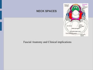
NECK SPACES
Fascial Anatomy and Clinical implications
contents
●Introduction
●Definition
●Classification
●Description of Fascia
●Spaces
●Pathways of spread
●Pathological organisms
●Management principles
●Conclusion
Introduction
●The infection in orofacial region does not spread haphazardly
through the loose connective tissue, but tends to accumulate in the
potential spaces around the head and neck
●Many of these spaces communicate with each other
●Knowledge of the spaces and potential area of communication assist
with not only the management but also assist us with potential
spread points
DEFINITATIONS:
●FASCIA: defined as a broad sheet of dense connective tissue,
whose function is to separate structure that must pass over each
other during movement such as muscles and glands and also
serve as pathway for course of vascular and neural structures
●Shapiro defined facial spaces as “potential spaces between
the layers of fascia”
●These areas are normally filled with loose connective tissue
which readily breaks down when invaded by infection
Classifications:
● Grodinsky and Holyoke in 1938
● Space 1 – the potential space superficial and deep to
platysma muscle
● Space 2. - the space behind the anterior laryer of deep
cervical fascia
● Space 3 – pretracheal space lies anterior to trachea
● Space 3a – lincoln”s highway
● Space 4 – danger space, potential space between the alar
and prevertebral fascia
Based on Clinical significance
● Face: - buccal, canine, masticatory and parotid
● Suprahyoid: - sublingual, submandicular, lateral pharyngeal
and peritonsillar
● Infrahyoid: - pretracheal
● Spaces of total neck :- retropharyngeal, parapharyngeal and
space of carotid sheath
Based on mode of involvement
● Direct involvement
Primary spaces : maxillary space
Mandibular spaces
● Indirect imvolvement: secondary spaces
FASCIA DESCRIBED UNDER:
●SUPERFICIAL CERVICAL FASCIA
Thin layer of connective tissue that lies between dermis
of the skin and investing layer of deep cervical fascia
Contains cutaneous nerves, blood and lymphatic
vessels, superficial lymph nodes and variable amount
of fat
Anteriolaterally it contains the platysma
VISCERAL LAYER
● Middle layer
Muscular division – surrounds the infrahyoid strap
muscles
Visceral division envelops the pharynx, larynx,
oesophagus, trachea and the thyroid gland
Passes inferiorly into the upper mediastinum where it
is continious with the fibrous pericardium and covers
the trachea and oesophagus
Above hyoid this layer continues on the posterior
pharyngeal wall as the buccopharyngeal fascia
Primary surgical significance of this layer is it must be
divided in midline in a surgical approach to trachea or
thyroid gland
DEEP LAYER
● Originates from the spinous processes of the cervical
vertebra and ligamentum nuchae
● At the transverse process of the cervical vertebra, it divides
into an anterior alar layer and posterior prevertebral layer
● Alar fascia extends from base of skull to the second thoracic
vertebra
● Prevertebral fascia lies just anterior to the vertebral bodies
and extends the entire length of vertebral column
FASCIA
●Deep cervical fascia consists of FOUR fascial layers
●Investing or superficial or anterior layer
●Pretracheal or middle layer
●Prevertebral or posterior layer
●Carotid sheath
●These layers support the viscera, muscles, vessels and deep
lymph nodes
●Provide the slipperiness that allows the structures in the neck
to move and pass over one another without difficulty (e.g.
when turning the head and swallowing)
●Layers form natural cleavage planes, allowing seperation of
tissues during surgery
Investing layer
Superficial layer of deep cervical layer
●Follows the “rule of Twos”
Envelops two muscles, two glands and forms two
spaces
Originates from the spinous porcess of the
vertebral column and spreads circumferentially
around the neck
Covers the sternocleidomastoid and trapezius
muscles
Encloses submandibular and parotid glands
Covers anterior bellies of the digastric and the
mylohyoid – forms floor of submandibular space
Investing layer
● Forms the space of the
posterior triangle
● and the suprasternal space
of burns in the midline
Middle layer
● Surrounds the infrahyoid
strap muscles, thyroid,
larynx, trachea and
oesophagus
● Below hyoid this layer
continues inferiorly to fuse
with pericardium
● Above hyoid this layer
continues on the posterior
pharyngeal wall as the
buccopharyngeal fascia
Deep (Prevertebral Fascia)
● Surrounds the prevertebral
muscle
● Anteriorly it divides to form
a thin alar layer and a
thicker prevertebral layer
● Between these two layer is
the “danger space”
extending from skull base
to the diaphragm
Carotid sheath
● Carotid sheath is a fascial layer that is associated with but is
anatomically separate from cervical fascial layers
● It receives contributions from all three layers of deep cervical
fascia
● Contains carotid artery, internal jugular vein, vagus nerve and
deep lymphatic chains
● It continues from the skull base through the neck along the
anterior surface of the prevertebral fascia and enters the chest
behind the clavicle
VISCERAL VASCULAR SPACE
● Potential space within the carotid sheath
● Contain little areolar tissue and is resistant to sperad of
infections
● Aka “lincoln's Highway”
● Extends from base of skull into the mediastimum
● Receives contributions from all three layes of deep fascia
● Can become secondarily involved by infection in any other
deep neck space by direct spread
SPACES SPANNING THE ENTIRE LENGTH OF NECK
● The superficial space is located between the superficial
fascia and the superficial layer of deep fascia.
● This potential space lies superficial and deep to the
platysma and contains loose areolar tissue, lymph nodes,
nerves and vessels – most significant of which is the
external jugular vein
● This space is most commonly involved with superficial
cellulitis of the neck, but if abscess formation does occur, it
will present with obvious fluctuance and typical signs of
infections
DEEP NECK SPACES
● Retropharyngeal space
● Danger space
● Prevertebral space
● Visceral Vascular space
RETROPHARYNGEAL SPACE
● LARGEST and most clinically interfascial space in the neck
because it is major pathway for spread of infection.
● Potential space consists of loose connective tissue between
visceral part of prevertebral layer of deep cervical fascia and
the buccopharyngeal fascia
● Occupies space posterior to the pharynx and oesophagus
● Anterior wall- buccopharyngeal fascia superiorly and the
visceral division of the middle fascial inferiorly
● Posterior wall is the alar layer of the deep fascia
● Lateral boundry is the carotid sheath
● Space extends from the base of skull to T1 T2 vetebra
DANGER SPACE
● Posterior to the retropharyngeal space
● Space between the alar layer and prevertebral layer of deep
fascia
● So named because it contains loose areolar tissue and
● offers little resistance to spread of infection
● Runs from the skull base to diaphragm
SUPRA HYOID SPACES
● Submandibular space
● Parapharyngeal space
● Peritonisllar space
● Masticator space
● Partoid space
SUBMANDIBULAR SPACE
● Bounded by the mandible
anteriorly and laterally
● Lingual mucosa superiorly
● Hyoid postero- inferiorly
● Superficial layer of deep
cervical fascia inferiorly
● Mylohyoid muscle divides space into a superior sublingual
space and submylohyoid space
● Sublingual space contains loose areolar tissue, the
hypoglossa and lingual nerves, the sublingual gland and
wharton's duct
● Submylohyoid space contains the anterior bellies of the
digastric and submandibular glands
● These two subdivisions freely communicate around the
posterior border of the mylyhyoid
PARAPHARYNGEAL SPACE
● Skull base superiorly
● Pterygo mandibular
raphe anteriorly
● Hyoid bone inferiorly
● Bucopharyngeal fascia
medially
PERITONSILLAR SPACE
● Medial – capsule of
palatine tonsil
● Lateral – superior
pharyngeal cronstrictor
● Superior – anterior
tonsillar pillar
● Inferior – posterior
tonsillar pillar
● Contains loose areolar
tissue adjacent to soft
palate
Masticator space
● Collective name for
submasseteric,
pterygomandibular,
superficial and deep
temporal spaces
● Muscles of mastication
are enclosed by
investing layer of fascia
● Contains mandibular
division of trigeminal
and internal maxillary
artery
Parotid space
● Formed by superficial
layer of deep fascia and
dense septa from
capsule into gland
● Contents: parotid lymph
nodes, facial nerve and
posterior facial vein
● Fascial envelope is
deficient on the supero-
medial surface of the
gland thus direct
communication btwn
this space and PPS
Direction of spread
● Tonsillitis – peritonsillar
abscess
● Spread to lateral
pharyngeal space
● To posterior pharyngeal
and prevertebral
spaces and chest
● Mediastinitis and
empyema
PPSParotid
space
Masticator
space
Submandibular
space
Retropharyngeal
space
Retropharyngeal abscess in children
● Most common deep neck abscess
● Retropharyngeal lymph nodes tend to involute with age
● Source of infections suppurative process in the nose,
nasopharynx, sinus and adenoids
● 96% of abscesses occur prior to 6 years of age
Common symptoms
● Fever
● Irritability
● Torticollis
● Trismus
● Cervical lymphadenopathy
● Posterior pharyngeal bulge
● Children often have poor
oral intake due to sore
throat initially
● Followed by dysphagia and
drooling
● Finally if not treated may
get laryngeal oedema and
respiratory distress
RA IN ADULTS
● Typically caused by:
Penetrating or blunt trauma
Instrumentation such as endoscopy
Intubation or NG placement
Extension of infection from an adjoining deep
neck space
Historically, the most common cause of
prevertebral abscess was the extension of a
tuberculous infection of a vertebral body (pott's
absces)
PARAPHARYNGEAL SPACE ABSCESS
● Infection in the pharynx, tonsils, adenoids, teeth, parotid or
lymph node chains
● Middle ear infections or mastoiditis
● Extension of infection from the nearby:
Peritonsillar space
submandibular space
retropharyngeal space
Masticator space
Signs and symptoms
● Differ depending on whether the prestyloid or poststyloid
compartment is involved
● In addition to fever, chills and malaise, anterior infection
often causes pain, dysphagia and significant trismus due to
medial pterygoid irritation
● Oedema in this area will cause medial bulging of the lateral
pharyngeal wall and the tonsils and there will be swelling at
the angle of mandible
● Posterior compartment infection may have no localizing
signs on examination
● Despite this patients do appear toxic “PUO”
● Involvement of neurovascular structures in this area may
lead to neuropathies, horner's syndrome, septic internal
jugular thrombosis or carotid artery rupture
● In recent years, submandibular space abscess has become
the most common deep neck space infection
● Seventy to 85% of these cases are odontogenic in origin,
the rest are caused by sialadenitis, lymphadenitis, flor of
mouth lacerations and infections and or mandibular
fractures
● Ludwig's angina is the prototypical submandibular space
infection
Ludwig's angina
● Cellulitic process of the submandibular space, not an
abscess
● Involvement of only the submandiblar space
● Gangrene with foul serosangious fluid on incision, no pus
● Involvement of the fascia, muscles and connective tissue
● There is direct spread of infection rather than spread by
lymphatics
Microbiology of deep neck infections:
● Mixed aerobic and anareobic organisms, often with
predominance of oral flora
● Gram positive cocci: Group A beta haemolytic sterptococci
species is most common
● A hemolytic streptococcal species (strept viridans, strept
pneumonae)
● Staph Aureus
● Fusobacterium nucleatum, bactreoids, spirtochaeta,
● Klebsiella and Neisseria species found together
● Ocassionaly H influenza and E coli also isolated
● Poly microbial culture in 90% of patients
● Aerobes in 100%
● Anaerobes in more than 50%
Management PRINCIPLES
●Airway protection
●Appropriate antimicrobial therapy
●Surgical drainage
CONCLUSION
Early recognition of orofacial infection and prompt
appropriate therapy is essential
A sound knowledge of anatomy of face and neck is
necessary to predict pathways of spread of
infection and drain the spaces adequately
THANK YOU......
Deep Neck Spaces
Deep Neck Spaces
Deep Neck Spaces
Deep Neck Spaces
Deep Neck Spaces
Deep Neck Spaces
Deep Neck Spaces

Mais conteúdo relacionado

Mais procurados

Spaces of middle ear and their surgical importance
Spaces of middle ear  and their surgical importanceSpaces of middle ear  and their surgical importance
Spaces of middle ear and their surgical importanceDr Soumya Singh
 
Surgical anatomy of facial nerve
Surgical anatomy of facial nerveSurgical anatomy of facial nerve
Surgical anatomy of facial nerveTasnia Mahmud
 
Anatomy of recurrent laryngeal nerveAnatomy of recurrent laryngeal nerveAnato...
Anatomy of recurrent laryngeal nerveAnatomy of recurrent laryngeal nerveAnato...Anatomy of recurrent laryngeal nerveAnatomy of recurrent laryngeal nerveAnato...
Anatomy of recurrent laryngeal nerveAnatomy of recurrent laryngeal nerveAnato...Ebtisam ~
 
Infratemporal fossa a systematic approach
Infratemporal fossa a systematic approachInfratemporal fossa a systematic approach
Infratemporal fossa a systematic approachAugustine raj
 
Embryology & anatomy of external ear
Embryology &  anatomy of external earEmbryology &  anatomy of external ear
Embryology & anatomy of external earDr. Pruthvi Raj S
 
Laryngeal framework surgery
Laryngeal framework  surgeryLaryngeal framework  surgery
Laryngeal framework surgeryDr Safika Zaman
 
Neck spaces anatomy and infections
Neck spaces anatomy and infectionsNeck spaces anatomy and infections
Neck spaces anatomy and infectionsAhmed Bahnassy
 
Mucosal folds and ventilation of middle ear
Mucosal folds and ventilation of middle ear  Mucosal folds and ventilation of middle ear
Mucosal folds and ventilation of middle ear AlkaKapil
 

Mais procurados (20)

Retropharyngeal space
Retropharyngeal spaceRetropharyngeal space
Retropharyngeal space
 
Parotidectomy
ParotidectomyParotidectomy
Parotidectomy
 
JNA
JNAJNA
JNA
 
Spaces of middle ear and their surgical importance
Spaces of middle ear  and their surgical importanceSpaces of middle ear  and their surgical importance
Spaces of middle ear and their surgical importance
 
Deep neck infection
Deep neck infection Deep neck infection
Deep neck infection
 
Sinus anatomy and variants
Sinus anatomy and variantsSinus anatomy and variants
Sinus anatomy and variants
 
Inverted papilloma
Inverted papillomaInverted papilloma
Inverted papilloma
 
Mucosal folds of the middle ear
Mucosal folds of the middle earMucosal folds of the middle ear
Mucosal folds of the middle ear
 
Surgical anatomy of facial nerve
Surgical anatomy of facial nerveSurgical anatomy of facial nerve
Surgical anatomy of facial nerve
 
Embryology nose and paranasal sinuses
Embryology nose and paranasal sinusesEmbryology nose and paranasal sinuses
Embryology nose and paranasal sinuses
 
Hypopharynx anatomy
Hypopharynx anatomyHypopharynx anatomy
Hypopharynx anatomy
 
Anatomy of recurrent laryngeal nerveAnatomy of recurrent laryngeal nerveAnato...
Anatomy of recurrent laryngeal nerveAnatomy of recurrent laryngeal nerveAnato...Anatomy of recurrent laryngeal nerveAnatomy of recurrent laryngeal nerveAnato...
Anatomy of recurrent laryngeal nerveAnatomy of recurrent laryngeal nerveAnato...
 
Infratemporal fossa a systematic approach
Infratemporal fossa a systematic approachInfratemporal fossa a systematic approach
Infratemporal fossa a systematic approach
 
Embryology & anatomy of external ear
Embryology &  anatomy of external earEmbryology &  anatomy of external ear
Embryology & anatomy of external ear
 
Laryngeal framework surgery
Laryngeal framework  surgeryLaryngeal framework  surgery
Laryngeal framework surgery
 
Thyroplasty
ThyroplastyThyroplasty
Thyroplasty
 
Radical neck dissection
Radical neck dissectionRadical neck dissection
Radical neck dissection
 
Neck spaces anatomy and infections
Neck spaces anatomy and infectionsNeck spaces anatomy and infections
Neck spaces anatomy and infections
 
Mucosal folds and ventilation of middle ear
Mucosal folds and ventilation of middle ear  Mucosal folds and ventilation of middle ear
Mucosal folds and ventilation of middle ear
 
Phonosurgery
PhonosurgeryPhonosurgery
Phonosurgery
 

Destaque

Odontogenic infections
Odontogenic infectionsOdontogenic infections
Odontogenic infectionsRida Waseem
 
Anatomy of neck spaces
Anatomy of neck spacesAnatomy of neck spaces
Anatomy of neck spacesWalid Rezk
 
Anatomy of Neck spaces & Infections
Anatomy of Neck spaces & InfectionsAnatomy of Neck spaces & Infections
Anatomy of Neck spaces & InfectionsDr Utkal Mishra
 
Spaces of head and neck and infections /certified fixed orthodontic courses b...
Spaces of head and neck and infections /certified fixed orthodontic courses b...Spaces of head and neck and infections /certified fixed orthodontic courses b...
Spaces of head and neck and infections /certified fixed orthodontic courses b...Indian dental academy
 
CT anatomy of Neck Spaces RV
CT anatomy of Neck Spaces RVCT anatomy of Neck Spaces RV
CT anatomy of Neck Spaces RVRoshan Valentine
 
Diagnostic Imaging of Deep Neck Spaces
Diagnostic Imaging of Deep Neck SpacesDiagnostic Imaging of Deep Neck Spaces
Diagnostic Imaging of Deep Neck SpacesMohamed M.A. Zaitoun
 
CT ANATOMY OF THE NECK SPACES
CT ANATOMY OF THE NECK SPACESCT ANATOMY OF THE NECK SPACES
CT ANATOMY OF THE NECK SPACEShazem youssef
 
Odontogenic infections (4)
Odontogenic infections (4)Odontogenic infections (4)
Odontogenic infections (4)Chelsea Mareé
 

Destaque (10)

SPACES OF HEAD AND NECK
SPACES OF HEAD AND NECKSPACES OF HEAD AND NECK
SPACES OF HEAD AND NECK
 
Odontogenic infections
Odontogenic infectionsOdontogenic infections
Odontogenic infections
 
Anatomy of neck spaces
Anatomy of neck spacesAnatomy of neck spaces
Anatomy of neck spaces
 
Anatomy of Neck spaces & Infections
Anatomy of Neck spaces & InfectionsAnatomy of Neck spaces & Infections
Anatomy of Neck spaces & Infections
 
Fascial spaces
Fascial spacesFascial spaces
Fascial spaces
 
Spaces of head and neck and infections /certified fixed orthodontic courses b...
Spaces of head and neck and infections /certified fixed orthodontic courses b...Spaces of head and neck and infections /certified fixed orthodontic courses b...
Spaces of head and neck and infections /certified fixed orthodontic courses b...
 
CT anatomy of Neck Spaces RV
CT anatomy of Neck Spaces RVCT anatomy of Neck Spaces RV
CT anatomy of Neck Spaces RV
 
Diagnostic Imaging of Deep Neck Spaces
Diagnostic Imaging of Deep Neck SpacesDiagnostic Imaging of Deep Neck Spaces
Diagnostic Imaging of Deep Neck Spaces
 
CT ANATOMY OF THE NECK SPACES
CT ANATOMY OF THE NECK SPACESCT ANATOMY OF THE NECK SPACES
CT ANATOMY OF THE NECK SPACES
 
Odontogenic infections (4)
Odontogenic infections (4)Odontogenic infections (4)
Odontogenic infections (4)
 

Semelhante a Deep Neck Spaces

DEEP NECK SPACES ANATOMY.pptx
DEEP NECK SPACES ANATOMY.pptxDEEP NECK SPACES ANATOMY.pptx
DEEP NECK SPACES ANATOMY.pptxegodoc222
 
Deep neck spaces and surgical anatomy
Deep neck spaces and surgical anatomyDeep neck spaces and surgical anatomy
Deep neck spaces and surgical anatomyHimanshu Mishra
 
DEEP NECK SPACES.pptx
DEEP NECK SPACES.pptxDEEP NECK SPACES.pptx
DEEP NECK SPACES.pptxANKIT PRAKASH
 
anatomy of pharynx.pptx
anatomy of pharynx.pptxanatomy of pharynx.pptx
anatomy of pharynx.pptxMeshwaOza
 
Fascial Spaces in the Head & Neck
Fascial Spaces in the Head & NeckFascial Spaces in the Head & Neck
Fascial Spaces in the Head & NeckDr Rayan Malick
 
Anatomy of Deep Neck spaces Radiology Pictorial representation
Anatomy of Deep Neck spaces Radiology Pictorial representationAnatomy of Deep Neck spaces Radiology Pictorial representation
Anatomy of Deep Neck spaces Radiology Pictorial representationsudhanracki
 
Neck spaces and mediastinum
Neck spaces and mediastinumNeck spaces and mediastinum
Neck spaces and mediastinumAhmed Bahnassy
 
DEEP NECK SPACE INFECTIONS - DR ADITYA GOEL (MS-ENT)
DEEP NECK SPACE INFECTIONS  - DR ADITYA GOEL (MS-ENT)DEEP NECK SPACE INFECTIONS  - DR ADITYA GOEL (MS-ENT)
DEEP NECK SPACE INFECTIONS - DR ADITYA GOEL (MS-ENT)Aditya Goel
 
Facial spaces and spread of odontogenic infection
Facial spaces and spread of odontogenic infectionFacial spaces and spread of odontogenic infection
Facial spaces and spread of odontogenic infectionLubna Abu Alrub,DDS
 
Facial spaces and spread of odontogenic infection
Facial spaces and spread of odontogenic infectionFacial spaces and spread of odontogenic infection
Facial spaces and spread of odontogenic infectionLubna Abu Alrub,DDS
 
Fascial Spaces of Neck(2).pptx
Fascial Spaces of Neck(2).pptxFascial Spaces of Neck(2).pptx
Fascial Spaces of Neck(2).pptxHtet Ko
 
DEEP NECK SPACES-1.pptx
DEEP NECK SPACES-1.pptxDEEP NECK SPACES-1.pptx
DEEP NECK SPACES-1.pptxManu Babu
 
Neck spaces .pptx
Neck spaces .pptxNeck spaces .pptx
Neck spaces .pptxIslah Raoof
 
Fascial Space infection
Fascial Space infection Fascial Space infection
Fascial Space infection Dr Yash Chaddha
 
Deep facial spaces of head and neck
Deep facial spaces of head and neckDeep facial spaces of head and neck
Deep facial spaces of head and neckTejal Ragji
 
Infratemporal fossa structure
Infratemporal fossa structure Infratemporal fossa structure
Infratemporal fossa structure FatemahIbrahim
 

Semelhante a Deep Neck Spaces (20)

Cervical fascia
Cervical fasciaCervical fascia
Cervical fascia
 
DEEP NECK SPACES ANATOMY.pptx
DEEP NECK SPACES ANATOMY.pptxDEEP NECK SPACES ANATOMY.pptx
DEEP NECK SPACES ANATOMY.pptx
 
Deep neck spaces and surgical anatomy
Deep neck spaces and surgical anatomyDeep neck spaces and surgical anatomy
Deep neck spaces and surgical anatomy
 
DEEP NECK SPACES.pptx
DEEP NECK SPACES.pptxDEEP NECK SPACES.pptx
DEEP NECK SPACES.pptx
 
Neck spaces (1).pptx
Neck spaces (1).pptxNeck spaces (1).pptx
Neck spaces (1).pptx
 
anatomy of pharynx.pptx
anatomy of pharynx.pptxanatomy of pharynx.pptx
anatomy of pharynx.pptx
 
Fascial Spaces in the Head & Neck
Fascial Spaces in the Head & NeckFascial Spaces in the Head & Neck
Fascial Spaces in the Head & Neck
 
Anatomy of Deep Neck spaces Radiology Pictorial representation
Anatomy of Deep Neck spaces Radiology Pictorial representationAnatomy of Deep Neck spaces Radiology Pictorial representation
Anatomy of Deep Neck spaces Radiology Pictorial representation
 
Neck spaces and mediastinum
Neck spaces and mediastinumNeck spaces and mediastinum
Neck spaces and mediastinum
 
DEEP NECK SPACE INFECTIONS - DR ADITYA GOEL (MS-ENT)
DEEP NECK SPACE INFECTIONS  - DR ADITYA GOEL (MS-ENT)DEEP NECK SPACE INFECTIONS  - DR ADITYA GOEL (MS-ENT)
DEEP NECK SPACE INFECTIONS - DR ADITYA GOEL (MS-ENT)
 
Facial spaces and spread of odontogenic infection
Facial spaces and spread of odontogenic infectionFacial spaces and spread of odontogenic infection
Facial spaces and spread of odontogenic infection
 
Facial spaces and spread of odontogenic infection
Facial spaces and spread of odontogenic infectionFacial spaces and spread of odontogenic infection
Facial spaces and spread of odontogenic infection
 
Fascial Spaces of Neck(2).pptx
Fascial Spaces of Neck(2).pptxFascial Spaces of Neck(2).pptx
Fascial Spaces of Neck(2).pptx
 
Secondary spaces
Secondary spacesSecondary spaces
Secondary spaces
 
DEEP NECK SPACES-1.pptx
DEEP NECK SPACES-1.pptxDEEP NECK SPACES-1.pptx
DEEP NECK SPACES-1.pptx
 
Neck spaces .pptx
Neck spaces .pptxNeck spaces .pptx
Neck spaces .pptx
 
Fascial Space infection
Fascial Space infection Fascial Space infection
Fascial Space infection
 
Deep facial spaces of head and neck
Deep facial spaces of head and neckDeep facial spaces of head and neck
Deep facial spaces of head and neck
 
Infratemporal fossa structure
Infratemporal fossa structure Infratemporal fossa structure
Infratemporal fossa structure
 
Anatomy of deep neck spaces
Anatomy of deep neck spacesAnatomy of deep neck spaces
Anatomy of deep neck spaces
 

Mais de sarita pandey

CASE presentation & TOPIC discussion
CASE presentation & TOPIC discussionCASE presentation & TOPIC discussion
CASE presentation & TOPIC discussionsarita pandey
 
Laryngeal paralysis final
Laryngeal paralysis finalLaryngeal paralysis final
Laryngeal paralysis finalsarita pandey
 
Paediatric ent emergencies
Paediatric ent emergenciesPaediatric ent emergencies
Paediatric ent emergenciessarita pandey
 
Facial nerve traumatic injury and repair
Facial nerve traumatic injury and repairFacial nerve traumatic injury and repair
Facial nerve traumatic injury and repairsarita pandey
 
Granulomatous diseases in ENT
Granulomatous diseases in ENTGranulomatous diseases in ENT
Granulomatous diseases in ENTsarita pandey
 
Anatomy & embryology of vestibular system
Anatomy & embryology of vestibular systemAnatomy & embryology of vestibular system
Anatomy & embryology of vestibular systemsarita pandey
 
Allergic rhinitis - presentation
Allergic rhinitis - presentationAllergic rhinitis - presentation
Allergic rhinitis - presentationsarita pandey
 
Complications of mastoiditis
Complications of mastoiditisComplications of mastoiditis
Complications of mastoiditissarita pandey
 
Immittance audiometry
Immittance audiometryImmittance audiometry
Immittance audiometrysarita pandey
 
Presbycusis, deafness of old age
Presbycusis, deafness of old agePresbycusis, deafness of old age
Presbycusis, deafness of old agesarita pandey
 

Mais de sarita pandey (12)

Fungalsinusitis
FungalsinusitisFungalsinusitis
Fungalsinusitis
 
CASE presentation & TOPIC discussion
CASE presentation & TOPIC discussionCASE presentation & TOPIC discussion
CASE presentation & TOPIC discussion
 
Laryngeal paralysis final
Laryngeal paralysis finalLaryngeal paralysis final
Laryngeal paralysis final
 
Paediatric ent emergencies
Paediatric ent emergenciesPaediatric ent emergencies
Paediatric ent emergencies
 
Facial nerve traumatic injury and repair
Facial nerve traumatic injury and repairFacial nerve traumatic injury and repair
Facial nerve traumatic injury and repair
 
Granulomatous diseases in ENT
Granulomatous diseases in ENTGranulomatous diseases in ENT
Granulomatous diseases in ENT
 
Anatomy & embryology of vestibular system
Anatomy & embryology of vestibular systemAnatomy & embryology of vestibular system
Anatomy & embryology of vestibular system
 
Allergic rhinitis - presentation
Allergic rhinitis - presentationAllergic rhinitis - presentation
Allergic rhinitis - presentation
 
Complications of mastoiditis
Complications of mastoiditisComplications of mastoiditis
Complications of mastoiditis
 
Epiglottitis
EpiglottitisEpiglottitis
Epiglottitis
 
Immittance audiometry
Immittance audiometryImmittance audiometry
Immittance audiometry
 
Presbycusis, deafness of old age
Presbycusis, deafness of old agePresbycusis, deafness of old age
Presbycusis, deafness of old age
 

Último

Measurement of Radiation and Dosimetric Procedure.pptx
Measurement of Radiation and Dosimetric Procedure.pptxMeasurement of Radiation and Dosimetric Procedure.pptx
Measurement of Radiation and Dosimetric Procedure.pptxDr. Dheeraj Kumar
 
VarSeq 2.6.0: Advancing Pharmacogenomics and Genomic Analysis
VarSeq 2.6.0: Advancing Pharmacogenomics and Genomic AnalysisVarSeq 2.6.0: Advancing Pharmacogenomics and Genomic Analysis
VarSeq 2.6.0: Advancing Pharmacogenomics and Genomic AnalysisGolden Helix
 
The next social challenge to public health: the information environment.pptx
The next social challenge to public health:  the information environment.pptxThe next social challenge to public health:  the information environment.pptx
The next social challenge to public health: the information environment.pptxTina Purnat
 
Introduction to Sports Injuries by- Dr. Anjali Rai
Introduction to Sports Injuries by- Dr. Anjali RaiIntroduction to Sports Injuries by- Dr. Anjali Rai
Introduction to Sports Injuries by- Dr. Anjali RaiGoogle
 
Presentació "Real-Life VR Integration for Mild Cognitive Impairment Rehabilit...
Presentació "Real-Life VR Integration for Mild Cognitive Impairment Rehabilit...Presentació "Real-Life VR Integration for Mild Cognitive Impairment Rehabilit...
Presentació "Real-Life VR Integration for Mild Cognitive Impairment Rehabilit...Badalona Serveis Assistencials
 
Case Report Peripartum Cardiomyopathy.pptx
Case Report Peripartum Cardiomyopathy.pptxCase Report Peripartum Cardiomyopathy.pptx
Case Report Peripartum Cardiomyopathy.pptxNiranjan Chavan
 
Presentation on General Anesthetics pdf.
Presentation on General Anesthetics pdf.Presentation on General Anesthetics pdf.
Presentation on General Anesthetics pdf.Prerana Jadhav
 
April 2024 ONCOLOGY CARTOON by DR KANHU CHARAN PATRO
April 2024 ONCOLOGY CARTOON by  DR KANHU CHARAN PATROApril 2024 ONCOLOGY CARTOON by  DR KANHU CHARAN PATRO
April 2024 ONCOLOGY CARTOON by DR KANHU CHARAN PATROKanhu Charan
 
Glomerular Filtration rate and its determinants.pptx
Glomerular Filtration rate and its determinants.pptxGlomerular Filtration rate and its determinants.pptx
Glomerular Filtration rate and its determinants.pptxDr.Nusrat Tariq
 
Biomechanics- Shoulder Joint!!!!!!!!!!!!
Biomechanics- Shoulder Joint!!!!!!!!!!!!Biomechanics- Shoulder Joint!!!!!!!!!!!!
Biomechanics- Shoulder Joint!!!!!!!!!!!!ibtesaam huma
 
Apiculture Chapter 1. Introduction 2.ppt
Apiculture Chapter 1. Introduction 2.pptApiculture Chapter 1. Introduction 2.ppt
Apiculture Chapter 1. Introduction 2.pptkedirjemalharun
 
call girls in Dwarka Sector 21 Metro DELHI 🔝 >༒9540349809 🔝 genuine Escort Se...
call girls in Dwarka Sector 21 Metro DELHI 🔝 >༒9540349809 🔝 genuine Escort Se...call girls in Dwarka Sector 21 Metro DELHI 🔝 >༒9540349809 🔝 genuine Escort Se...
call girls in Dwarka Sector 21 Metro DELHI 🔝 >༒9540349809 🔝 genuine Escort Se...saminamagar
 
SYNDESMOTIC INJURY- ANATOMICAL REPAIR.pptx
SYNDESMOTIC INJURY- ANATOMICAL REPAIR.pptxSYNDESMOTIC INJURY- ANATOMICAL REPAIR.pptx
SYNDESMOTIC INJURY- ANATOMICAL REPAIR.pptxdrashraf369
 
Big Data Analysis Suggests COVID Vaccination Increases Excess Mortality Of ...
Big Data Analysis Suggests COVID  Vaccination Increases Excess Mortality Of  ...Big Data Analysis Suggests COVID  Vaccination Increases Excess Mortality Of  ...
Big Data Analysis Suggests COVID Vaccination Increases Excess Mortality Of ...sdateam0
 
call girls in Connaught Place DELHI 🔝 >༒9540349809 🔝 genuine Escort Service ...
call girls in Connaught Place  DELHI 🔝 >༒9540349809 🔝 genuine Escort Service ...call girls in Connaught Place  DELHI 🔝 >༒9540349809 🔝 genuine Escort Service ...
call girls in Connaught Place DELHI 🔝 >༒9540349809 🔝 genuine Escort Service ...saminamagar
 
METHODS OF ACQUIRING KNOWLEDGE IN NURSING.pptx by navdeep kaur
METHODS OF ACQUIRING KNOWLEDGE IN NURSING.pptx by navdeep kaurMETHODS OF ACQUIRING KNOWLEDGE IN NURSING.pptx by navdeep kaur
METHODS OF ACQUIRING KNOWLEDGE IN NURSING.pptx by navdeep kaurNavdeep Kaur
 
call girls in green park DELHI 🔝 >༒9540349809 🔝 genuine Escort Service 🔝✔️✔️
call girls in green park  DELHI 🔝 >༒9540349809 🔝 genuine Escort Service 🔝✔️✔️call girls in green park  DELHI 🔝 >༒9540349809 🔝 genuine Escort Service 🔝✔️✔️
call girls in green park DELHI 🔝 >༒9540349809 🔝 genuine Escort Service 🔝✔️✔️saminamagar
 
PULMONARY EDEMA AND ITS MANAGEMENT.pdf
PULMONARY EDEMA AND  ITS  MANAGEMENT.pdfPULMONARY EDEMA AND  ITS  MANAGEMENT.pdf
PULMONARY EDEMA AND ITS MANAGEMENT.pdfDolisha Warbi
 
call girls in paharganj DELHI 🔝 >༒9540349809 🔝 genuine Escort Service 🔝✔️✔️
call girls in paharganj DELHI 🔝 >༒9540349809 🔝 genuine Escort Service 🔝✔️✔️call girls in paharganj DELHI 🔝 >༒9540349809 🔝 genuine Escort Service 🔝✔️✔️
call girls in paharganj DELHI 🔝 >༒9540349809 🔝 genuine Escort Service 🔝✔️✔️saminamagar
 
Report Back from SGO: What’s New in Uterine Cancer?.pptx
Report Back from SGO: What’s New in Uterine Cancer?.pptxReport Back from SGO: What’s New in Uterine Cancer?.pptx
Report Back from SGO: What’s New in Uterine Cancer?.pptxbkling
 

Último (20)

Measurement of Radiation and Dosimetric Procedure.pptx
Measurement of Radiation and Dosimetric Procedure.pptxMeasurement of Radiation and Dosimetric Procedure.pptx
Measurement of Radiation and Dosimetric Procedure.pptx
 
VarSeq 2.6.0: Advancing Pharmacogenomics and Genomic Analysis
VarSeq 2.6.0: Advancing Pharmacogenomics and Genomic AnalysisVarSeq 2.6.0: Advancing Pharmacogenomics and Genomic Analysis
VarSeq 2.6.0: Advancing Pharmacogenomics and Genomic Analysis
 
The next social challenge to public health: the information environment.pptx
The next social challenge to public health:  the information environment.pptxThe next social challenge to public health:  the information environment.pptx
The next social challenge to public health: the information environment.pptx
 
Introduction to Sports Injuries by- Dr. Anjali Rai
Introduction to Sports Injuries by- Dr. Anjali RaiIntroduction to Sports Injuries by- Dr. Anjali Rai
Introduction to Sports Injuries by- Dr. Anjali Rai
 
Presentació "Real-Life VR Integration for Mild Cognitive Impairment Rehabilit...
Presentació "Real-Life VR Integration for Mild Cognitive Impairment Rehabilit...Presentació "Real-Life VR Integration for Mild Cognitive Impairment Rehabilit...
Presentació "Real-Life VR Integration for Mild Cognitive Impairment Rehabilit...
 
Case Report Peripartum Cardiomyopathy.pptx
Case Report Peripartum Cardiomyopathy.pptxCase Report Peripartum Cardiomyopathy.pptx
Case Report Peripartum Cardiomyopathy.pptx
 
Presentation on General Anesthetics pdf.
Presentation on General Anesthetics pdf.Presentation on General Anesthetics pdf.
Presentation on General Anesthetics pdf.
 
April 2024 ONCOLOGY CARTOON by DR KANHU CHARAN PATRO
April 2024 ONCOLOGY CARTOON by  DR KANHU CHARAN PATROApril 2024 ONCOLOGY CARTOON by  DR KANHU CHARAN PATRO
April 2024 ONCOLOGY CARTOON by DR KANHU CHARAN PATRO
 
Glomerular Filtration rate and its determinants.pptx
Glomerular Filtration rate and its determinants.pptxGlomerular Filtration rate and its determinants.pptx
Glomerular Filtration rate and its determinants.pptx
 
Biomechanics- Shoulder Joint!!!!!!!!!!!!
Biomechanics- Shoulder Joint!!!!!!!!!!!!Biomechanics- Shoulder Joint!!!!!!!!!!!!
Biomechanics- Shoulder Joint!!!!!!!!!!!!
 
Apiculture Chapter 1. Introduction 2.ppt
Apiculture Chapter 1. Introduction 2.pptApiculture Chapter 1. Introduction 2.ppt
Apiculture Chapter 1. Introduction 2.ppt
 
call girls in Dwarka Sector 21 Metro DELHI 🔝 >༒9540349809 🔝 genuine Escort Se...
call girls in Dwarka Sector 21 Metro DELHI 🔝 >༒9540349809 🔝 genuine Escort Se...call girls in Dwarka Sector 21 Metro DELHI 🔝 >༒9540349809 🔝 genuine Escort Se...
call girls in Dwarka Sector 21 Metro DELHI 🔝 >༒9540349809 🔝 genuine Escort Se...
 
SYNDESMOTIC INJURY- ANATOMICAL REPAIR.pptx
SYNDESMOTIC INJURY- ANATOMICAL REPAIR.pptxSYNDESMOTIC INJURY- ANATOMICAL REPAIR.pptx
SYNDESMOTIC INJURY- ANATOMICAL REPAIR.pptx
 
Big Data Analysis Suggests COVID Vaccination Increases Excess Mortality Of ...
Big Data Analysis Suggests COVID  Vaccination Increases Excess Mortality Of  ...Big Data Analysis Suggests COVID  Vaccination Increases Excess Mortality Of  ...
Big Data Analysis Suggests COVID Vaccination Increases Excess Mortality Of ...
 
call girls in Connaught Place DELHI 🔝 >༒9540349809 🔝 genuine Escort Service ...
call girls in Connaught Place  DELHI 🔝 >༒9540349809 🔝 genuine Escort Service ...call girls in Connaught Place  DELHI 🔝 >༒9540349809 🔝 genuine Escort Service ...
call girls in Connaught Place DELHI 🔝 >༒9540349809 🔝 genuine Escort Service ...
 
METHODS OF ACQUIRING KNOWLEDGE IN NURSING.pptx by navdeep kaur
METHODS OF ACQUIRING KNOWLEDGE IN NURSING.pptx by navdeep kaurMETHODS OF ACQUIRING KNOWLEDGE IN NURSING.pptx by navdeep kaur
METHODS OF ACQUIRING KNOWLEDGE IN NURSING.pptx by navdeep kaur
 
call girls in green park DELHI 🔝 >༒9540349809 🔝 genuine Escort Service 🔝✔️✔️
call girls in green park  DELHI 🔝 >༒9540349809 🔝 genuine Escort Service 🔝✔️✔️call girls in green park  DELHI 🔝 >༒9540349809 🔝 genuine Escort Service 🔝✔️✔️
call girls in green park DELHI 🔝 >༒9540349809 🔝 genuine Escort Service 🔝✔️✔️
 
PULMONARY EDEMA AND ITS MANAGEMENT.pdf
PULMONARY EDEMA AND  ITS  MANAGEMENT.pdfPULMONARY EDEMA AND  ITS  MANAGEMENT.pdf
PULMONARY EDEMA AND ITS MANAGEMENT.pdf
 
call girls in paharganj DELHI 🔝 >༒9540349809 🔝 genuine Escort Service 🔝✔️✔️
call girls in paharganj DELHI 🔝 >༒9540349809 🔝 genuine Escort Service 🔝✔️✔️call girls in paharganj DELHI 🔝 >༒9540349809 🔝 genuine Escort Service 🔝✔️✔️
call girls in paharganj DELHI 🔝 >༒9540349809 🔝 genuine Escort Service 🔝✔️✔️
 
Report Back from SGO: What’s New in Uterine Cancer?.pptx
Report Back from SGO: What’s New in Uterine Cancer?.pptxReport Back from SGO: What’s New in Uterine Cancer?.pptx
Report Back from SGO: What’s New in Uterine Cancer?.pptx
 

Deep Neck Spaces

  • 1. NECK SPACES Fascial Anatomy and Clinical implications
  • 2. contents ●Introduction ●Definition ●Classification ●Description of Fascia ●Spaces ●Pathways of spread ●Pathological organisms ●Management principles ●Conclusion
  • 3. Introduction ●The infection in orofacial region does not spread haphazardly through the loose connective tissue, but tends to accumulate in the potential spaces around the head and neck ●Many of these spaces communicate with each other ●Knowledge of the spaces and potential area of communication assist with not only the management but also assist us with potential spread points
  • 4. DEFINITATIONS: ●FASCIA: defined as a broad sheet of dense connective tissue, whose function is to separate structure that must pass over each other during movement such as muscles and glands and also serve as pathway for course of vascular and neural structures ●Shapiro defined facial spaces as “potential spaces between the layers of fascia” ●These areas are normally filled with loose connective tissue which readily breaks down when invaded by infection
  • 5. Classifications: ● Grodinsky and Holyoke in 1938 ● Space 1 – the potential space superficial and deep to platysma muscle ● Space 2. - the space behind the anterior laryer of deep cervical fascia ● Space 3 – pretracheal space lies anterior to trachea ● Space 3a – lincoln”s highway ● Space 4 – danger space, potential space between the alar and prevertebral fascia
  • 6. Based on Clinical significance ● Face: - buccal, canine, masticatory and parotid ● Suprahyoid: - sublingual, submandicular, lateral pharyngeal and peritonsillar ● Infrahyoid: - pretracheal ● Spaces of total neck :- retropharyngeal, parapharyngeal and space of carotid sheath
  • 7. Based on mode of involvement ● Direct involvement Primary spaces : maxillary space Mandibular spaces ● Indirect imvolvement: secondary spaces
  • 8. FASCIA DESCRIBED UNDER: ●SUPERFICIAL CERVICAL FASCIA Thin layer of connective tissue that lies between dermis of the skin and investing layer of deep cervical fascia Contains cutaneous nerves, blood and lymphatic vessels, superficial lymph nodes and variable amount of fat Anteriolaterally it contains the platysma
  • 9. VISCERAL LAYER ● Middle layer Muscular division – surrounds the infrahyoid strap muscles Visceral division envelops the pharynx, larynx, oesophagus, trachea and the thyroid gland Passes inferiorly into the upper mediastinum where it is continious with the fibrous pericardium and covers the trachea and oesophagus Above hyoid this layer continues on the posterior pharyngeal wall as the buccopharyngeal fascia Primary surgical significance of this layer is it must be divided in midline in a surgical approach to trachea or thyroid gland
  • 10. DEEP LAYER ● Originates from the spinous processes of the cervical vertebra and ligamentum nuchae ● At the transverse process of the cervical vertebra, it divides into an anterior alar layer and posterior prevertebral layer ● Alar fascia extends from base of skull to the second thoracic vertebra ● Prevertebral fascia lies just anterior to the vertebral bodies and extends the entire length of vertebral column
  • 11. FASCIA ●Deep cervical fascia consists of FOUR fascial layers ●Investing or superficial or anterior layer ●Pretracheal or middle layer ●Prevertebral or posterior layer ●Carotid sheath ●These layers support the viscera, muscles, vessels and deep lymph nodes ●Provide the slipperiness that allows the structures in the neck to move and pass over one another without difficulty (e.g. when turning the head and swallowing) ●Layers form natural cleavage planes, allowing seperation of tissues during surgery
  • 12. Investing layer Superficial layer of deep cervical layer ●Follows the “rule of Twos” Envelops two muscles, two glands and forms two spaces Originates from the spinous porcess of the vertebral column and spreads circumferentially around the neck Covers the sternocleidomastoid and trapezius muscles Encloses submandibular and parotid glands Covers anterior bellies of the digastric and the mylohyoid – forms floor of submandibular space
  • 13. Investing layer ● Forms the space of the posterior triangle ● and the suprasternal space of burns in the midline
  • 14. Middle layer ● Surrounds the infrahyoid strap muscles, thyroid, larynx, trachea and oesophagus ● Below hyoid this layer continues inferiorly to fuse with pericardium ● Above hyoid this layer continues on the posterior pharyngeal wall as the buccopharyngeal fascia
  • 15. Deep (Prevertebral Fascia) ● Surrounds the prevertebral muscle ● Anteriorly it divides to form a thin alar layer and a thicker prevertebral layer ● Between these two layer is the “danger space” extending from skull base to the diaphragm
  • 16. Carotid sheath ● Carotid sheath is a fascial layer that is associated with but is anatomically separate from cervical fascial layers ● It receives contributions from all three layers of deep cervical fascia ● Contains carotid artery, internal jugular vein, vagus nerve and deep lymphatic chains ● It continues from the skull base through the neck along the anterior surface of the prevertebral fascia and enters the chest behind the clavicle
  • 17. VISCERAL VASCULAR SPACE ● Potential space within the carotid sheath ● Contain little areolar tissue and is resistant to sperad of infections ● Aka “lincoln's Highway” ● Extends from base of skull into the mediastimum ● Receives contributions from all three layes of deep fascia ● Can become secondarily involved by infection in any other deep neck space by direct spread
  • 18. SPACES SPANNING THE ENTIRE LENGTH OF NECK ● The superficial space is located between the superficial fascia and the superficial layer of deep fascia. ● This potential space lies superficial and deep to the platysma and contains loose areolar tissue, lymph nodes, nerves and vessels – most significant of which is the external jugular vein ● This space is most commonly involved with superficial cellulitis of the neck, but if abscess formation does occur, it will present with obvious fluctuance and typical signs of infections
  • 19. DEEP NECK SPACES ● Retropharyngeal space ● Danger space ● Prevertebral space ● Visceral Vascular space
  • 20. RETROPHARYNGEAL SPACE ● LARGEST and most clinically interfascial space in the neck because it is major pathway for spread of infection. ● Potential space consists of loose connective tissue between visceral part of prevertebral layer of deep cervical fascia and the buccopharyngeal fascia ● Occupies space posterior to the pharynx and oesophagus ● Anterior wall- buccopharyngeal fascia superiorly and the visceral division of the middle fascial inferiorly ● Posterior wall is the alar layer of the deep fascia ● Lateral boundry is the carotid sheath ● Space extends from the base of skull to T1 T2 vetebra
  • 21. DANGER SPACE ● Posterior to the retropharyngeal space ● Space between the alar layer and prevertebral layer of deep fascia ● So named because it contains loose areolar tissue and ● offers little resistance to spread of infection ● Runs from the skull base to diaphragm
  • 22. SUPRA HYOID SPACES ● Submandibular space ● Parapharyngeal space ● Peritonisllar space ● Masticator space ● Partoid space
  • 23. SUBMANDIBULAR SPACE ● Bounded by the mandible anteriorly and laterally ● Lingual mucosa superiorly ● Hyoid postero- inferiorly ● Superficial layer of deep cervical fascia inferiorly
  • 24. ● Mylohyoid muscle divides space into a superior sublingual space and submylohyoid space ● Sublingual space contains loose areolar tissue, the hypoglossa and lingual nerves, the sublingual gland and wharton's duct ● Submylohyoid space contains the anterior bellies of the digastric and submandibular glands ● These two subdivisions freely communicate around the posterior border of the mylyhyoid
  • 25. PARAPHARYNGEAL SPACE ● Skull base superiorly ● Pterygo mandibular raphe anteriorly ● Hyoid bone inferiorly ● Bucopharyngeal fascia medially
  • 26. PERITONSILLAR SPACE ● Medial – capsule of palatine tonsil ● Lateral – superior pharyngeal cronstrictor ● Superior – anterior tonsillar pillar ● Inferior – posterior tonsillar pillar ● Contains loose areolar tissue adjacent to soft palate
  • 27. Masticator space ● Collective name for submasseteric, pterygomandibular, superficial and deep temporal spaces ● Muscles of mastication are enclosed by investing layer of fascia ● Contains mandibular division of trigeminal and internal maxillary artery
  • 28. Parotid space ● Formed by superficial layer of deep fascia and dense septa from capsule into gland ● Contents: parotid lymph nodes, facial nerve and posterior facial vein ● Fascial envelope is deficient on the supero- medial surface of the gland thus direct communication btwn this space and PPS
  • 29. Direction of spread ● Tonsillitis – peritonsillar abscess ● Spread to lateral pharyngeal space ● To posterior pharyngeal and prevertebral spaces and chest ● Mediastinitis and empyema PPSParotid space Masticator space Submandibular space Retropharyngeal space
  • 30. Retropharyngeal abscess in children ● Most common deep neck abscess ● Retropharyngeal lymph nodes tend to involute with age ● Source of infections suppurative process in the nose, nasopharynx, sinus and adenoids ● 96% of abscesses occur prior to 6 years of age
  • 31. Common symptoms ● Fever ● Irritability ● Torticollis ● Trismus ● Cervical lymphadenopathy ● Posterior pharyngeal bulge ● Children often have poor oral intake due to sore throat initially ● Followed by dysphagia and drooling ● Finally if not treated may get laryngeal oedema and respiratory distress
  • 32. RA IN ADULTS ● Typically caused by: Penetrating or blunt trauma Instrumentation such as endoscopy Intubation or NG placement Extension of infection from an adjoining deep neck space Historically, the most common cause of prevertebral abscess was the extension of a tuberculous infection of a vertebral body (pott's absces)
  • 33. PARAPHARYNGEAL SPACE ABSCESS ● Infection in the pharynx, tonsils, adenoids, teeth, parotid or lymph node chains ● Middle ear infections or mastoiditis ● Extension of infection from the nearby: Peritonsillar space submandibular space retropharyngeal space Masticator space
  • 34. Signs and symptoms ● Differ depending on whether the prestyloid or poststyloid compartment is involved ● In addition to fever, chills and malaise, anterior infection often causes pain, dysphagia and significant trismus due to medial pterygoid irritation ● Oedema in this area will cause medial bulging of the lateral pharyngeal wall and the tonsils and there will be swelling at the angle of mandible
  • 35. ● Posterior compartment infection may have no localizing signs on examination ● Despite this patients do appear toxic “PUO” ● Involvement of neurovascular structures in this area may lead to neuropathies, horner's syndrome, septic internal jugular thrombosis or carotid artery rupture
  • 36. ● In recent years, submandibular space abscess has become the most common deep neck space infection ● Seventy to 85% of these cases are odontogenic in origin, the rest are caused by sialadenitis, lymphadenitis, flor of mouth lacerations and infections and or mandibular fractures ● Ludwig's angina is the prototypical submandibular space infection
  • 37. Ludwig's angina ● Cellulitic process of the submandibular space, not an abscess ● Involvement of only the submandiblar space ● Gangrene with foul serosangious fluid on incision, no pus ● Involvement of the fascia, muscles and connective tissue ● There is direct spread of infection rather than spread by lymphatics
  • 38. Microbiology of deep neck infections: ● Mixed aerobic and anareobic organisms, often with predominance of oral flora ● Gram positive cocci: Group A beta haemolytic sterptococci species is most common ● A hemolytic streptococcal species (strept viridans, strept pneumonae) ● Staph Aureus ● Fusobacterium nucleatum, bactreoids, spirtochaeta, ● Klebsiella and Neisseria species found together ● Ocassionaly H influenza and E coli also isolated ● Poly microbial culture in 90% of patients ● Aerobes in 100% ● Anaerobes in more than 50%
  • 39. Management PRINCIPLES ●Airway protection ●Appropriate antimicrobial therapy ●Surgical drainage
  • 40. CONCLUSION Early recognition of orofacial infection and prompt appropriate therapy is essential A sound knowledge of anatomy of face and neck is necessary to predict pathways of spread of infection and drain the spaces adequately

Notas do Editor

  1. A temporalis muscle B masseter muscle C lateral pterygoid D medial pterygoid E superficial temporal space f deep temporal space G submasseteric space, H pterygomandibular space I infratemporal space
  2. Lateral pharyngeal space to the contents of carotid sheath, int jug vein thrombosis, subacute bacterial endocarditis, pulm emboli, carotid artery thrombosis, CVA, horner syndrome etc