SlideShare a Scribd company logo
1 of 46
HRCT
Low attenuation pattern
DR SAKHER-ALKHADERI
CONSULTANT RADIOLOGIST AMC
Low Attenuation pattern
Emphysema
Lung cysts (LAM, LIP, Langerhans cell
histiocytosis)
Bronchiectasis
Honeycombing
Emphysema
Emphysema typically presents as
areas of low attenuation without
visible walls as a result of
parenchymal destruction.
EMPHYSEMA
Permanent, abnormal enlargement of air
spaces distal to the terminal bronchiole
and accompanied by the destruction of
the walls of the involved air spaces.
7
Most common type
Irreversible destruction of alveolar
walls in the centrilobular portion of
the lobule
Upper lobe predominance and
uneven distribution
Strongly associated with smoking.
Centrilobular (proximal or centriacinar)
emphysema
Centrilobular (proximal
or centriacinar)
emphysemaFound most commonly in the upper lobes
Manifests as multiple small areas of low attenuation without a
perceptible wall, producing a punched-out appearance.
Often the centrilobular artery is visible within the
centre of these lucencies.
9
Centrilobular emphysema due to smoking. The
periphery of the lung is spared (blue arrows).
Centrilobular artery (yellow arrows) is seen in the
center of the hypodense area.
PANLOBULAR EMPHYSEMA
Affects the whole secondary lobule
Lower lobe predominance
In alpha-1-antitrypsin deficiency, but
also seen in smokers with advanced
emphysema
PANLOBULAR
EMPHYSEMA
Affects the entire secondary pulmonary
lobule and is more pronounced in the lower
zones
Complete destruction of the entire
pulmonary lobule.
Results in an overall decrease in lung
attenuation and a reduction in size of
pulmonary vessels
12
PANLOBULAR
EMPHYSEMA
13
Panlobular emphysema
Paraseptal (distal
acinar) emphysema
Affects the peripheral parts of
the secondary pulmonary lobule
Produces subpleural lucencies.
15
Paraseptal emphysema
Cystic lung disease
Lung cysts are defined as radiolucent areas with a
wall thickness of less than 4mm.
There is proliferation of Langerhans cells in the bronchiolar and
bronchial epithelium, forming granulomas. It is postulated that as
these cellular granulomas evolve, peripheral fibrosis forms
resulting in traction on the central bronchiole which becomes
cyst-like 3
. This explains the presumed evolution from nodule,
through cavitating nodule and thick walled cysts, to the 'stable'
thin-walled cysts 3
CT :
nodules:more pronounced early in the disease
may range in number from a few to innumerable
1-10 mm in diameter (typically 1-5 mm 4
)
centrilobular distribution
usually irregular margin
surrounding lung parenchyma appears normal
may cavitate and become cysts
Cysts : more pronounced later in the disease
usually less than 10 mm in diameter
may measure up to 2-3 centimetres in size
the extreme bases may be preserved
usually thin-walled, but on occasion may be up to a few
millimetres thick
Other common findings include 1,3
:
ground-glass opacities:
emphysema
mosaic attenuation
Differential diagnoses
-granulomatous disease
Wegener granulomatosis
sarcoidosis
-metastases
-miliary tuberculosis-lymphangiomyomatosis (LAM)
The disease is characterised by the persistence of dilated lymphatics and
interstitial proliferation of abnormal smooth muscle that in turn can obstruct
venules, lymphatics, and small airways.
HRCT
thin walled cysts of variable sizes surrounded by normal lung parenchyma
can be seen throughout the lung
interlobular septal thickening
may show a dilated thoracic duct
haemorrhages may be seen as areas of increased attenuation
Differential diagnosis
For pulmonary manifestations, the primary differential to be considered
is Langerhans cell histiocytosis (LCH) which tends to happen in children
and young adults with history of heavy cigarette smoking. It has a mid to upper
lobe distribution with preservation of costophrenic angles. In addition, the cysts in
LCH tend to be more irregular in contour. LCH has much more
favourable prognosis compared with LAM.
Lymphangiomyomatosis complicated by pneumothorax
Lymphocytic interstitial pneumonia is a benign
lymphoproliferative disorder characterized by a
diffuse and exquisitely interstitial proliferation of
small lymphocytes and plasma cells (1).
Lymphocytic interstitial pneumonia occurs most
commonly in patients who have Sjögren
syndrome, autoimmune thyroid disease, acquired
immunodeficiency syndrome (AIDS), or
Castleman disease
LIP
Radiology findings :
ground-glass
attenuation, airspace
consolidation,
parenchymal nodules,
interlobular septal
Radiology findings :
ground-glass
attenuation, airspace
consolidation,
parenchymal nodules,
interlobular septal
Bronchiectasis
Bronchiectasis is defined as localized irreversible
bronchial dilatation.
bronchial wall thickening
lack of normal tapering with visibility of airways
in the peripheral lung
mucus retention in the bronchial lumen
associated atelectasis and sometimes air
trapping
Subtypes
According to macroscopic morphology, three types have been described, which
also represet a spectrum of severity 8
:
cylindrical bronchiectasis
bronchi have a uniform calibre, do not taper and have parallel walls (tram
track sign and signet ring sign)
commonest form 14
varicose bronchiectasis
relatively uncommon
beaded appearances where dilated bronchi have interspersed
sites of relative narrowing
cystic bronchiectasis
severe form with cyst-like bronchi that extend to the pleural
surface
air-fluid levels are commonly present
ABPA: glove-finger shadow due to mucoid impaction in central
bronchiectasis in a patient with asthma.
Signet-Ring Sign
A signet-ring sign represents an axial cut of a dilated
bronchus (ring) with its accompanying small artery
Tram Tracks
Bronchial dilation with lack of
tapering .Cystic bronchiectasis
HONEYCOMBING
Defined as - small cystic spaces with
irregularly thickened walls composed of
fibrous tissue.
Predominate in the peripheral and subpleural
lung regions
Subpleural honeycomb cysts typically occur
in several contiguous layers. D/D- paraseptal
emphysema in which subpleural cysts
40
Honeycombing
Honeycombing is defined by the presence of small cystic
spaces with irregularly thickened walls composed of
fibrous tissue.
Causes
Lower lobe predominance :
1. UIP or interstitial fibrosis
2. Connective tissue disorders
3. Hypersensitivity pneumonitis
4. Asbestosis
5. NSIP (rare)
Upper lobe predominance :
1. End stage sarcodosis
2. Radiation
3. Hypersensitivity Pneumonitis
4. End stage ARDS
42
Honeycombing
HRCT
showing
honeycobing
Honeycombing and traction bronchiectasis in
UIP.
Typical UIP with honeycombing and traction
bronchiectasis in a patient with idiopathic
pulmonary fibrosis (IPF)
THE END

More Related Content

What's hot

lung hrct patterns
lung hrct patterns lung hrct patterns
lung hrct patterns
Satish Naga
 
Clinical Applications of Chest Sonography
Clinical Applications of Chest SonographyClinical Applications of Chest Sonography
Clinical Applications of Chest Sonography
Gamal Agmy
 
Mosaic pattern
Mosaic patternMosaic pattern
Mosaic pattern
Gamal Agmy
 
chest radiography: Collapse
chest radiography: Collapsechest radiography: Collapse
chest radiography: Collapse
Nikhil Murkey
 
CT ANATOMY OF NORMAL MEDIASTINUM MADE EASY
CT ANATOMY OF NORMAL MEDIASTINUM MADE EASYCT ANATOMY OF NORMAL MEDIASTINUM MADE EASY
CT ANATOMY OF NORMAL MEDIASTINUM MADE EASY
DrNikrish Hegde
 

What's hot (20)

Ct diffuse lung disease
Ct diffuse lung diseaseCt diffuse lung disease
Ct diffuse lung disease
 
lung hrct patterns
lung hrct patterns lung hrct patterns
lung hrct patterns
 
HRCT Chest - By Dr. Tinku Joseph
HRCT Chest - By Dr. Tinku JosephHRCT Chest - By Dr. Tinku Joseph
HRCT Chest - By Dr. Tinku Joseph
 
The solitary lung nodule. A diagnostic dilemma.
The solitary lung nodule. A diagnostic dilemma.  The solitary lung nodule. A diagnostic dilemma.
The solitary lung nodule. A diagnostic dilemma.
 
Technical aspect of hrct; normal lung anatomy & hrct findings of lung disease
Technical aspect of hrct; normal lung anatomy & hrct findings of lung diseaseTechnical aspect of hrct; normal lung anatomy & hrct findings of lung disease
Technical aspect of hrct; normal lung anatomy & hrct findings of lung disease
 
Clinical Applications of Chest Sonography
Clinical Applications of Chest SonographyClinical Applications of Chest Sonography
Clinical Applications of Chest Sonography
 
Ct in pulmonology
Ct in pulmonologyCt in pulmonology
Ct in pulmonology
 
HRCT Chest
HRCT ChestHRCT Chest
HRCT Chest
 
Interstitial lung disease Radiology
Interstitial lung disease RadiologyInterstitial lung disease Radiology
Interstitial lung disease Radiology
 
Normal chest ct
Normal chest ctNormal chest ct
Normal chest ct
 
Mosaic pattern
Mosaic patternMosaic pattern
Mosaic pattern
 
Imaging in solitary pulmonary nodule ppt
Imaging in solitary pulmonary nodule pptImaging in solitary pulmonary nodule ppt
Imaging in solitary pulmonary nodule ppt
 
Hrct thorax- A-Z
Hrct thorax- A-ZHrct thorax- A-Z
Hrct thorax- A-Z
 
Hrct chest in interstitial lung diseases
Hrct chest in interstitial lung diseasesHrct chest in interstitial lung diseases
Hrct chest in interstitial lung diseases
 
chest radiography: Collapse
chest radiography: Collapsechest radiography: Collapse
chest radiography: Collapse
 
Solitary pulmonary nodule (SPN)
Solitary pulmonary nodule (SPN)Solitary pulmonary nodule (SPN)
Solitary pulmonary nodule (SPN)
 
CT ANATOMY OF NORMAL MEDIASTINUM MADE EASY
CT ANATOMY OF NORMAL MEDIASTINUM MADE EASYCT ANATOMY OF NORMAL MEDIASTINUM MADE EASY
CT ANATOMY OF NORMAL MEDIASTINUM MADE EASY
 
Imaging of pulmonary embolism
Imaging of pulmonary embolismImaging of pulmonary embolism
Imaging of pulmonary embolism
 
Diffuse Parenchymal Lung Disease : Radiological Approach
Diffuse Parenchymal Lung Disease : Radiological ApproachDiffuse Parenchymal Lung Disease : Radiological Approach
Diffuse Parenchymal Lung Disease : Radiological Approach
 
HRCT Reticular pattern
HRCT Reticular pattern HRCT Reticular pattern
HRCT Reticular pattern
 

Viewers also liked

Secondary Peritoneal Disease
Secondary Peritoneal DiseaseSecondary Peritoneal Disease
Secondary Peritoneal Disease
Naglaa Mahmoud
 
Respiratory Complication Of Rheumatic Disease
Respiratory Complication Of Rheumatic DiseaseRespiratory Complication Of Rheumatic Disease
Respiratory Complication Of Rheumatic Disease
drmomusa
 
Primary Immunodeficiency Diseases
Primary Immunodeficiency DiseasesPrimary Immunodeficiency Diseases
Primary Immunodeficiency Diseases
guestd0256b8
 
Smoking Related Interstitial Lung Diseases
Smoking Related Interstitial Lung DiseasesSmoking Related Interstitial Lung Diseases
Smoking Related Interstitial Lung Diseases
Gamal Agmy
 

Viewers also liked (20)

Secondary Peritoneal Disease
Secondary Peritoneal DiseaseSecondary Peritoneal Disease
Secondary Peritoneal Disease
 
HRCT Nodular pattern
HRCT Nodular pattern HRCT Nodular pattern
HRCT Nodular pattern
 
Imaging Of Peritoneal Pathology
Imaging Of Peritoneal Pathology  Imaging Of Peritoneal Pathology
Imaging Of Peritoneal Pathology
 
Smoking related ILD
Smoking related ILDSmoking related ILD
Smoking related ILD
 
Immune Mechanisms of Inflamation in SJIA
Immune Mechanisms of Inflamation in SJIAImmune Mechanisms of Inflamation in SJIA
Immune Mechanisms of Inflamation in SJIA
 
Respiratory Complication Of Rheumatic Disease
Respiratory Complication Of Rheumatic DiseaseRespiratory Complication Of Rheumatic Disease
Respiratory Complication Of Rheumatic Disease
 
Understanding Chest X Rays - Dr Khan Health Blogs
Understanding Chest X Rays - Dr Khan Health BlogsUnderstanding Chest X Rays - Dr Khan Health Blogs
Understanding Chest X Rays - Dr Khan Health Blogs
 
Smoking related interstitial lung diseases
Smoking related interstitial lung diseasesSmoking related interstitial lung diseases
Smoking related interstitial lung diseases
 
Primary Immunodeficiency Diseases
Primary Immunodeficiency DiseasesPrimary Immunodeficiency Diseases
Primary Immunodeficiency Diseases
 
Scleroderma: State of the Art Management
Scleroderma: State of the Art ManagementScleroderma: State of the Art Management
Scleroderma: State of the Art Management
 
Hrct iv
Hrct ivHrct iv
Hrct iv
 
Local Allergic Rhinitis
Local Allergic RhinitisLocal Allergic Rhinitis
Local Allergic Rhinitis
 
Presentation1.pptx, radiological imaging of bronchiectasis.
Presentation1.pptx, radiological imaging of bronchiectasis.Presentation1.pptx, radiological imaging of bronchiectasis.
Presentation1.pptx, radiological imaging of bronchiectasis.
 
Pin prick test
Pin prick testPin prick test
Pin prick test
 
Organizing pneumonia
Organizing  pneumoniaOrganizing  pneumonia
Organizing pneumonia
 
Nasal and Bronchial Provocation Tests
Nasal and Bronchial Provocation TestsNasal and Bronchial Provocation Tests
Nasal and Bronchial Provocation Tests
 
Smoking Related Interstitial Lung Diseases
Smoking Related Interstitial Lung DiseasesSmoking Related Interstitial Lung Diseases
Smoking Related Interstitial Lung Diseases
 
Patch Testing: A to Z
Patch Testing: A to ZPatch Testing: A to Z
Patch Testing: A to Z
 
Imaging: BOOP
Imaging: BOOPImaging: BOOP
Imaging: BOOP
 
Bowel wall thickening at ct
Bowel wall thickening at ctBowel wall thickening at ct
Bowel wall thickening at ct
 

Similar to HRCT Low attenuation pattern

Approach to cystic lung disease on hrct
Approach to cystic lung disease on hrctApproach to cystic lung disease on hrct
Approach to cystic lung disease on hrct
AhmadAbunaglah
 
Radiological signs in chest medicine Part 1
Radiological signs in chest medicine Part 1Radiological signs in chest medicine Part 1
Radiological signs in chest medicine Part 1
Gamal Agmy
 
Presentation1.pptx, radiological imaging of copd.
Presentation1.pptx, radiological imaging of copd.Presentation1.pptx, radiological imaging of copd.
Presentation1.pptx, radiological imaging of copd.
Abdellah Nazeer
 
presentation1-140523194630-phpapp01.pptx
presentation1-140523194630-phpapp01.pptxpresentation1-140523194630-phpapp01.pptx
presentation1-140523194630-phpapp01.pptx
Oluseyi7
 

Similar to HRCT Low attenuation pattern (20)

Radiological Presentation of Pulmonary Pathology
Radiological Presentation of Pulmonary PathologyRadiological Presentation of Pulmonary Pathology
Radiological Presentation of Pulmonary Pathology
 
Approach to cystic lung disease on hrct
Approach to cystic lung disease on hrctApproach to cystic lung disease on hrct
Approach to cystic lung disease on hrct
 
Differential diagnosis of cavitary lung lesions
Differential diagnosis of cavitary lung lesionsDifferential diagnosis of cavitary lung lesions
Differential diagnosis of cavitary lung lesions
 
Patterns in HRCT chest
Patterns in HRCT chest Patterns in HRCT chest
Patterns in HRCT chest
 
hrct.pptx high resolution ct patterns
hrct.pptx high resolution ct patterns hrct.pptx high resolution ct patterns
hrct.pptx high resolution ct patterns
 
Abnormal Chest xray
Abnormal Chest xray Abnormal Chest xray
Abnormal Chest xray
 
Abnormal chest xray with multiple chest views
Abnormal chest xray with multiple chest viewsAbnormal chest xray with multiple chest views
Abnormal chest xray with multiple chest views
 
HRCT evaluation of nodular lung diseases and CT guided biopsy of nodular lung...
HRCT evaluation of nodular lung diseases and CT guided biopsy of nodular lung...HRCT evaluation of nodular lung diseases and CT guided biopsy of nodular lung...
HRCT evaluation of nodular lung diseases and CT guided biopsy of nodular lung...
 
Radiological signs in chest medicine Part 1
Radiological signs in chest medicine Part 1Radiological signs in chest medicine Part 1
Radiological signs in chest medicine Part 1
 
Interstitial lung-diseases
Interstitial lung-diseases Interstitial lung-diseases
Interstitial lung-diseases
 
SPL.pptx
SPL.pptxSPL.pptx
SPL.pptx
 
Radiological imaging of copd
Radiological imaging of copdRadiological imaging of copd
Radiological imaging of copd
 
Hrct
HrctHrct
Hrct
 
BRONCHIECTASIS.pptx
BRONCHIECTASIS.pptxBRONCHIECTASIS.pptx
BRONCHIECTASIS.pptx
 
Role of hrct in interstitial lung diseases pk upload
Role of hrct in interstitial lung diseases pk uploadRole of hrct in interstitial lung diseases pk upload
Role of hrct in interstitial lung diseases pk upload
 
abnormal chest xray ppt
abnormal chest xray ppt abnormal chest xray ppt
abnormal chest xray ppt
 
Cystic Lung Diseases
Cystic Lung DiseasesCystic Lung Diseases
Cystic Lung Diseases
 
Presentation1.pptx, radiological imaging of copd.
Presentation1.pptx, radiological imaging of copd.Presentation1.pptx, radiological imaging of copd.
Presentation1.pptx, radiological imaging of copd.
 
presentation1-140523194630-phpapp01.pptx
presentation1-140523194630-phpapp01.pptxpresentation1-140523194630-phpapp01.pptx
presentation1-140523194630-phpapp01.pptx
 
Bronchiectasis airway disease
Bronchiectasis  airway diseaseBronchiectasis  airway disease
Bronchiectasis airway disease
 

More from Sakher Alkhaderi

Imaging of Focal Lung Lesions
Imaging of Focal Lung Lesions Imaging of Focal Lung Lesions
Imaging of Focal Lung Lesions
Sakher Alkhaderi
 

More from Sakher Alkhaderi (15)

Imaging of Acute Diverticulitis
Imaging of Acute Diverticulitis  Imaging of Acute Diverticulitis
Imaging of Acute Diverticulitis
 
Imaging of Acute Appendicitis
Imaging of Acute Appendicitis Imaging of Acute Appendicitis
Imaging of Acute Appendicitis
 
CT Imaging of Bowel Wall Thickening
CT Imaging  of Bowel Wall Thickening CT Imaging  of Bowel Wall Thickening
CT Imaging of Bowel Wall Thickening
 
Imaging of Acute Abdomen
Imaging of Acute Abdomen Imaging of Acute Abdomen
Imaging of Acute Abdomen
 
CT ABDOMEN ANATOMY
 CT ABDOMEN ANATOMY CT ABDOMEN ANATOMY
CT ABDOMEN ANATOMY
 
Imaging of Aortic Dissection
Imaging of Aortic DissectionImaging of Aortic Dissection
Imaging of Aortic Dissection
 
Imaging of Focal Lung Lesions
Imaging of Focal Lung Lesions Imaging of Focal Lung Lesions
Imaging of Focal Lung Lesions
 
CT OF THE MEDIASTINUM
CT OF THE MEDIASTINUMCT OF THE MEDIASTINUM
CT OF THE MEDIASTINUM
 
CT CHEST ANATOMY
CT CHEST ANATOMYCT CHEST ANATOMY
CT CHEST ANATOMY
 
Basics of MRI
Basics of MRIBasics of MRI
Basics of MRI
 
Brain Tumors
Brain TumorsBrain Tumors
Brain Tumors
 
CT Imaging of Cerebral Ischemia and Infarction
CT Imaging of Cerebral Ischemia and Infarction CT Imaging of Cerebral Ischemia and Infarction
CT Imaging of Cerebral Ischemia and Infarction
 
Cerebral Hemorrhage and Traumatic Brain Injury
Cerebral Hemorrhage and Traumatic Brain InjuryCerebral Hemorrhage and Traumatic Brain Injury
Cerebral Hemorrhage and Traumatic Brain Injury
 
Brain CT Anatomy and Basic Interpretation Part II
Brain CT Anatomy and Basic Interpretation Part IIBrain CT Anatomy and Basic Interpretation Part II
Brain CT Anatomy and Basic Interpretation Part II
 
Brain CT Anatomy and Basic Interpretation Part I
Brain CT Anatomy and Basic Interpretation Part IBrain CT Anatomy and Basic Interpretation Part I
Brain CT Anatomy and Basic Interpretation Part I
 

Recently uploaded

👉 Chennai Sexy Aunty’s WhatsApp Number 👉📞 7427069034 👉📞 Just📲 Call Ruhi Colle...
👉 Chennai Sexy Aunty’s WhatsApp Number 👉📞 7427069034 👉📞 Just📲 Call Ruhi Colle...👉 Chennai Sexy Aunty’s WhatsApp Number 👉📞 7427069034 👉📞 Just📲 Call Ruhi Colle...
👉 Chennai Sexy Aunty’s WhatsApp Number 👉📞 7427069034 👉📞 Just📲 Call Ruhi Colle...
rajnisinghkjn
 
Pune Call Girl Service 📞9xx000xx09📞Just Call Divya📲 Call Girl In Pune No💰Adva...
Pune Call Girl Service 📞9xx000xx09📞Just Call Divya📲 Call Girl In Pune No💰Adva...Pune Call Girl Service 📞9xx000xx09📞Just Call Divya📲 Call Girl In Pune No💰Adva...
Pune Call Girl Service 📞9xx000xx09📞Just Call Divya📲 Call Girl In Pune No💰Adva...
Sheetaleventcompany
 
Kolkata Call Girls Service ❤️🍑 9xx000xx09 👄🫦 Independent Escort Service Kolka...
Kolkata Call Girls Service ❤️🍑 9xx000xx09 👄🫦 Independent Escort Service Kolka...Kolkata Call Girls Service ❤️🍑 9xx000xx09 👄🫦 Independent Escort Service Kolka...
Kolkata Call Girls Service ❤️🍑 9xx000xx09 👄🫦 Independent Escort Service Kolka...
Sheetaleventcompany
 
Dehradun Call Girls Service {8854095900} ❤️VVIP ROCKY Call Girl in Dehradun U...
Dehradun Call Girls Service {8854095900} ❤️VVIP ROCKY Call Girl in Dehradun U...Dehradun Call Girls Service {8854095900} ❤️VVIP ROCKY Call Girl in Dehradun U...
Dehradun Call Girls Service {8854095900} ❤️VVIP ROCKY Call Girl in Dehradun U...
Sheetaleventcompany
 
Premium Call Girls Nagpur {9xx000xx09} ❤️VVIP POOJA Call Girls in Nagpur Maha...
Premium Call Girls Nagpur {9xx000xx09} ❤️VVIP POOJA Call Girls in Nagpur Maha...Premium Call Girls Nagpur {9xx000xx09} ❤️VVIP POOJA Call Girls in Nagpur Maha...
Premium Call Girls Nagpur {9xx000xx09} ❤️VVIP POOJA Call Girls in Nagpur Maha...
Sheetaleventcompany
 
Cara Menggugurkan Kandungan Dengan Cepat Selesai Dalam 24 Jam Secara Alami Bu...
Cara Menggugurkan Kandungan Dengan Cepat Selesai Dalam 24 Jam Secara Alami Bu...Cara Menggugurkan Kandungan Dengan Cepat Selesai Dalam 24 Jam Secara Alami Bu...
Cara Menggugurkan Kandungan Dengan Cepat Selesai Dalam 24 Jam Secara Alami Bu...
Cara Menggugurkan Kandungan 087776558899
 
Difference Between Skeletal Smooth and Cardiac Muscles
Difference Between Skeletal Smooth and Cardiac MusclesDifference Between Skeletal Smooth and Cardiac Muscles
Difference Between Skeletal Smooth and Cardiac Muscles
MedicoseAcademics
 

Recently uploaded (20)

👉 Chennai Sexy Aunty’s WhatsApp Number 👉📞 7427069034 👉📞 Just📲 Call Ruhi Colle...
👉 Chennai Sexy Aunty’s WhatsApp Number 👉📞 7427069034 👉📞 Just📲 Call Ruhi Colle...👉 Chennai Sexy Aunty’s WhatsApp Number 👉📞 7427069034 👉📞 Just📲 Call Ruhi Colle...
👉 Chennai Sexy Aunty’s WhatsApp Number 👉📞 7427069034 👉📞 Just📲 Call Ruhi Colle...
 
Call Girls Rishikesh Just Call 9667172968 Top Class Call Girl Service Available
Call Girls Rishikesh Just Call 9667172968 Top Class Call Girl Service AvailableCall Girls Rishikesh Just Call 9667172968 Top Class Call Girl Service Available
Call Girls Rishikesh Just Call 9667172968 Top Class Call Girl Service Available
 
Pune Call Girl Service 📞9xx000xx09📞Just Call Divya📲 Call Girl In Pune No💰Adva...
Pune Call Girl Service 📞9xx000xx09📞Just Call Divya📲 Call Girl In Pune No💰Adva...Pune Call Girl Service 📞9xx000xx09📞Just Call Divya📲 Call Girl In Pune No💰Adva...
Pune Call Girl Service 📞9xx000xx09📞Just Call Divya📲 Call Girl In Pune No💰Adva...
 
ANATOMY AND PHYSIOLOGY OF RESPIRATORY SYSTEM.pptx
ANATOMY AND PHYSIOLOGY OF RESPIRATORY SYSTEM.pptxANATOMY AND PHYSIOLOGY OF RESPIRATORY SYSTEM.pptx
ANATOMY AND PHYSIOLOGY OF RESPIRATORY SYSTEM.pptx
 
Kolkata Call Girls Service ❤️🍑 9xx000xx09 👄🫦 Independent Escort Service Kolka...
Kolkata Call Girls Service ❤️🍑 9xx000xx09 👄🫦 Independent Escort Service Kolka...Kolkata Call Girls Service ❤️🍑 9xx000xx09 👄🫦 Independent Escort Service Kolka...
Kolkata Call Girls Service ❤️🍑 9xx000xx09 👄🫦 Independent Escort Service Kolka...
 
ANATOMY AND PHYSIOLOGY OF REPRODUCTIVE SYSTEM.pptx
ANATOMY AND PHYSIOLOGY OF REPRODUCTIVE SYSTEM.pptxANATOMY AND PHYSIOLOGY OF REPRODUCTIVE SYSTEM.pptx
ANATOMY AND PHYSIOLOGY OF REPRODUCTIVE SYSTEM.pptx
 
Chandigarh Call Girls Service ❤️🍑 9809698092 👄🫦Independent Escort Service Cha...
Chandigarh Call Girls Service ❤️🍑 9809698092 👄🫦Independent Escort Service Cha...Chandigarh Call Girls Service ❤️🍑 9809698092 👄🫦Independent Escort Service Cha...
Chandigarh Call Girls Service ❤️🍑 9809698092 👄🫦Independent Escort Service Cha...
 
❤️Chandigarh Escorts Service☎️9814379184☎️ Call Girl service in Chandigarh☎️ ...
❤️Chandigarh Escorts Service☎️9814379184☎️ Call Girl service in Chandigarh☎️ ...❤️Chandigarh Escorts Service☎️9814379184☎️ Call Girl service in Chandigarh☎️ ...
❤️Chandigarh Escorts Service☎️9814379184☎️ Call Girl service in Chandigarh☎️ ...
 
Independent Bangalore Call Girls (Adult Only) 💯Call Us 🔝 7304373326 🔝 💃 Escor...
Independent Bangalore Call Girls (Adult Only) 💯Call Us 🔝 7304373326 🔝 💃 Escor...Independent Bangalore Call Girls (Adult Only) 💯Call Us 🔝 7304373326 🔝 💃 Escor...
Independent Bangalore Call Girls (Adult Only) 💯Call Us 🔝 7304373326 🔝 💃 Escor...
 
tongue disease lecture Dr Assadawy legacy
tongue disease lecture Dr Assadawy legacytongue disease lecture Dr Assadawy legacy
tongue disease lecture Dr Assadawy legacy
 
Dehradun Call Girls Service {8854095900} ❤️VVIP ROCKY Call Girl in Dehradun U...
Dehradun Call Girls Service {8854095900} ❤️VVIP ROCKY Call Girl in Dehradun U...Dehradun Call Girls Service {8854095900} ❤️VVIP ROCKY Call Girl in Dehradun U...
Dehradun Call Girls Service {8854095900} ❤️VVIP ROCKY Call Girl in Dehradun U...
 
Call Girls Bangalore - 450+ Call Girl Cash Payment 💯Call Us 🔝 6378878445 🔝 💃 ...
Call Girls Bangalore - 450+ Call Girl Cash Payment 💯Call Us 🔝 6378878445 🔝 💃 ...Call Girls Bangalore - 450+ Call Girl Cash Payment 💯Call Us 🔝 6378878445 🔝 💃 ...
Call Girls Bangalore - 450+ Call Girl Cash Payment 💯Call Us 🔝 6378878445 🔝 💃 ...
 
Premium Call Girls Nagpur {9xx000xx09} ❤️VVIP POOJA Call Girls in Nagpur Maha...
Premium Call Girls Nagpur {9xx000xx09} ❤️VVIP POOJA Call Girls in Nagpur Maha...Premium Call Girls Nagpur {9xx000xx09} ❤️VVIP POOJA Call Girls in Nagpur Maha...
Premium Call Girls Nagpur {9xx000xx09} ❤️VVIP POOJA Call Girls in Nagpur Maha...
 
7 steps How to prevent Thalassemia : Dr Sharda Jain & Vandana Gupta
7 steps How to prevent Thalassemia : Dr Sharda Jain & Vandana Gupta7 steps How to prevent Thalassemia : Dr Sharda Jain & Vandana Gupta
7 steps How to prevent Thalassemia : Dr Sharda Jain & Vandana Gupta
 
Circulatory Shock, types and stages, compensatory mechanisms
Circulatory Shock, types and stages, compensatory mechanismsCirculatory Shock, types and stages, compensatory mechanisms
Circulatory Shock, types and stages, compensatory mechanisms
 
💰Call Girl In Bangalore☎️63788-78445💰 Call Girl service in Bangalore☎️Bangalo...
💰Call Girl In Bangalore☎️63788-78445💰 Call Girl service in Bangalore☎️Bangalo...💰Call Girl In Bangalore☎️63788-78445💰 Call Girl service in Bangalore☎️Bangalo...
💰Call Girl In Bangalore☎️63788-78445💰 Call Girl service in Bangalore☎️Bangalo...
 
❤️Call Girl Service In Chandigarh☎️9814379184☎️ Call Girl in Chandigarh☎️ Cha...
❤️Call Girl Service In Chandigarh☎️9814379184☎️ Call Girl in Chandigarh☎️ Cha...❤️Call Girl Service In Chandigarh☎️9814379184☎️ Call Girl in Chandigarh☎️ Cha...
❤️Call Girl Service In Chandigarh☎️9814379184☎️ Call Girl in Chandigarh☎️ Cha...
 
Cara Menggugurkan Kandungan Dengan Cepat Selesai Dalam 24 Jam Secara Alami Bu...
Cara Menggugurkan Kandungan Dengan Cepat Selesai Dalam 24 Jam Secara Alami Bu...Cara Menggugurkan Kandungan Dengan Cepat Selesai Dalam 24 Jam Secara Alami Bu...
Cara Menggugurkan Kandungan Dengan Cepat Selesai Dalam 24 Jam Secara Alami Bu...
 
Difference Between Skeletal Smooth and Cardiac Muscles
Difference Between Skeletal Smooth and Cardiac MusclesDifference Between Skeletal Smooth and Cardiac Muscles
Difference Between Skeletal Smooth and Cardiac Muscles
 
Call girls Service Phullen / 9332606886 Genuine Call girls with real Photos a...
Call girls Service Phullen / 9332606886 Genuine Call girls with real Photos a...Call girls Service Phullen / 9332606886 Genuine Call girls with real Photos a...
Call girls Service Phullen / 9332606886 Genuine Call girls with real Photos a...
 

HRCT Low attenuation pattern

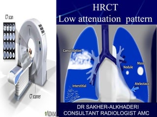
  • 1. HRCT Low attenuation pattern DR SAKHER-ALKHADERI CONSULTANT RADIOLOGIST AMC
  • 2. Low Attenuation pattern Emphysema Lung cysts (LAM, LIP, Langerhans cell histiocytosis) Bronchiectasis Honeycombing
  • 3.
  • 4. Emphysema Emphysema typically presents as areas of low attenuation without visible walls as a result of parenchymal destruction.
  • 5.
  • 6.
  • 7. EMPHYSEMA Permanent, abnormal enlargement of air spaces distal to the terminal bronchiole and accompanied by the destruction of the walls of the involved air spaces. 7
  • 8. Most common type Irreversible destruction of alveolar walls in the centrilobular portion of the lobule Upper lobe predominance and uneven distribution Strongly associated with smoking. Centrilobular (proximal or centriacinar) emphysema
  • 9. Centrilobular (proximal or centriacinar) emphysemaFound most commonly in the upper lobes Manifests as multiple small areas of low attenuation without a perceptible wall, producing a punched-out appearance. Often the centrilobular artery is visible within the centre of these lucencies. 9
  • 10. Centrilobular emphysema due to smoking. The periphery of the lung is spared (blue arrows). Centrilobular artery (yellow arrows) is seen in the center of the hypodense area.
  • 11. PANLOBULAR EMPHYSEMA Affects the whole secondary lobule Lower lobe predominance In alpha-1-antitrypsin deficiency, but also seen in smokers with advanced emphysema
  • 12. PANLOBULAR EMPHYSEMA Affects the entire secondary pulmonary lobule and is more pronounced in the lower zones Complete destruction of the entire pulmonary lobule. Results in an overall decrease in lung attenuation and a reduction in size of pulmonary vessels 12
  • 15. Paraseptal (distal acinar) emphysema Affects the peripheral parts of the secondary pulmonary lobule Produces subpleural lucencies. 15
  • 17. Cystic lung disease Lung cysts are defined as radiolucent areas with a wall thickness of less than 4mm.
  • 18.
  • 19.
  • 20. There is proliferation of Langerhans cells in the bronchiolar and bronchial epithelium, forming granulomas. It is postulated that as these cellular granulomas evolve, peripheral fibrosis forms resulting in traction on the central bronchiole which becomes cyst-like 3 . This explains the presumed evolution from nodule, through cavitating nodule and thick walled cysts, to the 'stable' thin-walled cysts 3 CT : nodules:more pronounced early in the disease may range in number from a few to innumerable 1-10 mm in diameter (typically 1-5 mm 4 ) centrilobular distribution usually irregular margin surrounding lung parenchyma appears normal may cavitate and become cysts
  • 21. Cysts : more pronounced later in the disease usually less than 10 mm in diameter may measure up to 2-3 centimetres in size the extreme bases may be preserved usually thin-walled, but on occasion may be up to a few millimetres thick Other common findings include 1,3 : ground-glass opacities: emphysema mosaic attenuation Differential diagnoses -granulomatous disease Wegener granulomatosis sarcoidosis -metastases -miliary tuberculosis-lymphangiomyomatosis (LAM)
  • 22.
  • 23.
  • 24. The disease is characterised by the persistence of dilated lymphatics and interstitial proliferation of abnormal smooth muscle that in turn can obstruct venules, lymphatics, and small airways. HRCT thin walled cysts of variable sizes surrounded by normal lung parenchyma can be seen throughout the lung interlobular septal thickening may show a dilated thoracic duct haemorrhages may be seen as areas of increased attenuation Differential diagnosis For pulmonary manifestations, the primary differential to be considered is Langerhans cell histiocytosis (LCH) which tends to happen in children and young adults with history of heavy cigarette smoking. It has a mid to upper lobe distribution with preservation of costophrenic angles. In addition, the cysts in LCH tend to be more irregular in contour. LCH has much more favourable prognosis compared with LAM.
  • 26.
  • 27.
  • 28.
  • 29. Lymphocytic interstitial pneumonia is a benign lymphoproliferative disorder characterized by a diffuse and exquisitely interstitial proliferation of small lymphocytes and plasma cells (1). Lymphocytic interstitial pneumonia occurs most commonly in patients who have Sjögren syndrome, autoimmune thyroid disease, acquired immunodeficiency syndrome (AIDS), or Castleman disease LIP Radiology findings : ground-glass attenuation, airspace consolidation, parenchymal nodules, interlobular septal Radiology findings : ground-glass attenuation, airspace consolidation, parenchymal nodules, interlobular septal
  • 30.
  • 31.
  • 32. Bronchiectasis Bronchiectasis is defined as localized irreversible bronchial dilatation. bronchial wall thickening lack of normal tapering with visibility of airways in the peripheral lung mucus retention in the bronchial lumen associated atelectasis and sometimes air trapping
  • 33. Subtypes According to macroscopic morphology, three types have been described, which also represet a spectrum of severity 8 : cylindrical bronchiectasis bronchi have a uniform calibre, do not taper and have parallel walls (tram track sign and signet ring sign) commonest form 14 varicose bronchiectasis relatively uncommon beaded appearances where dilated bronchi have interspersed sites of relative narrowing cystic bronchiectasis severe form with cyst-like bronchi that extend to the pleural surface air-fluid levels are commonly present
  • 34.
  • 35. ABPA: glove-finger shadow due to mucoid impaction in central bronchiectasis in a patient with asthma.
  • 36. Signet-Ring Sign A signet-ring sign represents an axial cut of a dilated bronchus (ring) with its accompanying small artery
  • 37.
  • 39. Bronchial dilation with lack of tapering .Cystic bronchiectasis
  • 40. HONEYCOMBING Defined as - small cystic spaces with irregularly thickened walls composed of fibrous tissue. Predominate in the peripheral and subpleural lung regions Subpleural honeycomb cysts typically occur in several contiguous layers. D/D- paraseptal emphysema in which subpleural cysts 40
  • 41. Honeycombing Honeycombing is defined by the presence of small cystic spaces with irregularly thickened walls composed of fibrous tissue.
  • 42. Causes Lower lobe predominance : 1. UIP or interstitial fibrosis 2. Connective tissue disorders 3. Hypersensitivity pneumonitis 4. Asbestosis 5. NSIP (rare) Upper lobe predominance : 1. End stage sarcodosis 2. Radiation 3. Hypersensitivity Pneumonitis 4. End stage ARDS 42
  • 44. Honeycombing and traction bronchiectasis in UIP.
  • 45. Typical UIP with honeycombing and traction bronchiectasis in a patient with idiopathic pulmonary fibrosis (IPF)

Editor's Notes

  1. Results from imbalance between proteolytic and antiproteolytic enzymes ,the balance is shifted toward proteolysis by smoking or enzymatic deficiencies, such as a1-antiprotease deficiency .
  2. The yellow arrows indicates the pulmonary vessels