Understanding Leprosy (Hansen's Disease
•Download as PPTX, PDF•
1 like•795 views

Oldest disease known to mankind First described in ancient Indian texts as “Kustha roga” attributed ] to curse from God Leper : Greek “scaly” Hansen’s Disease – 1873 Norwegian Armauer Hansen discovered that leprosy is caused by bacterium - Mycobacterium leprae Albert Neisser (1879) – stained the organism with fuchsin & gentian violet ( AFB )

Recommended
More Related Content
What's hot
What's hot (20)
Similar to Understanding Leprosy (Hansen's Disease
Similar to Understanding Leprosy (Hansen's Disease (20)
More from NCRIMS, Meerut
More from NCRIMS, Meerut (20)
Recently uploaded
Recently uploaded (20)
Understanding Leprosy (Hansen's Disease
- 2. Introduction & History •Oldest disease known to mankind •First described in ancient Indian texts as “Kustha roga” attributed to curse from God •Leper : Greek “scaly” Hansen’s Disease – 1873 Norwegian Armauer Hansen discovered that leprosy is caused by bacterium - Mycobacterium leprae Albert Neisser (1879) – stained the organism with fuchsin & gentian violet ( AFB )
- 3. Leprosy is a social disease associated with stigma
- 5. Morphology • They are Gram positive and acid fast 5% H2 SO4. but to lesser extant than tubercle bacilli. • The acid-fast bacilli are arranged singly, in parallel bundles (like rolls of cigarettes in a packet) or in globular masses. • The bacilli are slender, slightly curved or straight rods, 1-8 um X 0.2-0.5 um in size.
- 6. Cultivation • M. leprae is found only in cases of human infection. • They have not yet been grown ion artificial media or tissue culture. • The generation time of leprae bacillus is found to be 12-20 days on an average. • When nasal scrapping from lepromatous leprosy containing lepra bacilli are inoculated intradermally into foot pad of mouse and kept at low temperature (20oC), a granulomatous lesion develops at the site of injection in 1-6 months. • When nine band armadillo is inoculated with lepra bacilli, generalized infection develops with extensive multiplication of the bacilli.
- 7. Resistance: •M. leprae can survive in warm humid environment for 9-16 days, • 46 days in moist soil, •2 hours in sunlight and •for about 30 minutes in UV light. BacteriaResidesin CoolerPartsoftheBody Skin Peripheral Nerves
- 9. Transmission Nasal/oral Droplets Dermal Inoculations
- 10. Contraction of disease • It is likely to spread through direct skin contact (damaged skin) and air dispersement of infectious aerosols from coughing or sneezing of infected patient. • Contracting the disease depends on how susceptible the person is to the disease, how long you are exposed, and environmental conditions. • Only 10 to 29% of people exposed to bacilli actually develop leprosy.
- 11. Slow, chronic &progressive Granulomatous disease of Peripheral nerves,skin and Muco- cutaneous tissues (Nasal mucosa). It affects Skin, Lungs,liver, testes ,bones. Leprosy Pathogenesis
- 12. Pathogenesis • The principal target cell of lepra bacilli are Schwann cell and the resulting nerve damage causes manifestation of leprosy, which include muscle paralysis. • A non-specific or Indeterminate skin lesion is the First sign of disease.
- 13. Tuberculoid Borderline Tuberculoid Borderline laptomatous Lepromatous Classification • Clinical manifestaion is determined by cellular immune response of the individual to the bacilli. There are two major forms (polar forms) of leprosy: 1. Lepromatous leprosy: 2. Tuberculoid leprosy 3. Many patients occupy an intermediate position on the spectrum, which may be classified as borderline lepromatous (BL) which may turn into any of the polar forms.
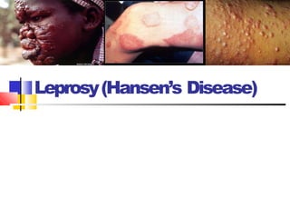
- 14. 1. Lepromatous or nodular type: • It is a generalized form and found in host with low resistance. • The bacteria disseminate hematogenously although, the lesions are superficial. (MULTIBACILLARY) • Nodular Lesions are large, diffuse and granulomatous. • Facial disfigurement is common which is due to extensive collagen destruction • leading to thickening of the loose skin of the forehead, lips and ears, resulting the classic leonine facies. Leonine face
- 16. • Localized form of disease and found in patient with high degree of resistance where cell mediated immunity is intact Skin lesions: •The skin lesions are few and characterized by non- elevated macular, hyperpigmented, anasthetic patches on the trunk, face and limbs. •Bacilli are scanty or absent (Paucibacillary). M. leprae invades sensory nerves leading to patchy anaesthesia in the lesion. Localized anaesthesia often leads to injury and severe bacterial infection of hand and feet, sometimes producing deformities. Infectivity is low. Lepromin test is positive. Patient shows a delayed type hypersensitivity and disease progression is slow . Tuberculoid leprosy
- 20. Symptoms Tuberculoid Leprosy Symptoms • Severe pain • Muscle weakness • Skin stiffness and dryness • Loss of fingers and toes • Eye problems • Blindeness • Enlarged nerves • Thickened skin on face • Nasal stuffiness • Bloody nose • Laryngitis • Collapsing of the nose • Swelling of the lymph nodes in the groin and armpits • Scarring of the testes that leads to infertility • Enlargement of male breasts There is two ways leprosy is presented: Lepromatous Leprosy Symptom
- 21. Tuberculoid Lepromatous CMI to M leprae +++ --- Lepra bacilli in tissue -/+ ++++ Lepromin test +++ --- CD4/CD8 > 1 <1 Plasma cells in lymphoid tissue + ++++ Lymphocytic ifiltration of lesion ++++ --- Lesion Scaly Nodular
- 23. MechanismofNerveDamage Scollard, DM et al. 2006. “The continuing challenges of leprosy.” Clinical microbiology reviews 19, no. 2: 338-81. 1. EntryThrough BloodVessels 2. Inflam matory Response 3. Demyelination
- 24. SensoryLoss Paralysis Deformities OutcomesofNerveDamage Brought to you by
- 27. Lepromin test •Detects delayed hypersensitivity •Described by Mitsuda (1919) •Prepared from boiled bacilli-rich lepromatous lesions •Biphasic reaction: •Fernandez reaction: erythema & induration in 24-48 h •Mitsuda reaction: indurated skin nodule, may ulcerate; starts in 1-2 weeks, peaks in 4 weeks
- 28. Lepromin test •Not used for diagnosis • To classify the lesions (+ve in tuberculoid, -ve in lepromatous) •To assess prognosis & response to treatment (+ve reaction – good prognosis)
- 29. •Lab. Diagnosis Specimens : 1. Scrapings from Lesion ,Nasal mucosa and ear lobule Z-N staining. Acid fast bacilli Live bacilli : Solid, uniformly stained. Dead bacilli :Fragmented and granular.
- 30. 1. Bacteriological index: 2. Morphological index(% of uniformly stained bacilli) = Uniformly stained bacilli X 100 Total number of bacilli • Bacteriological index ranges from 1 to 6+ as shown below: • 1-10 bacilli in 100 fields = 1+ • 1-10 bacilli in 10 fields = 2+ • 1-10 bacilli per field = 3+ • 10-100 bacilli per field = 4+ • 100-1,000 bacilli per filed = 5+ • More than 1,000 bacilli, clumps and groups in every field = 6+
- 31. 2. Skin & Nerve biopsy. 3. Lepromin test : To know prognosis. Not for diagnosis. 5. Serological test : ELISA . 6. Molecular diagnosis: Identifying DNA codes for 65 & 18-kDa M.leprae proteins.
- 32. Multibacillary or Lepromatous Laprorosy
- 33. TREATMENT
- 35. 1960’s:Rifampicinand ClofazimineDiscovered Rifampicin (Rifampin): InhibitRNA synthesis Clofazimine: Anti- inflam matory
- 36. 1981:WHO ProposesMulti- DrugTherapy (MDT) Combinationof DAPSONE, RIFAMPICIN, and CLOFAZIMINE + +
- 39. Treatment Type of leprosy Drug Dose Frequency Total duration Paucibacillary Rifampicin Dapsone 600 mg 100 mg Monthly Daily 6 months Multibacillary Rifampicin Dapsone Clofazimine 600 mg 100 mg 300 mg + 50 mg Monthly Daily Monthly Daily 2 years or more