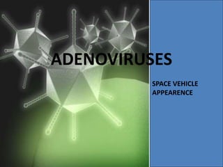
DNA Viruses: Adenoviruses and Parvoviruses
•Transferir como PPTX, PDF•
3 gostaram•341 visualizações

Adenoviruses are non-enveloped DNA viruses that commonly cause respiratory illness. There are multiple serotypes that can cause pharyngitis, conjunctivitis, or pneumonia. Adenoviruses are transmitted through direct contact or respiratory droplets. Laboratory diagnosis involves virus isolation in cell culture or PCR detection of viral DNA. There is no vaccine or antiviral treatment available.

Recomendados
Mais conteúdo relacionado
Mais procurados
Mais procurados (20)
Semelhante a DNA Viruses: Adenoviruses and Parvoviruses
Semelhante a DNA Viruses: Adenoviruses and Parvoviruses (20)
Mais de NCRIMS, Meerut
Mais de NCRIMS, Meerut (20)
Último
Último (20)
DNA Viruses: Adenoviruses and Parvoviruses
- 2. DNA VIRUSES All are DS except Parvoviridae All replicate in the nucleus except Poxviridae Icosahedral Complex 1- Poxviridae Naked Enveloped non-enveloped 1- Parvoviridae 1- Herpesviridae 2- Papillomaviridae 2- Hepadnaviridae 3- Adenoviridae
- 3. DNA viruses first isolated from adenoidal tissue in 1953 Family Adenoviridae Genus Mastadenovirus
- 4. Morphology • Non-enveloped, 70-90nm in size. • Capsid is icosahedral, comprised of 252 capsomeres -240 are hexons and 12 pentons at the vertices. • Each penton has a projecting fiber with a terminal knob. Together, this complex is toxic to cells, causing rounding and death of cells through inhibition of protein synthesis. • The fiber proteins determine target cell specificity. • Genome is made of linear double- stranded (ds) DNA
- 5. CLASSIFICATION • Family Adenoviridae : 2 genera – Aviadenovirus : adenoviruses of birds; – Mastadenovirus : infect mammals; • 47 serotypes of human origin;
- 6. DISEASE ASSOCIATIONS Serotype Disease At risk 1-7 Acute febrile pharyngitis Children 3,7,14 Pharyngoconjunctival fever Young children 8,9,37 Epidemic keratoconjunctivitis (shipyard eye) Adults 3,4,11 Acute follicular conjunctivitis Any age 40,41 Diarrhoea Infants, young children 11,21 Hemorrhagic cystitis Children
- 7. Pathogenesis: • Adenoviruses spread by: – direct contact, – respiratory droplets – feco-oral route.
- 8. Pathogenesis: • Adenoviruses infect and replicate in the epithelial cells of the: – pharynx, – conjunctiva, – urinary bladder – small intestine. They usually do not spread beyond the regional lymph nodes EXCEPT IN THE IMMUNE COMPROMISED HOST.
- 9. Pathogenesis: • The virus has a tendency to become latent in lymphoid tissue, • The virus can be reactivated by immunosuppression.
- 10. Clinical Syndromes: • Adenoviruses cause primary infection in: – children – less commonly adults. • Several distinct clinical syndromes are associated with Adenovirus infection.
- 11. CLINICAL SYNDROMES A. Respiratory diseases: B. Eye infections: C. Gastrointestinal disease D. Other diseases: E. Adenoviral infections of the immune compromised host
- 12. A. Respiratory diseases: • The most important etiological association of adenoviruses is with the respiratory diseases. • They are responsible for 5% of acute respiratory diseases in: – young children – and much less in adults.
- 13. A. Respiratory diseases: Four different syndromes of respiratory infection have been linked to Adenoviruses. • Acute febrile pharyngitis: – most commonly seen in infants and young children, – symptoms include cough, stuffy nose, fever and sore throat. • Pharyngo conjunctival fever: – symptoms are similar to those of acute febrile pharyngitis but conjunctivitis is also present. – It tends to occur in outbreaks such as at children's summer camps (swimming pool conjunctivitis).
- 14. A. Respiratory diseases: • Acute respiratory disease: – is characterized by pharyngitis, fever, cough and malaise. – It occurs in an epidemic form among young recruits under conditions of overcrowding • Pneumonia: a complication of acute respiratory disease in both children and adults.
- 15. NOTE Outbreaks & epidemic adenovirus infections • Pharyngo conjunctival fever: – outbreaks – in children's summer camps (swimming pool conjunctivitis). • Acute respiratory disease: – occurs in an epidemic form – among young recruits • Epidemic keratoconjunctivitis:
- 16. B. Eye infections: • Mild conjunctivitis: – can occur as a part of respiratory pharyngeal syndromes. – Complete recovery with no lasting sequelae is the common outcome. – Can occur sporadically or in outbreaks. • Epidemic keratoconjunctivitis: • a highly contagious and a more serious disease occurring mainly in adults. • Corneal involvement may be followed by various degrees of visual disability.
- 17. C. Gastrointestinal disease: 1. No disease association 1. Many Adenoviruses replicate in intestinal cells and are present in the stools without being associated with GIT disease. 2. Infantile gastroenteritis 1. Two serotypes (40, 41) have been etiologically associated with infantile gastroenteritis.
- 18. NOTE 1. The enteric Adenoviruses are very difficult to cultivate. 2. Lab diagnosis depend on direct detection
- 19. D. Other diseases: • Acute haemorrhagic cystitis: – types 11, 21 may cause acute haemorrhagic cystitis in children especially boys.
- 20. E. Adenoviral infections of the immune compromised host • The most common clinical manifestations are: • pneumonia, • hepatitis • gastroenteritis.
- 21. LABORATORY DIAGNOSIS • Specimens : from throat, eye, urine, feces. • Isolation of virus: – Inoculation into cell cultures; – human embryonic kidney/HeLa/HEp2 – CPE : cell rounding and aggregation into grape like clusters; – Other tests : HA, Neutralization, CF
- 22. • Serology : rise in titre of antibodies in paired sera; (single specimen – not useful) • Electron Microscopy : for stool to see virus • Immunofluorescence : antigen detection in Nasopharyngeal /occular specimens
- 23. Prevention and control • Careful hand washing is the easiest way to prevent infection. • Disinfection of Environmental surfaces with hypochlorites. • The risk of water borne outbreaks of conjunctivitis can be minimized by chlorination of swimming pools. • Epidemic keratoconjunctivitis can be controlled by strict asepsis during eye examination.
- 24. Parvoviruses
- 25. Introduction • Smallest DNA virus • Belong to Family Parvoviridae • Icosahedral in shape • Non enveloped • Single stranded DNA • Human parvovirus B19 is only pathogenic in man • Shows tropism for erythroid progenitor cells
- 26. Classification Consists of 3 genera Dependovirus also called adeno associated virus no disease in humans Parvovirus cause leucopenia in canine animals Erythrovirus consist of B19virus cause Flu, erthyematous infection, infection in pregnant, chronic viral infection leucopenia aplastic anemia, Thrombocytopenia
- 27. Pathogenesis • Virus shows tropism for RBC’s & endothelial cells of blood vessels • Virus enters nasopharynx----spreads in the blood cause viremia-----then infect mitotically active erythroid precussor cells in bone marrow------ infection established-------virus infects nucleus of the cells ----replicates----kill erythroid cells-- --lead to flu like symptoms--- virus shed in oral and respiratory secretions and can cross the placenta • Antibodies IgM & IgG appear in due course of time and form immune complexes---deposit of immune complexes lead to artharlgia & rash
- 29. HumanparvovirusB19 • Discovered in the serum of asymptomatic blood donors. • Associated with: • A very common exanthematous disease of children (Fifth disease) • Aplastic crisis (in patients with chronic hemolytic anemia) • Hydrops fetalis 5th .
- 30. Erythema infectiosum or fifth disease Gloves and sock syndrome
- 31. Human Parvo virus infection
- 32. Clinical syndromes of B19 virus Contd…. Chronic B19 infection • Infection occurs in immunocompromised patients such as HIV, patients receiving chemotherapy, Transplant patients • Patient develops chronic anemia, leucopenia, thrombocytopenia
- 33. Laboratory Diagnosis • Demonstration of specific IgM & IgG Ab • In pregnant woman serum positive for IgM & IgG indicates infection within 1-4 weeks. • Positive IgG and negative IgM indicates past infection • PCR to demonstrate B19 genome
- 34. Treatment & Prevention • No specific therapy is available • Supportive treatment only • No vaccine available • Phase I trial are on
- 35. Human papilloma virus Human papillomavirus is the most common sexually transmitted infection. Most sexually active men and women being exposed to the virus at some point during their lifetime
- 36. Structure • small, non-enveloped, icosahedral Double stranded DNA viruses that have a diameter of 52–55 nm. • single double-stranded DNA molecule of about 8000 base-pairs. • a protein capsid composed of 72 pentameric capsomers. Structure
- 37. Structure Classification • the International Committee on the Taxonomy of Viruses (ICTV) as two separate families — Papillomaviridae and Polyomaviridae. • HPV is divided into High risk HPV and Low risk HPV. Low- risk types cause warts and high-risk types can cause cancer.
- 38. Structure Pathogenesis • HPV infection is limited to the basal cells of stratified epithelium cells and induce within those cells through micro-abrasions leading to characteristic cytoplasmic vacuole. • The virus cannot bind to live tissue Most warts are benign and do not progress to malignancy. However, HPV infection is associated with carcinoma of the uterine cervix and penis. Immunosuppressed patients have more extensive warts, and women infected with human immunodeficiency virus (HIV) have a very high rate of carcinoma of the cervix.
- 39. Symptoms • HPV may not cause symptoms at once, but they can appear years later. Some types can lead to warts, while others can cause cancer. • Warts • Common symptoms of some types of HPV are warts, especially genital warts. • Genital warts may appear as a small bump, cluster of bumps. They commonly affect the vulva in women, or possibly the cervix, and the penis or scrotum in men. They may also appear around the anus and in the groin. • They can range in size and appearance and be large, small, flat, or cauliflower shaped, • and may be white or flesh tone. • HPV6 and HPV11 are common causes of genital warts and laryngeal papillomatosis.
- 40. Symptoms
- 41. Symptoms Other warts associated with HPV include common warts, plantar, and flat warts. • Common warts - rough, raised bumps most commonly found on the hands, fingers, and elbows. • Plantar warts - described as hard, grainy growths on the feet; they most commonly appear on the heels or balls of the feet. • Flat warts - generally affect children, adolescents, and young adults; they appear as flat-topped, slightly raised lesions that are darker than normal skin color and are most commonly found on the face, neck, or areas that have been scratched. Common warts on the dorsal surface of the hand Planter wart
- 42. Plane wart
- 44. Epidermodysplasia Verruciformis (Tree branches apperance)
- 45. PERIUNGUAL WARTS
- 47. INTERNAL WARTS
- 48. CERVICAL WARTS
- 50. Cervical – HPV infection Cervical cancer is second most frequent cancer in women world wide 5,00,000 cases present with cervical cancer Major leading cause of deaths related to malignancy in the Devloping world Several cases associated with HPV infections.
- 51. Symptoms: Cervical Cancer • As in many cancers, there may be no signs or symptoms of cervical cancer until it has progressed to a dangerous stage. However, if symptoms do occur they may include: • Abnormal vaginal bleeding • Pain in very advanced stages • Any bleeding from the vagina other than during menstruation. • Abnormal vaginal discharge
- 52. Transmission • HPV is transmitted through intimate skin-to-skin contact. having vaginal, anal, or oral sex with someone who has the virus. • It is most commonly spread during vaginal or anal sex. • common that nearly all men and women get it at some point in their lives. • can be passed even when an infected person has no signs or symptoms. • can develop symptoms years after being infected, making it hard to know when you first became infected. In most cases, HPV goes away on its own and does not cause any health problems. Butwhen HPV does not go away, it can cause health problems like genital warts and cancer.
- 53. Diagnosis The traditional methods of viral diagnosis such as electron microscopy, cell culture, and certain immunological methods are not suitable for HPV detection. HPV cannot be cultured in cell cultures. The important methods to diagnose HPV infection are: • Biopsy • DNA test (PCR, Southern Blot Hybridization, In Situ Hybridization) • Pap smear
- 54. Diagnosis PCR-based methods: HPV DNA can be amplified selectively by a series of reactions that lead to an exponential and reproducible increase in viral sequences present in the biological specimen. serological assays: ELISA or western blot analysis
- 55. Diagnosis Biopsy: If the biopsy results show pre-cancer (dysplasia) or cancer, then treatment is recommended. The dysplasia may be mild, moderate, or severe. PAP smear or PAP test: It is a screening test. Apart from premalignant and malignant changes, viral infections like HPV infection and Herpes can also be detected.
- 56. Treatment There is currently no specific treatment for HPV infection. However warts can be treated. • Salicylic acid. Over-the-counter treatments that contain salicylic acid work by removing layers of a wart a little at a time. • Imiquimod (Aldara, Zyclara). This prescription cream might enhances immune system's ability to fight HPV. Common side effects include redness and swelling at the application site. • Podofilox (Condylox). Another topical prescription, podofilox works by destroying genital wart tissue. Podofilox may cause pain and itching where it's applied. • Trichloroacetic acid. This chemical treatment burns off warts on the palms, soles and genitals. It might cause local irritation.
- 57. Treatment Surgical and other procedures If medications don't work the following methods can be use to remove warts • Freezing with liquid nitrogen (cryotherapy) • Burning with an electrical current (electrocautery) • Surgical removal • Laser surgery "condom use may reduce the risk for genital human papillomavirus (HPV) infection"
- 58. Vaccination Three vaccines are available to prevent infection by some HPV types. All protect against initial infection with HPV types 16 and 18, which cause most of the HPV-associated cancer cases. Gardasil: protects against HPV types 6 and 11, which cause 90% of genital warts. quadrivalent vaccine Cervarix: bivalent, and is prepared from virus-like particles (VLP) of The capsid. Gardasil 9: nonavalent, it has the potential to prevent about 90% of cervical, vulvar, vaginal, and anal cancers.
- 59. Prevention • Vaccination • Avoid skin-to-skin contact by not having sex with strangers. • Use condoms and/or dental dams every time you have vaginal, anal, or oral sex. • Though condoms and dental dams are not as effective against HPV as they are against other STDs like chlamydia and HIV, safer sex can lower your chances of getting HPV.
Notas do Editor
- Infantile gastroenteritis: Watery diarrohea lasting for 8-2 days with fever and vomiting
- Acute haemorrhagic cystitis: inflammation of bladder(dysuria, hematuria, hemorrhage)