Best Rate (Guwahati ) Call Girls Guwahati ⟟ 8617370543 ⟟ High Class Call Girl...
RDP_UPDATED_HAP-II_NERVOUS SYSTEM_ BRAIN.pdf
1. HAP-II_NERVOUS SYTEM
PREPARED BY: Mrs. Rishita Patel_ IICP
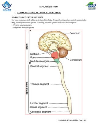
1. NERVOUS SYSTEM (CNS - BRAIN & CIRCULATION)
DIVISIONS OF NERVOUS SYSTEM
Nervous system controls all the activities of the body. It is quicker than other control system in the
body, namely endocrine system. Primarily, nervous system is divided into two parts:
1. Central nervous system
2. Peripheral nervous system.
3. HAP-II_NERVOUS SYTEM
PREPARED BY: Mrs. Rishita Patel_ IICP
Parts of Brain
Brain consists of three major divisions: 1. Prosencephalon 2. Mesencephalon 3. Rhombencephalon
INTRODUCTION
Neuron or nerve cell is defined as the structural and functional unit of nervous system.
Neuron is similar to any other cell in the body, having nucleus and all the organelles in cytoplasm.
However, it is different from other cells by two ways:
1. Neuron has branches or processes called axon and dendrites
2. Neuron does not have centrosome. So, it cannot undergo division.
„ CLASSIFICATION OF NEURON
Neurons are classified by three different methods.
A. Depending upon the number of poles
B. Depending upon the function
C. Depending upon the length of axon.
„ DEPENDING UPON THE NUMBER OF POLES
Based on the number of poles from which the nerve fibers arise, neurons are divided into three
types:
1. Unipolar neurons
2. Bipolar neurons
3. Multipolar neurons.
4. HAP-II_NERVOUS SYTEM
PREPARED BY: Mrs. Rishita Patel_ IICP
1. Unipolar Neurons
Unipolar neurons are the neurons that have only one pole. From a single pole, both axon
and dendrite arise. This type of nerve cells is present only in embryonic stage in human
beings.
2. Bipolar Neurons
Neurons with two poles are known as bipolar neurons. Axon arises from one pole and
dendrites arise from the other pole.
3. Multipolar Neurons
Multipolar neurons are the neurons which have many poles. One of the poles gives rise to
axon and all other poles give rise to dendrites.
DEPENDING UPON THE FUNCTION
On the basis of function, nerve cells are classified into two types:
1. Motor or efferent neurons
2. Sensory or afferent neurons.
1. Motor or Efferent Neurons: Motor or efferent neurons are the neurons which carry the
motor impulses from central nervous system to peripheral effector organs like muscles, glands,
blood vessels, etc. Generally, each motor neuron has a long axon and short dendrites.
2. Sensory or Afferent Neurons: Sensory or afferent neurons are the neurons which carry the
sensory impulses from periphery to central nervous system. Generally, each sensory neuron
has a short axon and long dendrites.
5. HAP-II_NERVOUS SYTEM
PREPARED BY: Mrs. Rishita Patel_ IICP
DEPENDING UPON THE LENGTH OF AXON
Depending upon the length of axon, neurons are divided into two types:
1. Golgi type I neurons
2. Golgi type II neurons.
1. Golgi Type I Neurons Golgi type I neurons have long axons. Cell body of these neurons is in
different parts of central nervous system and their axons reach the remote peripheral organs.
2. Golgi Type II Neurons Neurons of this type have short axons. These neurons are present in
cerebral cortex and spinal cord.
STRUCTURE OF NEURON
Neuron is made up of three parts: 1. Nerve cell body 2. Dendrite 3. Axon.
Dendrite and axon form the processes of neuron.
Dendrites are short processes and the axons are long processes.
Dendrites and axons are usually called nerve fibers.
„ NERVE CELL BODY
Nerve cell body is also known as soma or perikaryon. It is irregular in shape. Like any other cell,
it is constituted by a mass of cytoplasm called neuroplasm, which is covered by a cell membrane.
The cytoplasm contains a large nucleus, Nissl bodies, neurofibrils, mitochondria and Golgi
apparatus. Nissl bodies and neurofibrils are found only in nerve cell and not in other cells.
Nucleus
Each neuron has one nucleus, which is centrally placed in the nerve cell body. Nucleus has one or
two prominent nucleoli. Nucleus does not contain centrosome. So, the nerve cell cannot multiply
like other cells.
6. HAP-II_NERVOUS SYTEM
PREPARED BY: Mrs. Rishita Patel_ IICP
Nissl Bodies
Nissl bodies or Nissl granules are small basophilic granules found in cytoplasm of neurons and are
named after the discoverer. These bodies are present in soma and dendrite but not in axon and
axon hillock. Nissl bodies are called tigroid substances, since these bodies are responsible for
tigroid or spotted appearance of soma after suitable staining.
Dendrites are distinguished from axons by the presence of Nissl granules under microscope.
Nissl bodies are membranous organelles containing ribosomes. So, these bodies are concerned
with synthesis of proteins in the neurons.
Proteins formed in soma are transported to the axon by axonal flow. Number of Nissl bodies varies
with the condition of the nerve. During fatigue or injury of the neuron, these bodies fragment and
disappear by a process called chromatolysis. Granules reappear after recovery from fatigue or after
regeneration of nerve fibers.
Neurofibrils
Neurofibrils are thread-like structures present in the form of network in the soma and the nerve
processes. Presence of neurofibrils is another characteristic feature of the neurons. The neurofibrils
consist of microfilaments and microtubules.
Mitochondria
Mitochondria are present in soma and in axon. As in other cells, here also mitochondria form the
powerhouse of the nerve cell, where ATP is produced.
7. HAP-II_NERVOUS SYTEM
PREPARED BY: Mrs. Rishita Patel_ IICP
Golgi Apparatus
Golgi apparatus of nerve cell body is similar to that of other cells. It is concerned with processing
and packing of proteins into granules.
DENDRITE
Dendrite is the branched process of neuron and it is branched repeatedly. Dendrite may be present
or absent. If present, it may be one or many in number. Dendrite has Nissl granules and
neurofibrils. Dendrite transmits impulses towards the nerve cell body. Usually, the dendrite is
shorter than axon.
AXON
Axon is the longer process of nerve cell. Each neuron has only one axon. Axon arises from axon
hillock of the nerve cell body and it is devoid of Nissl granules. Axon extends for a long distance
away from the nerve cell body. Length of longest axon is about 1 meter. Axon transmits impulses
away from the nerve cell body.
Organization of Nerve:
Each nerve is formed by many bundles or groups of nerve fibers. Each bundle of nerve fibers is
called a fasciculus.
Coverings of Nerve
The whole nerve is covered by tubular
sheath, which is formed by a areolar
membrane. This sheath is called
epineurium. Each fasciculus is covered
by perineurium and each nerve fiber
(axon) is covered by endoneurium.
8. HAP-II_NERVOUS SYTEM
PREPARED BY: Mrs. Rishita Patel_ IICP
Internal Structure of Axon –
Axis Cylinder Axon has a long central core of cytoplasm called axoplasm.
Axoplasm is covered by the tubular sheath like membrane called axolemma.
Axolemma is the continuation of the cell membrane of nerve cell body. Axoplasm along with
axolemma is called the axis cylinder of the nerve fiber.
Axoplasm contains mitochondria, neurofibrils and axoplasmic vesicles. Because of the absence
of Nissl bodies in the axon, proteins necessary for the nerve fibers are synthesized in the soma and
not in axoplasm.
After synthesis, the protein molecules are transported from soma to axon, by means of axonal flow.
Some neurotransmitter substances are also transported by axonal flow from soma to axon. Axis
cylinder of the nerve fiber is covered by a membrane called neurilemma.
Non-myelinated Nerve
Fiber Nerve fiber described above is the non-myelinated nerve fiber, which is not covered by
myelin sheath
FIGURE: A. Myelinated nerve fiber; B. Non-myelinated nerve fiber.
9. HAP-II_NERVOUS SYTEM
PREPARED BY: Mrs. Rishita Patel_ IICP
Myelinated Nerve Fiber
Nerve fiber which is insulated by myelin sheath is called myelinated nerve fibers.
„ MYELIN SHEATH
Myelin sheath is a thick lipoprotein sheath that insulates the myelinated nerve fiber. Myelin
sheath is not a continuous sheath. It is absent at regular intervals. The area where myelin sheath
is absent is called node of Ranvier. Segment of the nerve fiber between two nodes is called
internode. Myelin sheath is responsible for white color of nerve fibers.
Chemistry of Myelin Sheath
Myelin sheath is formed by concentric layers of proteins, alternating with lipids. The lipids are
cholesterol, lecithin and cerebroside (sphingomyelin).
Formation of Myelin Sheath –
Myelinogenesis Formation of myelin sheath around the axon is called the myelinogenesis. It is
formed by Schwann cells in neurilemma. In the peripheral nerve, the myelinogenesis starts at 4th
month of intrauterine life. It is completed only in the second year after birth.
Functions of Myelin Sheath
1. Faster conduction
Myelin sheath is responsible for faster conduction of impulse through the nerve fibers. In
myelinated nerve fibers, the impulses jump from one node to another node. This type of
transmission of impulses is called saltatory conduction.
2. Insulating capacity
Myelin sheath has a high insulating capacity. Because of this quality, myelin sheath
restricts the nerve impulse within single nerve fiber and prevents the stimulation of
neighboring nerve fibers.
„ NEURILEMMA
Neurilemma is a thin membrane, which surrounds the axis cylinder. It is also called neurilemmal
sheath or sheath of Schwann.
It contains Schwann cells, which have flattened and elongated nuclei.
Cytoplasm is thin and modified to form the thin sheath of neurilemma. One nucleus is present in
each internode of the axon.
Nucleus is situated between myelin sheath and neurilemma. In non-myelinated nerve fiber, the
neurilemma surrounds axolemma continuously. In myelinated nerve fiber, it covers the myelin
10. HAP-II_NERVOUS SYTEM
PREPARED BY: Mrs. Rishita Patel_ IICP
sheath. At the node of Ranvier (where myelin sheath is absent), neurilemma invaginates and runs
up to axolemma in the form of a finger-like process.
Functions of Neurilemma
In non-myelinated nerve fiber, the neurilemma serves as a covering membrane. In myelinated
nerve fiber, it is necessary for the formation of myelin sheath (myelinogenesis). Neurilemma is
absent in central nervous system. So, the neuroglial cells called oligodendroglia are responsible
for myelinogenesis in central nervous system.
Classification of Nerve Fibers
BASIS OF CLASSIFICATION
Nerve fibers are classified by six different methods.
The basis of classification differs in each method.
Different methods of classification are listed in.
Different methods to classify nerve fibers Classification of nerve fibers
1. Depending upon structure
2. Depending upon distribution
3. Depending upon origin
4. Depending upon function
5. Depending upon secretion of neurotransmitter
6. Depending upon diameter and conduction of impulse (Erlanger Gasser classification)
1. DEPENDING UPON STRUCTURE
Based on structure, nerve fibers are classified into two types:
i. Myelinated Nerve Fibers
Myelinated nerve fibers are the nerve fibers that are covered by myelin sheath.
ii. Non-myelinated Nerve Fibers
Nonmyelinated nerve fibers are the nerve fibers which are not covered by myelin sheath.
2.DEPENDING UPON DISTRIBUTION
Nerve fibers are classified into two types, on the basis of distribution:
i. Somatic Nerve Fibers Somatic nerve fibers supply the skeletal muscles of the body.
ii. Visceral or Autonomic Nerve Fibers Autonomic nerve fibers supply the various
internal organs of the body.
3. DEPENDING UPON ORIGIN On the basis of origin, nerve fibers are divided into two types:
i. Cranial Nerve Fibers Nerve fibers arising from brain are called cranial nerve fibers.
11. HAP-II_NERVOUS SYTEM
PREPARED BY: Mrs. Rishita Patel_ IICP
ii. Spinal Nerve Fibers Nerve fibers arising from spinal cord are called spinal nerve fibers.
4.DEPENDING UPON FUNCTION
Functionally, nerve fibers are classified into two types:
I: Sensory Nerve Fibers
Sensory nerve fibers carry sensory impulses from different parts of the body to the central
nervous system. These nerve fibers are also known as afferent nerve fibers.
II: Motor Nerve Fibers
Motor nerve fibers carry motor impulses from central nervous system to different parts of the
body. These nerve fibers are also called efferent nerve fibers.
5.DEPENDING UPON SECRETION OF NEUROTRANSMITTER
Depending upon the neurotransmitter substance secreted, nerve fibers are divided into two
types:
I: Adrenergic Nerve Fibers Adrenergic nerve fibers secrete noradrenaline.
II: Cholinergic Nerve Fibers Cholinergic nerve fibers secrete acetylcholine.
6. DEPENDING UPON DIAMETER AND CONDUCTION OF IMPULSE (ERLANGER-
GASSER CLASSIFICATION) Erlanger and Gasser classified the nerve fibers into three major
types, on the basis of diameter (thickness) of the fibers and velocity of conduction of impulses:
i. Type A nerve fibers
ii. Type B nerve fibers
iii. Type C nerve fibers.
Among these fibers, type A nerve fibers are the thickest fibers and type C nerve fibers are the
thinnest fibers. Type C fibers are also known as Type IV fibers. Except type C fibers, all the nerve
fibers are myelinated.
Type A nerve fibers are divided into four types:
a. Type A alpha or Type I nerve fibers
b. Type A beta or Type II nerve fibers
c. Type A gamma nerve fibers
d. Type A delta or Type III nerve fibers.
Velocity of Impulse Velocity of impulse through a nerve fiber is directly proportional to the
thickness of the fiber.
12. HAP-II_NERVOUS SYTEM
PREPARED BY: Mrs. Rishita Patel_ IICP
PROPERTIES OF NERVE FIBERS
EXCITABILITY
Excitability is defined as the physiochemical change that occurs in a tissue when stimulus is
applied. Stimulus is defined as an external agent, which produces excitability in the tissues.
Chronaxie is an important parameter to determine the condition of nerve fiber. Clinically, the
damage of nerve fiber is determined by measuring the chronaxie. It is measured by chronaxie
meter. Nerve fibers have a low threshold for excitation than the other cells.
Response Due to Stimulation of Nerve Fiber When a nerve fiber is stimulated, based on the
strength of stimulus, two types of response develop:
1. Action potential or nerve impulse
Action potential develops in a nerve fiber when it is stimulated by a stimulus with adequate
strength. Adequate strength of stimulus, necessary for producing the action potential in a
nerve fiber is known as threshold or minimal stimulus. Action potential is propagated.
2. Electrotonic potential or local potential
When the stimulus with subliminal strength is applied, only electrotonic potential develops
and the action potential does not develop. Electrotonic potential is non propagated.
Cathelectrotonic and Anelectrotonic Potentials
While recording electrical potential in a nerve fiber, two electrodes, namely cathode and anode are
used. The potential change that is produced at cathode is called cathelectrotonic potential. The
potential that is developed at anode is known as anelectrotonic potential. Only the cathelectrotonic
potential can be transformed into electrotonic potential or action potential.
Properties of Action Potential Properties of action potential are given.
„ ELECTROTONIC POTENTIAL OR LOCAL POTENTIAL
Electrotonic potential or local potential is a non-propagated local response that develops in the
nerve fiber when a subliminal stimulus is applied.
Subliminal or subthreshold stimulus does not produce action potential. But, it alters the resting
membrane potential and produces slight depolarization for about 7 mV. This slight depolarized
state is called electrotonic potential. Firing level is reached only if depolarization occurs up to 15
mV. Then only action potential can develop. Electrotonic potential is a graded potential
Properties of Electrotonic Potential
1. Electrotonic potential is non-propagated
2. It does not obey all-or-none law. If the intensity of the stimulus is increased gradually every
time, there is increase in the amplitude till the firing level is reached, i.e. at 15 mV.
13. HAP-II_NERVOUS SYTEM
PREPARED BY: Mrs. Rishita Patel_ IICP
GENERATION OF ACTION POTENTIAL
An action potential (AP) or impulse is a sequence of rapidly occurring events that decrease and
reverse the membrane potential and then eventually restore it to the resting state. An action
potential has two main phases: a depolarizing phase and a repolarizing phase.
During the depolarizing phase, the negative membrane potential becomes less negative, reaches
zero, and then becomes positive.
During the repolarizing phase, the membrane potential is restored to the resting state of -70 mV.
Following the repolarizing phase there may be an after-hyperpolarizing phase, during which the
membrane potential temporarily become more negative than the resting level.
The period of time after an action potential begins during which an excitable cell cannot generate
another action potential in response to a normal threshold stimulus is called the refractory
period. During the absolute refractory period, even a very strong stimulus cannot initiate a
second action potential. This period coincides with the period of Na+ channel activation and
inactivation.
The relative refractory period is the period of time during which a second action potential can
be initiated, but only by a larger-than-normal stimulus. It coincides with the period when the
voltage-gated K+ channels are still open after inactivated Na+ channels have returned to their
resting state.
14. HAP-II_NERVOUS SYTEM
PREPARED BY: Mrs. Rishita Patel_ IICP
AP GENERATION:
Depolarizing Phase
When a depolarizing graded potential or some other stimulus causes the membrane of the axon to
depolarize to threshold, voltage-gated Na+ channels open rapidly. Both the electrical and the
chemical gradients favor inward movement of Na+, and the resulting in rush of Na+ causes the
depolarizing phase of the action potential.
The inflow of Na+ changes the membrane potential from -55 mV to +30 mV. At the peak of the
action potential, the inside of the membrane is 30 mV more positive than the outside.
Repolarizing Phase Shortly after the activation gates of the voltage-gated Na+ channels open, the
inactivation gates close. Now the voltage-gated Na+ channel is in an inactivated state. In addition
to opening voltage-gated Na+ channels, a threshold level depolarization also opens voltage-gated
K+ channels. Because the voltage-gated K+ channels open more slowly, their opening occurs at
about the same time the voltage-gated Na+ channels are closing. The slower opening of voltage-
gated K+ channels and the closing of previously open voltage-gated Na+ channels produce the
repolarizing phase of the action potential.
After-hyperpolarizing Phase While the voltage-gated K+ channels are open, outflow of K+ may
be large enough to cause an after-hyperpolarizing phase of the action potential. During this phase,
the voltage-gated K+ channels remain open and the membrane potential becomes even more
negative (about -90 mV). As the voltage-gated K+ channels close, the membrane potential returns
to the resting level of -70 mV. Unlike voltage-gated Na+ channels, most voltage-gated K+
channels do not exhibit an inactivated state. Instead, they alternate between closed (resting) and
open (activated) states.
16. HAP-II_NERVOUS SYTEM
PREPARED BY: Mrs. Rishita Patel_ IICP
CONDUCTIVITY
Conductivity is the ability of nerve fibers to transmit the impulse from the area of stimulation to
the other areas. Action potential is transmitted through the nerve fiber as nerve impulse. Normally
in the body, the action potential is transmitted through the nerve fiber in only one direction.
However, in experimental conditions when, the nerve is stimulated, the action potential travels
through the nerve fiber in either direction.
MECHANISM OF CONDUCTION OF ACTION POTENTIAL
Depolarization occurs first at the site of stimulation in the nerve fiber. It causes depolarization of
the neighboring areas. Like this, depolarization travels throughout the nerve fiber. Depolarization
is followed by repolarization.
CONDUCTION THROUGH MYELINATED NERVE FIBER –
SALTATORY CONDUCTION
Saltatory conduction is the form of conduction of nerve impulse in which, the impulse jumps from
one node to another. Conduction of impulse through a myelinated nerve fiber is about 50 times
faster than through a non-myelinated fiber. It is because the action potential jumps from one node
to another node of Ranvier instead of travelling through the entire nerve fiber.
Mechanism of Saltatory Conduction
Myelin sheath is not permeable to ions. So, the entry of sodium from extracellular fluid into nerve
fiber occurs only in the node of Ranvier, where the myelin sheath is absent. It causes depolarization
in the node and not in the internode. Thus, depolarization occurs at successive nodes. So, the action
potential jumps from one node to another. Hence, it is called saltatory conduction (saltare =
jumping).
17. HAP-II_NERVOUS SYTEM
PREPARED BY: Mrs. Rishita Patel_ IICP
Mode of conduction through nerve fibers A. Non-myelinated nerve fiber: continuous
conduction. B. Myelinated nerve fiber: saltatory conduction (impulse jumps from node to
node). AP = Action potential.
ADAPTATION
While stimulating a nerve fiber continuously, the excitability of the nerve fiber is greater in the
beginning. Later the response decreases slowly and finally the nerve fiber does not show any
response at all. This phenomenon is known as adaptation or accommodation. Cause for Adaptation
18. HAP-II_NERVOUS SYTEM
PREPARED BY: Mrs. Rishita Patel_ IICP
When a nerve fiber is stimulated continuously, depolarization occurs continuously. Continuous
depolarization inactivates the sodium pump and increases the efflux of potassium ions.
INFATIGABILITY
Nerve fiber cannot be fatigued, even if it is stimulated continuously for a long time. The reason is
that nerve fiber can conduct only one action potential at a time. At that time, it is completely
refractory and does not conduct another action potential.
ALL-OR-NONE LAW
All-or-none law states that when a nerve is stimulated by a stimulus it gives maximum response
or does not give response at all. Refer Chapter 90 for more details on all-or-none law.
PROTECTION OF THE CNS
The brain and spinal cord are protected (surrounded) by bones, membranes, and fluid.
A. Bones
1. The brain is encased by eight skull bones
2. The spinal cord is encased by 26 bones that make up the vertebral column
B. Meninges
Three membranes around the
brain and spinal cord are called
"meninges"
a. Dura mater = "tough
mother" outermost covering
b. Arachnoid Mater =
"spider mother" due to spider-
web-like
appearance; subarachnoid
space = contains cerebrospinal
fluid
c. Pia Mater = "tender
mother" innermost covering
19. HAP-II_NERVOUS SYTEM
PREPARED BY: Mrs. Rishita Patel_ IICP
The Central Nervous System (BRAIN)
A. The brain is the largest and most complex portion of the nervous system. It occupies the
cranial cavity and is composed of one hundred billion multipolar neurons.
B. Cerebrum = the largest portion of the brain, which is divided into two cerebral
hemispheres
1. Anatomy of Cerebrum
a. Conscious thought, memory storage and processing, sensory processing,
and the regulation of skeletal muscle contractions.
b. The surface of the cerebrum is highly folded and covered with a superficial
layer of gray matter called the cerebral cortex.
a. Fissures = deep grooves
1. longitudinal fissure separates the two cerebral
hemispheres.
2. transverse fissure (separates cerebrum from cerebellum)
b. Sulci (sulcus) = shallow depressions that separate the folds or
wrinkles
1. central sulcus (separates frontal lobe from parietal lobe)
2. lateral sulcus (separates frontal and parietal lobes from
temporal lobe)
c. Gyri (gyrus) = elevated regions that increase surface area
1. precentral gyrus (motor cortex)
2. postcentral gyrus (somatosensory cortex)
c. Each cerebral hemisphere is further subdivided into lobes.
There are five lobes: the frontal, parietal, occipital, temporal, and insula
(deep).
d. The gyrus immediately anterior to the central sulcus is called the precentral
gyrus while the gyrus immediately posterior the central sulcus is called the
postcentral gyrus.
e. Gray matter = neuron cell bodies and unmyelinated axons.
f. White matter = myelinated axons. For example the corpus callosum
provides the major pathway for communication between the two
20. HAP-II_NERVOUS SYTEM
PREPARED BY: Mrs. Rishita Patel_ IICP
hemispheres of the cerebral cortex
2. Important features of cerebrum
a. Cerebrum functions include conscious thought, memory storage and
processing, sensory processing, and the regulation of skeletal muscle
contractions.
b. The two hemispheres are not equal in function.
i. Right brain = analyzes sensory information and relates the body to
the sensory environment; interpretive centers in this hemisphere
enable you to identify familiar objects by touch, sight, smell, taste,
or feel. Right brained individuals are often more artistic, musically
inclined, or attuned to their emotions.
ii. Left brain = possesses the general interpretive and speech centers
and is important in language-based skills; important in reading,
writing, speaking, math, and logic.
c. The cerebral cortex was analyzed microscopically and divided into 52 areas
called Brodmann’s areas which align with the functional differences
within the cortex.
21. HAP-II_NERVOUS SYTEM
PREPARED BY: Mrs. Rishita Patel_ IICP
Lateral view of the cerebral cortex showing the principal gyri and
sulci.
d. Motor areas of Cerebral Cortex
i. Primary Motor Cortex (Area 4) = precentral gyrus of the frontal
lobe
1. Possesses large neurons called pyramidal cells
2. Conscious control of skilled voluntary movements of
skeletal muscles.
3. Motor areas of the precentral gyrus have been spatially
mapped = somatotopic organization.
ii. Premotor Cortex
1. Regions that control learned motor skills that are
repeated/patterned (such as playing an instrument).
Sometimes called called muscle memory.
iii. Broca’s Area (area 44/45) = motor speech center
1. Production of language, or controlling muscles responsible
for speech
2. Present in only one hemisphere (typically the left)
iv. Frontal Eye Field
1. Controls the voluntary movements of the eye
2. Has no role in the interpretation of visual stimuli.
e. Sensory areas of Cerebral Cortex
i. Somatosensory Cortex = located within the parietal lobe
1. Primary somatosensory cortex = postcentral gyrus of
parietal lobe
a. Neurons receive touch information (i.e. temperature,
pressure, vibration, pain, etc.) from the somatic
sensory receptors of the skin and from proprioceptors
in skeletal muscle.
b. Allow you to interpret what body region is being
stimulated.
2. Somatosensory association area
a. Integrates and analyzes the somatic sensory inputs
form the primary somatosensory cortex and
memories of previous experience to produce an
understanding about what is being felt.
b. Allows you to recognize the cold, flat, round thing in
your pocket is a quarter.
ii. Visual Cortex = located within the occipital lobe
22. HAP-II_NERVOUS SYTEM
PREPARED BY: Mrs. Rishita Patel_ IICP
1. Primary visual cortex receives light information from the
retina of the eye such as color, form, texture, and movement.
2. Visual association area integrates and analyzes the visual
information coming from the primary visual cortex and past
experiences to interpret what the image is or means (a face,
a flower, a car, a stop sign).
iii. Auditory Cortex = located within the temporal lobes
1. Primary auditory cortex (areas 22) receives sound
information from the ear such as pitch, rhythm, and volume.
2. Auditory association area integrates and analyzes the
information from the primary auditory cortex and past
experiences to interpret what the sound stimulus is or means
(a scream, music, a fire alarm, thunder, etc.)
iv. Other sensory areas:
1. Olfactory cortex = perception of odors or smells.
2. Gustatory cortex = perception of taste.
3. Visceral sensory cortex = visceral sensations (upset
stomach, full bladder, urge to defecate, etc.)
23. HAP-II_NERVOUS SYTEM
PREPARED BY: Mrs. Rishita Patel_ IICP
f. Interpretive areas of Cerebral Cortex
i. Prefrontal Cortex = also called the anterior association area
1. Involved with intellect, complex learning abilities, recall,
and personality
2. Necessary for abstract ideas, judgment, reasoning, planning,
concern for others and a conscience. Tumors in this area
frequently lead to personality disorders.
ii. General Interpretive Areas = also called the posterior association
area
1. Parts of the temporal, occipital, and parietal lobes of one
hemisphere (usually the left)
2. Seamlessly integrates sensory and motor information with
emotions.
3. Wernicke’s area = understanding written and spoken
language
g. Basal Nuclei = There are islands of gray matter located deep within the
white matter of the cerebrum.
i. The function of these islands of gray matter is to subconsciously
control large automatic skeletal muscle contractions (arm
swinging when walking), play in a role in maintaining attention, and
to produce dopamine.
ii. Disorder of the basal nuclei result in too much or too little
movements as exemplified by Huntington’s (too much) and
Parkinson’s (too little).
h. Limbic System
i. The limbic system is our emotional brain.
1. The amygdala (amygdaloid body) recognizes angry and
fear, assesses danger, and elicits fear responses associated
with “fight or flight”.
2. The cingulate gyrus plays a role in expressing our emotions
through gestures and in resolving mental conflicts when
frustrated.
ii. The limbic system also holds the hippocampus region which (along
with the amygdala) plays a role in formation of memories.
C. Diencephalon = serves as the structural and functional link between the cerebral
hemispheres and the rest of the central nervous system.
24. HAP-II_NERVOUS SYTEM
PREPARED BY: Mrs. Rishita Patel_ IICP
1. The diencephalon is covered by the cerebrum and is not visible by external
examination.
2. The diencephalon is further subdivided into the:
a. Thalamus
i. Contains important relay and processing centers for sensory
information. Sometimes called the gatekeeper or relay station.
b. Hypothalamus
i. Its homeostatic roles include:
1. Autonomic control center=influences BP, HR, GI motility
2. Body temperature regulation and initiates sweating and
shivering
3. Regulates food intake
4. Regulates water balance through the thirst mechanism
c. Epithalamus
i. pineal gland secretes melatonin and regulates the sleep-wake cycles.
D. Cerebellum
1. The cerebellum functions in the coordinates subconsciously skilled muscle
movements, posture, equilibrium (i.e. balance)
2. The cerebellum is separated from the cerebrum by the transverse fissure.
3. The cerebellum also possesses fold-like “leaf-like” wrinkles called folia
4. The two cerebellar hemispheres are separated by the vermis
5. The white matter of the cerebellum is called the arbor vitae and is surrounded by
gray matter fold-like “leaf-like” wrinkles called folia of the cerebellar cortex.
25. HAP-II_NERVOUS SYTEM
PREPARED BY: Mrs. Rishita Patel_ IICP
E. Brain stem
1. Subdivided into:
a. Midbrain
i. Corpora quadrigemina – “four twin bodies”
1. Superior colliculi – visual reflexes; head-eye coordination
2. Inferior colliculi – auditory reflexes and startle reflex centers
ii. Substantia nigra – produces dopamine, and regulates the activity
of the basal nuclei of the brain, lack of dopamine has been linked to
Parkinson’s disease
b. Pons
i. “bridge”
ii. contains respiratory centers that help to maintain the normal
rhythm of breathing (pneumotaxic area)
c. Medulla oblongata
i. Cardiac center = Controls the force and rate of heart contraction
ii. Vasomotor center = Regulates blood pressure by regulating the
smooth muscle of
blood vessels (vasoconstriction = BP increase; vasodilation = BP
decrease)
iii. Respiratory center = Regulates the rate and depth of breathing
iv. Regulates visceral reflexes such as vomiting, hiccupping,
swallowing, coughing, and sneezing.
26. HAP-II_NERVOUS SYTEM
PREPARED BY: Mrs. Rishita Patel_ IICP
2. Reticular Activating System (RAS) - Reticular Formation
= extends through the medulla oblongata, pons and midbrain.
a. Maintains cerebral cortical awareness, regulates consciousness
b. Controls brains alertness; inhibited = sleep, alcohol, tranquilizers
c. Filters out repetitive or weak stimuli
13.2 Circulation and the Central Nervous System
A. Meninges
1. The meninges are specialized membranes surrounding the brain and spinal cord and
provide physical support and shock absorption.
2. The spinal meninges consist of three layers:
- The tough fibrous dura mater “tough mother” is the outermost covering
- The arachnoid mater “spider mother” is the middle meningeal
layer. The subarachnoid space extends between the arachnoid mater and
the pia mater is filled with cerebro-spinal fluid (CSF). CSF acts as a shock
absorber and a diffusion medium for dissolved gases, nutrients, chemical
messengers, and waste products
- The pia mater “delicate mother” is the innermost layer and firmly bound
to the underlying neural tissue.
B. The brain possesses four, fluid-filled chambers called ventricles.
1. The ventricles contain CSF. CSF is formed by the choroid plexus, an intricate
network of capillaries, and CSF is absorbed by the arachnoid villi (granulations,
trabeculae).
2. The ventricles are lined with ependymal cells which whip their cilia to circulate the
CSF.
3. There are four ventricles:
a. There are two lateral ventricles (right and left) each within one of the
cerebral hemispheres.
b. The third ventricle is located in the diencephalon.
c. The fourth ventricle begins in the metencephalon and extends into the
superior portion of the medulla oblongata. It then narrows and is continuous
with the central canal of the spinal cord.
4. The third ventricle communicates with the fourth ventricle via the cerebral
aqueduct.