Selaginella: features, morphology ,anatomy and reproduction.
Doppler in Obs
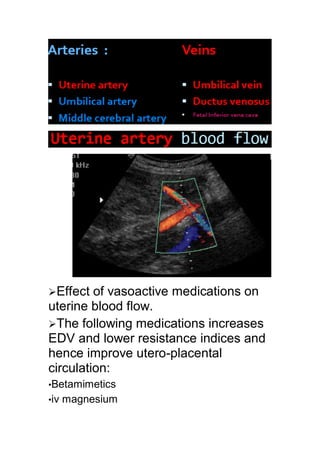
1. Effect of vasoactive medications on
uterine blood flow.
The following medications increases
EDV and lower resistance indices and
hence improve utero-placental
circulation:
•Betamimetics
•iv magnesium
3. Persistence of Bilateral diastolic
notch after 24 w of gestation is
abnormal Increased Risk of –
preeclampsia
- placental abruption
- Existing or impending IUGR -
Increased rates of prematurity,
caesarean section, and low birth
weight Start treatment Aspirin -
Vitamins C/E - Low molecular
weight heparin
Previous or present history of
preeclampsia or any other maternal
disease like: - Maternal collagen
vascular disease - Maternal
hypertension - DM with vasculopathy
Previous child with IUGR
4. Unexplained high maternal alpha
fetoprotein level
High HCG levels.
Very high level of fetal care
•In high risk pregnancy not
in low risk pregnancy
•The test is considered
positive in cases of: -
Bilateral persistent diacrotic
notch - PI more than 95TH
Percentile
•Screening has high negative
predictive value : If PI of
both ut. arteries are normal
the patient is likely will not
develop preeclampsia or IUGR
6. 50% of cases
Easily detected with CDS
No pathologic significance if the
CTG is normal
7. Up to 70% of the placental
tertiary villi should be
affected to show changes in
umbilical a. waveform
When 40% of the villi are
affected, IUGR will be
present
8. When pathology is seen in
umbilical artery examine all the
remaining vessels :
- MCA
- UMBILICAL VEIN
- DUCTUS VENOSUS
Detailed examination to exclude anomalies
and chromosome abnormalities
In normal pregnancy:
9. {progressive increase in end-
diastolic velocity
{growth& dilatation of the
umbilical circulation}: Resistance
index falls.
In IUGR and/or PET:
> 0.72 is outside the normal
limits from 26 w.
In IUGR and/or PET:reduced,
then absent (AED) or reversed
(RED) in severe cases
Absent or reversed: Fetal
distress is almost
certain:Immediate BPP or NST
or Delivery may be indicated
Should be <3.
10. Small increases in S/D= 3-5:
IUGR.
Not strictly useful: 1. low
sensitivity. 2. Gestation age
dependent
Every 14 days.in SFGA with
normal Doppler
More frequent: severe SGA
Twice weekly:
abnormal Um A D
(PI or RI > +2 SDs above mean
for gesage) and end–diastolic
velocities present
Daily: AED/RED
NORMAL FLOW
11. ABSENT DIASTOLIC FLOW
REVERSED FLOW
Comparing FHR monitoring, FBP
and umbilical artery Doppler: only
umbilical artery Doppler had value in
12. predicting poor perinataloutcomes in
SGA
In preterm SGA: limited
accuracy: should not be used
In term SGA: Normal Um A D,
an abnormal MCA D (PI < 5th
centile) has moderate predictive
value for acidosis at birth: used
to time delivery.
13. Enhanced fetal cardiac output
and
Decrease in blood viscosity:
Increased blood flow velocity
preferentially shunt blood to brain
fastermost pronounced MCA
PSV
More sensitive for predicting f
anemia than the ΔOD450
Alternative to serial
amniocenteses
Excellent noninvasive tool for
the monitoring of f anemia.
14.
15. CPR = RI mca / RI pl = more than 1
It is very sensitive predictor of fetal
growth 80%
MCA is more sensitive to
hypoxia than umbilical
artery.
MCA response to fetal
hypoxia is instant.
16. High systole in MCA →
fetal anemia
High diastole in MCA →
brain sparing effect in fetal
hypoxia
Brain sparing : High diastolic
flow, decrease PI
When O2 deficit is HIGH PI
tends to rise ,which presumably
reflects development of brain
edema.
Reversal in MCA : cerebral
edema
In growth retarded fetus :
the disappearance of the brain
sparing effect
17. The presence of reversed MCA
flow is
a critical event for the fetus and
precedes fetal death
Moderate predictive value
used in: preterm SGA with
abnormal Um A D and to time
delivery
- 50 – 60 % oxygen rich blood from
placenta to right atrium via umbilical vein
DV sampling in 1st trimester
is only indicated if NT is
abnormal
18. DV sampling in IUGR fetus
is indicated if umbilical, MCA
or both are abnormal
when Tricuspid insufficiency occurs in cases of
[anemia – congenital defects-placental
insufficiency ] dysfunction of cardiac
hemodynamics will happen …… LEAD TO steady
decline in diastolic flow ……then Diastolic reverse
flow …..associated with abnormal CTG &
increased perinatal mortality
In cases of congenital heart
disease
Volume overload on heart induces
venous pulsations
19. Single pulsations is a sign of cardiac
decompensation
Double pulsations is a sign of severe
cardiac insufficiency