2. 1126 LINGUAL FLAP RETRACTION FOR THIRD MOLAR REMOVAL
either be bone removal distal to the third molar or
where the crown of the third molar would need to be
sectioned. The technique was not used when teeth
could be elevated whole, either without any bone
removal, or with buccal bone removal only. In some
cases the decision to use lingual retraction was made
intraoperatively if crown sectioning or distal bone
removal was found to be necessary. Patients were
studied for access and ease of tooth removal and also
for the presence of any postoperative lingual or
chorda tympani nerve involvement. All patients were
questioned 1 week postoperatively regarding lingual
nerve sensory impairment or taste impairment. Pa-
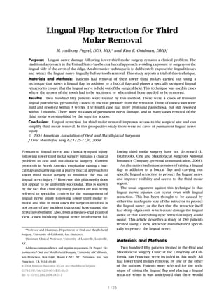
tients reporting a change were tested with von Freys FIGURE 2. The double-ended Walters lingual retractor (KLS-Martin,
order no. 92-380-00) with a Molt bone curette (above) and a Ward’s
hairs.5 Human Research Committee approval was ob- periosteal elevator (below).
tained for this study. Pogrel and Goldman. Lingual Flap Retraction for Third Molar
In the technique used, an incision was made down Removal. J Oral Maxillofac Surg 2004.
the external oblique ridge of the mandible approxi-
mately 1.5 to 2 cm in length down to the distobuccal
line angle of the lower second molar, and then a
releasing incision was made into the buccal sulcus
(Fig 1). A buccal flap was raised and an appropriate
buccal retractor placed (usually a Minnesota-type re-
tractor). The lingual flap was then raised by means of
a Molt or Ward’s periosteal elevator (ie, a spoon-
shaped elevator). Once an adequate lingual flap was
raised, a Walter’s lingual retractor was placed (KLS-
Martin LP, Jacksonville, FL). This is a double-ended
instrument (one end for the left side, one end for the
right side) shaped to fit the lingual contour of the
mandible of the third molar region, broad enough to
protect the whole area in which bone may be re-
moved, and also has a small lip on it that engages the
medial oblique ridge and prevents the retractor from
migrating too deeply (Figs 2, 3).
Results
In the 250 patients studied there were no cases of
permanent lingual nerve injury. There were 4 cases of
transient lingual nerve injury consisting of sensation
only (not taste). These 4 cases were tested with von
Frey’s hairs,5 and in 3 of the cases the difference in
von Frey’s hairs between the normal side and the
abnormal side was 3 von Frey’s hairs or less,6 denot-
ing a minor loss of sensation. In all 3 of these cases the
paresthesia resolved within 21 days. In the fourth
case, the difference in feeling with von Frey’s hairs
between the normal and abnormal side was 7 hairs,
which indicates a more substantial loss of sensation,
and in this case normal sensation did not return for 2
months. On review of this case there appeared to be
FIGURE 1. Diagram of the standard incision used to raise buccal
and lingual flaps (right side).
nothing abnormal about the case that might have
Pogrel and Goldman. Lingual Flap Retraction for Third Molar given this result. Ease of access was confirmed and is
Removal. J Oral Maxillofac Surg 2004. shown in the clinical photograph which shows that
3. POGREL AND GOLDMAN 1127
FIGURE 3. A, Lingual retractor in place viewed from the buccal side showing excellent access to the tooth and surrounding bone. B, Lingual
retractor in place viewed from the lingual aspect showing excellent coverage of the lingual tissues and the lip (arrow), which engages the internal
oblique ridge and prevents the retractor migrating inferiorly. Also note the contour of the retractor that follows the contour of the lingual plate in the
third molar region.
Pogrel and Goldman. Lingual Flap Retraction for Third Molar Removal. J Oral Maxillofac Surg 2004.
both the crown of the tooth and the bone surround- This study suggests a transient lingual paresthesia
ing the third molar are fully displayed both buccally rate of 1.6% and 0% permanent lingual nerve damage,
and lingually (Fig 4). With this retractor in place no using this particular lingual retraction technique.
additional tongue retractor is required. Figure 5 is
interesting in that it does show the results after the
Discussion
third molar was removed in 1 case and shows that
there has been a minor fracture of the lingual plate Since the 1980s most protocols for removal of third
with loss of a small piece of the superior lingual plate molars have recommended a buccally based flap with
(arrowed). It can be envisaged that if a lingual retrac- buccal retraction only and removal of teeth with a
tor had not been placed in this region, and this small drill from the buccal approach. It is recommended
flake of bone had been displaced, it could have dam- that impacted teeth are sectioned multiply to remove
aged a highly placed lingual nerve. This may show the all fragments without the necessity of removing distal
additional value of lingual retraction in that it not only or lingual bone.1-3 A major reason for this is an at-
protects the lingual nerve from damage from instru- tempt to avoid lingual nerve damage while removing
mentation, but also from minor fractures of the lin- the teeth. It has been realized since the mid 1980s
gual plate. that in around 15% to 20% of patients the lingual
FIGURE 5. View of the lingual plate after tooth removal. Note the
FIGURE 4. Lingual retractor in place clinically showing excellent small fracture of the lingual plate (arrow) that could have inadvertently
visualization and access to bone and tooth removal. Also note the lip damaged a lingual nerve that was superiorly positioned, had the
of the retractor engaging the internal oblique ridge. lingual retractor not been in place.
Pogrel and Goldman. Lingual Flap Retraction for Third Molar Pogrel and Goldman. Lingual Flap Retraction for Third Molar
Removal. J Oral Maxillofac Surg 2004. Removal. J Oral Maxillofac Surg 2004.
4. 1128 LINGUAL FLAP RETRACTION FOR THIRD MOLAR REMOVAL
nerve lies in an abnormally high position and may be clearly where one is drilling, and the lingual nerve is
level with or superior to the crest of the lingual protected.4,11,16,37,38 Robinson and Smith39 stated that
plate.7-10 To avoid damage to a lingual nerve in this permanent lingual nerve injury often results from di-
aberrant position, it has been recommended that all rect damage from a rotating bur. In a more recent
incisions are made well to the buccal side of the ridge, article, Robinson et al40 found that the affected nerves
and only a buccal flap is elevated and no attempt is were always found trapped in scar tissue and some-
made to elevate a lingual flap. Similarly, all bone times expanded to form a neuroma. Complete divi-
removal is recommended to be carried out from the sion of the nerves was evident in approximately 50%,
buccal side and, following removal of the tooth, ex- and small fragments of metal were sometimes found
treme care should be taken in removing follicular embedded within the epineurium of scar tissue, pre-
remnants on the lingual side of the socket, and if the sumably having been shaved from the lingual retrac-
socket is to be sutured postoperatively, there should tor (Howarth’s elevator, a narrow elevator used in
only be very superficial sutures on the lingual side. All much of Europe) during the initial operation. The
the above are recommended to avoid damage to the technique described in this article is not the same as
lingual nerve. Historical studies have shown variable the lingual split technique where lingual bone is de-
results for lingual nerve damage following removal of liberately removed with a chisel and the tooth deliv-
lower third molars with temporary lingual nerve dam- ered lingually.41 The technique described in this arti-
age ranging from 0% to 22%, and permanent damage cle involves lingual retraction only, but with bone
ranging from 0% to 2% of all lower third molar remov- removal from the buccal side with a drill. The weak-
als, depending on a number of factors, including the ness of this technique in the past has been that the
techniques used.11-32 Several studies, however, have retraction itself was often provided by a periosteal
tended to show that since these policies were elevator such as a Howarth’s elevator, which is poorly
adopted in the late 1980s, there has been no signifi- designed for the purpose in that it is too narrow to
cant decrease in the incidence of lingual nerve dam-
protect the whole of the lingual nerve, has sharp
age following third molar removal, which has vari-
edges that themselves can damage the nerve, and is
ously been estimated between 0.2% and 1.6% of all
incorrectly shaped to fit the lingual aspect of the
lower third molars having some degree of change of
mandible.12 Nevertheless, studies with this technique
lingual nerve sensation postoperatively.13-15,18,19,33,34
are variable, but do tend to show that approximately
Although it is known that most cases recover sponta-
11% of patients may have some form of transient
neously, and one study shows that 83% of lingual
paresthesia of the tongue, but there are virtually no
nerve injuries resolve spontaneously,35 there does not
cases of permanent nerve involvement.11,15,22,25 This
appear to be any significant decrease in the number of
cases of permanent lingual nerve involvement. Cer- would suggest that there may be some traction inju-
tainly if medicolegal experience is any guide, the ries of the nerve, but they are still mild and transient.
number of cases has certainly not decreased (L. Esta- Another issue has been that the Howarth or similar
brooks, Oral and Maxillofacial Surgeons National In- elevator can be placed or can migrate too deeply and
surance Company, personal communication, 2003). A can go down far enough to affect the mylohyoid
recent study in California (where replies were ob- nerve and cause a paresthesia (usually temporary)
tained from 86% of all practicing oral and maxillofa- over the appropriate area innervated by the mylohy-
cial surgeons in California), where the vast majority of oid nerve.42 Because of problems with the Howarth
practitioners use a buccal approach, showed that 53% elevator, the issue of lingual nerve retraction has be-
of oral and maxillofacial surgeons were aware of a come controversial.39,43-50
case of lingual nerve damage that had occurred with To circumvent these problems, a new lingual re-
them during the preceding year (temporary and per- tractor (KLS Martin LP, No. 92-380-00) and compli-
manent). Additionally, 46% of all the oral and maxil- mentary periosteal elevators were designed in the mid
lofacial surgeons studied were aware of having caused 1990s by Dr Hugh Walters, a consultant oral and
a permanent lingual nerve injury following third mo- maxillofacial surgeon in the United Kingdom (since
lar removal during their professional lifetime. In 57% deceased), which appears to eliminate these potential
of the cases, the practitioner involved had no idea of problems.45 The right- and left-sided periosteal eleva-
the actual cause of the injury.36 An alternative tech- tors are designed to be used with the lingual retractor
nique is to practice elevation and retraction of a to initially retract the lingual flap. In fact, we have
lingual flap, and the placement of a retractor down found the periosteal elevators awkward to use be-
the lingual periosteum of the mandible to improve cause the actual working tip is at right angles to the
access to the area and also to protect the lingual handle, making its moment of force inefficient. A
nerve. Bone can still be removed with the drill in the straight periosteal elevator is much more efficient and
normal way, but with better access one can see more effective and an elevator with a spoon-shaped tip,
5. POGREL AND GOLDMAN 1129
such as a straight Molt bone curette or Ward’s eleva- from drills, instruments, or inadvertent lingual plate
tor, adapts just as well to the shape of the lingual or tooth fracture or displacement. These may be re-
plate. The elevator itself is then placed, and it is broad sponsible for the cases of permanent nerve involve-
enough to protect the whole aspect of the lingual ment seen following lower third molar removal. It
nerve in the third molar region, has no sharp edges on may be appropriate to re-examine the techniques and
it, and has a notch that fits into the internal oblique principles of lingual flap elevation and retraction.
ridge of the mandible and prevents the instrument
from slipping too deeply and possibly involving the
mylohyoid nerve. It allows excellent access to the References
third molar and protects the lingual tissue from drills, 1. Alling RD, Alling C: Part 1, Mandibular third molars buccal-
instruments, or lingual plate fractures. An initial study occlusal approaches, in Alling CC, Helfrick JF, Alling RD (eds):
using this retractor showed that in the first 100 cases Impacted Teeth. Philadelphia, PA, Saunders, 1993, pp 150-202
2. Khanuja A, Powers MP: Surgical management of impacted
there was only 1 patient with a transient paresthesia teeth, in Fonseca RJ (ed): Oral and Maxillofacial Surgery. Vol 1.
that resolved within 3 weeks.51 Philadelphia, PA, Saunders, 2000, pp 259-268
Conceptually, the idea of lingual nerve identifica- 3. Peterson LJ: Principles of management of impacted teeth, in
Peterson LJ, Indresano AT, Marciani RD, et al (eds): Principles
tion and retraction mirrors more closely what hap- of Oral and Maxillofacial Surgery, vol 1. Philadelphia, PA, Lip-
pens in other surgical procedures, whereby if one pincott, 1992, pp 103-117
wishes to avoid damaging a certain structure, one first 4. Gulicher D, Gerlach KL: Sensory impairment of the lingual and
inferior alveolar nerves following removal of impacted mandib-
identifies the structure and retracts it out of the way, ular third molars. Int J Oral Maxillofac Surg 30:306, 2001
before proceeding with the surgery. As stated by 5. Weinstein S: Tactile sensitivity in the phalanges. Percept Motor
Seward,52 “In no other surgical situation is it consid- Skills 14:351, 1962
6. Pogrel MA: The results of microneurosurgery of the inferior
ered appropriate to operate close to a valuable nerve alveolar and lingual nerve. J Oral Maxillofac Surg 60:485, 2002
and not to identify it and protect it from damage.” 7. Kiesselbach JE, Chamberlain JE: Clinical and anatomic obser-
Additionally, in oral and maxillofacial surgery, such a vatory on the relationship of the lingual nerve to the mandib-
ular third molar. J Oral Maxillofac Surg 42:565, 1984
technique would actually improve access to the third 8. Pogrel MA, Renaut A, Ammar A: The relationship of the lingual
molar region, enabling bone and tooth removal to nerve to the mandibular third molar region: An anatomic study.
proceed more accurately, and would prevent damage J Oral Maxillofac Surg 53:1178, 1995
9. Miloro M, Halkias LE, Slone HW, et al: Assessment of the lingual
to the lingual nerve by a drill, chisel, or by any frag- nerve in the third molar region using magnetic resonance
ments of bone and tooth becoming displaced during imaging. J Oral Maxillofac Surg 55:134, 1997
surgery. 10. Behnia H, Kheradvar A, Shahrokhi M: An anatomic study of the
lingual nerve in the third molar region. J Oral Maxillofac Surg
However, meta-analysis has failed to show any dif- 58:649, 2000
ference in permanent lingual nerve injury rates 11. Schultze-Mosgau S, Reich RH: Assessment of inferior alveolar
whether a lingual retractor was used or not.53 It is also and lingual nerve disturbances after dentoalveolar surgery, and
of recovery of sensitivity. Int J Oral Maxillofac Surg 22:214,
recognized that the only 2 controlled trials to date 1993
between lingual retraction and protection and a 12. Rood JP: Permanent damage to inferior alveolar and lingual
purely buccal approach showed no difference in lin- nerves during the removal of impacted mandibular third mo-
lars: Comparison of two methods of bone removal. Br Dent J
gual nerve injury rates. However, these studies were 172:108, 1992
confined to 300 patients each,54,55 and all other stud- 13. Rud J: The split bone technique for removal of impacted
ies have involved a relatively small number of pa- mandibular third molars. J Oral Surg 28:416, 1970
14. Van Gool AV, Ten Bosch JJ, Boering G: Clinical consequences
tients. Because it is now realized that permanent lin- of complaints and complications after removal of the mandib-
gual nerve involvement may occur in approximately 1 ular third molar. Int J Oral Surg 6:29, 1977
in 10,000 lower third molar removals in California,36 15. Bruce RA, Frederickson GC, Small GS: Age of patients and
morbidity associated with mandibular third molar surgery.
it is acknowledged that such a study could only show J Am Dent Assoc 101:240, 1980
a statistically significant difference if many thousands 16. Rood JP: Lingual split technique. Damage to inferior alveolar
of patients were included in a study. Indeed, it would and lingual nerves during removal of impacted mandibular
third molars. Br Dent J 154:402, 1983
involve 30,000 patients to arrive at a statistically sig- 17. Hochwald DA, Davis WH, Martinoff J: Modified distolingual
nificant absolute risk reduction. splitting technique for removal of impacted mandibular third
Lingual flap elevation and lingual flap retraction molars: incidence of postoperative sequelae. Oral Surg Oral
Med Oral Pathol 56:9, 1983
with a broad retractor may be suitable techniques to 18. Rud J: Re-evaluation of the lingual split bone techniques for
re-evaluate with regard to lower third molar removal. removal of impacted mandibular third molars. J Oral Maxillo-
Although the actual raising of the lingual flap and facial Surg 42:114, 1984
19. Goldberg MH, Nemarich AN, Marco WP: Complications after
placement of a lingual retractor can theoretically mandibular third molar surgery: A statistical analysis of 500
cause a traction injury to the lingual nerve, it appears consecutive procedures in private practice. J Am Dent Assoc
from the present study that these are transient and of 111:277, 1995
20. Sisk AL, Hammer WB, Shelton DW, et al: Complications follow-
no long-term consequence. Conversely, this tech- ing removal of impacted third molars: The role of the experi-
nique may protect the nerve from inadvertent damage ence of the surgeon. J Oral Maxillofac Surg 44:855, 1986
6. 1130 LINGUAL FLAP RETRACTION FOR THIRD MOLAR REMOVAL
21. Wofford DT, Miller RI: Prospective study of dysesthesia follow- 38. Seward GR, Harris M, McGowan DA: Surgical removal of roots
ing odontectomy of impacted mandibular third molars. J Oral of impacted teeth, in Seward GR, Harris M, McGowan DA
Maxillofac Surg 45:15, 1987 (eds): Killey & Kay’s Outline of Oral Surgery (ed 2). Bristol,
22. Mason DA: Lingual nerve damage following lower third molar Wright, 1987, pp 61-62
surgery. Int J Oral Maxillofac Surg 17:290, 1988 39. Robinson PP, Smith KG: Lingual nerve damage during lower
23. Middlehurst RJ, Barker GR, Rood JP: Postoperative morbidity third molar removal: A comparison of two surgical methods. Br
with mandibular third molar surgery: a comparison of two Dent J 180:456, 1996
techniques. J Oral Maxillofac Surg 46:474, 1988 40. Robinson PP, Loescher AR, Smith KG: A prospective, quantita-
24. Blackburn CW, Bramley PA: Lingual nerve damage associated tive study on the clinical outcome of lingual nerve repair. Br J
with the removal of lower third molars. Br Dent J 167:103, Oral Maxillofac Surg 38:255, 2000
1989 41. Ward TG: The split bone technique for the removal of lower
25. Von Arx DP, Simpson MT: The effect of dexamethasone on third molars. Br Dent J 101:297, 1956
neurapraxia following third molar surgery. Br J Oral Maxillofac 42. Tier GA, Rees RT, Rood JP: The sensory nerve supply to the
Surg 27:477, 1989 tongue: A clinical reappraisal. Br Dent J 157:354, 1984
26. Carmichael FA, McGowan DA: Incidence of nerve damage 43. Greenwood M, Langton SG, Rood JP: A comparison of broad
following third molar removal: A West of Scotland Oral Surgery and narrow retractors for lingual nerve protection during
Research Group study. Br J Oral Maxillofac Surg 30:78, 1992 lower third molar surgery. Br J Oral Maxillofac Surg 32:114,
27. Absi EG, Shepherd JP: A comparison of morbidity following the 1994
removal of lower third molars by the lingual split and surgical 44. To EW, Chan FF: Lingual nerve retractor. Br J Oral Maxillofac
bur methods. Int J Oral Maxillofac Surg 22:149, 1993 Surg 32:125, 1994
28. Chiapasco M, De Cicco L, Marrone G: Side effects and compli- 45. Walters H: Reducing lingual nerve damage in third molar sur-
cations associated with third molar surgery. Oral Surg Oral Med gery: A clinical audit of 1350 cases. Br Dent J 178:140, 1995
Oral Pathol 76:412, 1993 46. Walters H: Lingual nerve damage during lower third molar
29. Malden NJ, Maidment YG: Lingual nerve injury subsequent to removal: A comparison of two surgical methods. Br Dent J
wisdom teeth removal–A 5-year retrospective audit from a high 181:163, 1996
street dental practice. Br Dent J 193:203, 2002 47. Mason DA: “To retract or not to retract.” Br Dent J 168:94,
30. Renton T, McGurk M: Evaluation of factors predictive of lingual 1990
nerve injury in third molar surgery. Br J Oral Maxillofac Surg 48. Robinson P: To retract or not to retract. Br Dent J 168:5, 1990
39:423, 2001 49. Shotts N: It ain’t what you do... Br Dent J 181:121, 1996
31. Valmaseda-Castellon E, Berini-Aytes L, Gay-Escoda C: Lingual 50. Christian C: Retractors have their detractors. The Dentist Nov:
nerve damage after third lower molar surgical extraction. Oral 42, 1992
Surg Oral Med Oral Pathol Oral Radiol Endod 90:567, 2000 51. Goldman KE, Pogrel MA: Lingual flap retraction in third molar
32. Bataineh AB: Sensory nerve impairment following mandibular surgery. J Oral Maxillofac Surg 56:82, 1998 (suppl 4)
third molar surgery. J Oral Maxillofac Surg 59:1012, 2001 52. Seward GR: Protecting the lingual nerve from damage. Br J Oral
33. Schwartz LJ: Lingual anesthesia following mandibular odontec- Maxillofac Surg 39:76, 2001
tomy. J Oral Surg 31:918, 1973 53. Pichler JW, Beirne OR: Lingual flap retraction and prevention
34. Fielding AF, Rachieze DP, Frazier G: Lingual nerve paresthesia of lingual nerve damage associated with third molar surgery: A
following third molar surgery: A retrospective clinical study. systematic review of the literature. Oral Surg Oral Med Oral
Oral Surg Oral Med Oral Pathol 84:345, 1997 Pathol Oral Radiol Endod 91:395, 2001
35. Alling CC: Dysesthesia of the lingual and inferior alveolar 54. Gargallo-Albiol J, Buenechea-Imaz R, Gay-Escoda C: Lingual
nerves following third molar surgery. J Oral Maxillofac Surg nerve protection during surgical removal of lower third molars.
44:454, 1986 a prospective randomised study. Int J Oral Maxillofac Surg
36. Robert RC, Pogrel MA: Nerve damage and lower third molars. 29:268, 2000
J Oral Maxillofac Surg 61:54, 2003 (suppl 1) 55. Chossegros C, Guyot L, Cheynet F, et al: Is lingual nerve
37. Moss CE, Wake MJ: Lingual access for third molar surgery: A protection necessary for lower third molar germectomy? A
20-year retrospective audit. Br J Oral Maxillofac Surg 37:255, prospective study of 300 procedures. Int J Oral Maxillofac Surg
1999 31:620, 2002