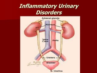
2. URINARY TRACT INFECTION
The urinary tract infection may be broadly classified
as upper and lower urinary tract infections.
The patient may have both an upper and a lower
urinary tract infection. The frequency of urinary
tract infections varies with age and sex and may be
acute or chronic.
3. Risk Factors For Urinary Tract
Infection
Inability or failure to empty the bladder
completely
Obstructed urinary flow ,from congenital
anomalies, from urethral strictures ,
contracture of the bladder neck, bladder
tumors , calculi in the ureters or kidneys
compression of the ureters and neurologic
abnormalities.
4. Contributing conditions as:
diabetes mellitus
Pregnancy
Neurologic disorders
Gout
Urinary stasis
Inflammations or abrasions in the urethral
mucosa
Instrumentations of the urinary tract
Immunosuppressant's
5. PATHOPHYSIOLOGY OF URINARY
TRACT INFECTION
Urethrovesical Reflux” back flow of urine “ with:
Coughing
Sneezing
Straining
Routes of infection:
Ascending infection e.g.
Because the female urethra is short, also several
Studies show that sexual intercourse is the major
precipitating factor of UTI in women.
6. Clinical Manifestations of UTI
1. Urgency
2. Dysuria
3. Slight to gross hematuria
4. Bacteriuria and positive urine cultures as
the basis for diagnosing lower urinary
tract infections.
7. Diagnostic findings of UTI
Urine cultures
Testing methods
– Leukocyte esterase test is positive “WBCs in urine”
– STD”sexual transmitted disease” may be performed
Computerized Tomography ”C.T.” to detect pyelonephritis,
abscess
Ultrasonography to detect obstruction, abscess, tumors,
cysts.
Intravenous pyelography to detect strictures or stones.
8. Specific Nursing Care for UTI
1. The medication “anti bacterial” must be
given on a time on a regular schedule.
2. The nurse must follow complete aseptic
technique if instrumentation is indicated.
3. Sitz bath may provide to relieve pain or
itching.
9. Pyelonephritis
Definition:
It is an bacterial infections that involves both the
parenchyma and the pelvis of the kidney, it may
affect one or both kidneys.
It is frequently secondary to ureterovesical reflux
It may be acute or chronic when it is chronic the
kidneys are scarred, contracted and non-functioning
10. Clinical Findings of Acute
peylonephritis
A.Symptoms :
1. Chills, moderate to high fever.
2. Constant loin pain unilateral or bilateral.
3. Symptoms of cystitis :
- frequency
- nocturia
- urgency
- dysuria
4. Nausea, vomiting and diarrhea are common.
5. Young children complain of abdominal discomfort.
B.Signs :
1. The patient appears quite ill.
2. Intermittent chills with fever ranging 38.5 : 40C.
3. Tachycardia (90 beat/m : 140 beat/m).
4. Abdominal distention.
11. Specific Nursing Care for
peylonephritis
1.Health promotion and maintenance measures should be
applied.
2.Early treatment for cystitis to prevent ascending infections.
3.Encourage the patient to drink at least 2000 ml of fluid
everyday.
4.Antibiotic therapy according to results of urine cultures.
5.Serial urine cultures and other evaluation studies must be
continued.
13. Images of chronic pyelonephritis
Stag horn stone x-ray film for renal
calculi causing
chronic pyelonephritis
14. Pathology of Chronic Pyelonephritis
The kidney shows atrophy of variable degree
depending upon the severity of the
involvement. In minimal involvement, the
kidney shows scarring in the renal surfaces
while in extensive involvement, there is a
fibrosis specially in the pelvic mucosa.
15. Clinical manifestations of chronic
peylonephritis
It does not have symptoms of infection
Fatigue
headache
Poor appetite
Polyuria
Excessive thirst
Weight loss
17. Specific Nursing Care for chronic
Pyelonephritis
1.The nurse must instruct the patient to continue
antibiotic and antimicrobial therapy even after
symptoms resolve.
2.Encourage the patient to drink 3 liters/day of fluids
unless otherwise instructed.
3.Monitor urinary output and report if there is oliguria
or intake more than output.
18. 4.Weighing daily and instruct the patient to
report immediately about weight gain.
5. Teach the patient measures to prevent
infection and early seek for medical advice
if there are signs of urinary infection.
6.Continue with medical follow-up and get
follow-up urine cultures as instructed.
19. Interstitial Cystitis
It is a Chronic inflammatory condition of
bladder wall, frequently remained undiagnosed
It can be occur at any age , in both genders
Almost 90% of the affected patients are
women why?
20. Pathology o f chronic cystitis
In chronic cystitis, the bladder mucosa
becomes move edematous, erythematous
and friable. It may lead to ulceration of the
bladder mucosa then fibrosis and becomes
inelastic and thick.
21. Clinical manifestations of chronic
cystitis
Severe ,irritable voiding at day and night
Frequency
Nocturia
Urgency
Pain “ suprapubic pressure
Irritable bowl syndrome
Chronic tension type headache
22. Treatment of chronic cystitis
Anti-microbial therapy based on culture
and sensitivity testing.
Appropriate correction of contributing
factors when possible.
23. Primary Glomerular Diseases
A variety of diseases can affect the glomerular
capillaries, including acute and chronic
glomerulonephritis
Acute Glomerulonephritis
It is an inflammation of the glomerular
capillaries
It is primarily occurs with children but it can
occurs at any age.
24. Clinical Manifestations
Clear hematuria” either micro/macroscopic”
RBCs and protein plugs or casts “indicate glomerular
injury”
Proteinuria due to increased permeability of the
glomerular membrane
BUN, creatinine
urine output
Headache, malaise, flank pain
Some degree of edema
Hypertension in 75% of the cases
in old age ; circulatory overload
25. Assessment and Diagnostic
Findings
A: Kidney: large, swollen, and congested
ASOT: Anti Streptolysin O Titre due to
streptococcal infection
D: Kidney: biopsy
If the patient improves ,urine increases and
urinary protein diminish
If not, dialysis will be needed for survival
27. Medical Management
Treating symptoms
Treating complications
Treat streptococcal infection by penicillin
Corticosteroids and immunosuppressant for
rapidly progressive acute glomerulonephritis
protein and salt in diet in case of edema
and hypertension
Diuretics to control hypertension
28. Nursing Management
carbohydrate in diet to provide energy
Fluid balance chart carefully
Daily weighing to patient
Fluid intake according loss considering
insensible loss
Teach patient how to care him/her self at
home
Care of edema
Care of Skin
29. Chronic Glomerulonephritis
Repeated attacks of acute Glomerulonephritis
due to:
Hypertensive nephrosclerosis
Hyperlipidemia
Glomerular sclerosis
Clinically:
the kidneys shrinks
reduce its size
It has rough and irregular surface
Thickened renal artery
Glomerular damage
ESRD
30. Clinical Manifestations
Most of cases has no symptoms until hypertension
or BUN/ creatinine elevation can be detected
The disease may be discovered during routine eye
examination
The first indication might be :
Severe Nose Bleeds
Stroke
Seizure
General symptoms as:
Loss of weight
Increase irritability
Headache
Dizziness
Nocturia
GIT disturbances
Swollen feet specially at night
31. The patient appears poorly nourished
Blood pressure may be normal or severely
elevated
Mucous membranes are pale because of
anemia
Peripheral neuropathy occurs late in the
disease
32. Assessment and Diagnostic
Findings
A:
Chest x-ray shows
Cardiomegaly *Pulmonary edema
Distended neck veins
Crackles can be heared in the lungs
D:
Urine analysis …
specific gravity is 1.010 * Proteinuria
Urinary casts due to glomerular damage
33. Impaired nerve conduction due to
uremia
Blood chemistry…
Hyperkalemia
Anemia”lack of erythropoiesis”
Hypoalbuminemia due to protein
loss
Increased phosphorus and
decreased calcium in blood
34. Medical Management
Treat hypertension
Restrict sodium and water
Monitor weight daily
Diuretics to overcome fluid overload
Increase protein in diet
Initiation of dialysis as early as possible “benefits”:
Optimal physical condition
Minimize risk of complications
Prevent fluid and electrolyte imbalances
35. Nursing Management
Observe signs of fluid and electrolyte
imbalances
Report changes in fluid and electrolyte status
, cardiac and neurologic status.
Emotional support to alleviate anxiety
Teach patient self care
36. Nephrotic Syndrome
It is a primary glomerular disease characterized by
the following:
Marked increase in protein in the urine
Decrease in albumin in the blood
Edema
High serum cholesterol
37. Pathophysiology of nephrotic
syndrome
Damage glomerular capillary
membrane
Loss of plasma protein ”albumin”
Stimulate synthesis of
lipoproteins
hyperlipedemia
hypoalbuminemia
Activation of renin-angiotensin
system
Sodium retention
Edema
41. Management of Nephrotic
Syndrome
Diuretics for edema
Immunosupprsant medications
Low salt diet
Protein in diet around 0.8 gm g/kg/day
Patients with nephrotic syndrome need instructions towards:
Dietary regimen
Referral system
medications