2. Optic Atrophy
Optic atrophy refers to the ophthalmoscopic
appearance of the optic disc that may result from
damage to any portion of the ganglion cells from their
cell bodies to their synapse at the lateral geniculate
body.
It represents permanent loss of retinal ganglion cell
axon in conjunction with retinal ganglion cell death.
3. Optic atrophy
• It should be considered a pathologic end point
that is clinically discernible but does not imply
cause.
4. Optic atrophy
• Optic atrophy sets in 4-6 weeks after axonal damage.
• Severe damage with chalky white optic disc is easily
identified.
• Milder forms are identified by:
- comparison of colour of the 2 discs.
- evaluation of surface vasculature of the disc.
- evaluation of the peripapillary nerve fibre layer.
6. Primary optic atrophy
• Primary optic atrophy results from the loss of
optic nerve fibres with otherwise minimum
disturbance of optic nerve head
microanatomy.
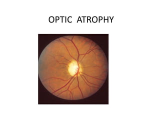
7. Primary optic atrophy
• Ophthalmoscopic features:
-Pale to white disc.
-Clearly defined disc
margins.
-Decrease in capillaries on
the disc.
-Arteriolar narrowing.
8. Primary Optic atrophy
• Results from any injury to the retinal ganglion cell
or its axon , anywhere in its course.
• Common causes:
-AION( Ant ischaemic optic neuropathy)
-Compressive lesions of the optic nerve
-Other ischaemic ,inflammatory and lesions
- Less common- PION
Trauma
Granulomatous inflammation of the
optic nerve
9. Secondary optic atrophy
• It represents disorganised appearance of the
optic disc usually as a result of severe disc
edema/ papilledema, severe inflammation at
the optic nerve head ( papillitis/neuroretinitis)
or, long standing severe orbital inflammation.
11. Consecutive optic atrophy
Optic atrophy due to destruction of ganglion
cells resulting from the extensive diseases of
the retina is called consecutive optic atrophy.
Examples:
Pigmentary retinal dystrophy
Central retinal artery occlusion
15. Optic atrophies: comparison
Fundus Primary Secondary Consecutive Glaucomatous
Disc
colour
Pale/
White
gray Waxy Pale/
White
Margin Sharp Blurred Sharp/blurred Sharp
Cup unremark
able
full unremarkable Deep,
cavernous
BV on
the disc
decreased decreased decreased decreased
Retina Normal May show
gliosis
Retinal
disease
Normal
20. Toxic/nutritional optic neuropathies
• These include a number of conditions in which
optic nerve fibres are damaged due to
exposure to exogenous poisons or nutritional
deficiencies.
• Many of them have similar clinical picture
because of their common pathways.
22. Symptoms
• In the early stages symptoms and signs are minimal
• Subtle depression of VF within 10⁰ fixation region
,noticed on Amsler's grid.
• With progression visual acuity and colour vision
• suffers.
• Central field defect
• Fundus: Normal initially/optic atrophy finally.
24. Diagnosis
• History of exposure to toxin , drug intake,
substance abuse.
• Blood test for serum B12 ,transketolase for
thiamine deficiency.
• Neuroimaging
26. Course and prognosis
• Untreated deficiency optic neuropathy can
lead to profound visual loss.
• Correction of deficiency reverses the deficit
within few months.