

Recommended
More Related Content
What's hot
What's hot (20)
Viewers also liked
Viewers also liked (20)
Similar to 23204925
Similar to 23204925 (20)
More from radgirl
Recently uploaded
Recently uploaded (20)
23204925
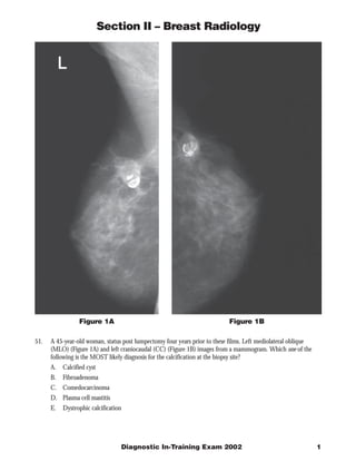
- 1. Section II – Breast Radiology Figure 1A Figure 1B 51. A 45-year-old woman, status post lumpectomy four years prior to these films. Left mediolateral oblique (MLO) (Figure 1A) and left craniocaudal (CC) (Figure 1B) images from a mammogram. Which one of the following is the MOST likely diagnosis for the calcification at the biopsy site? A. Calcified cyst B. Fibroadenoma C. Comedocarcinoma D. Plasma cell mastitis E. Dystrophic calcification Diagnostic In-Training Exam 2002 1
- 2. Section II – Breast Radiology Question #51 Findings: The mediolateral oblique (MLO)/craniocaudal (CC) view demonstrates calcifications that are curvilinear in shape. These conform to the biopsy site. These calcifications are most consistent with dystrophic deposition at the biopsy site. Rationales: A) Incorrect. Calcifications can be deposited in the wall of benign cysts. These calcifications are usually very thin and under 1mm in thickness when seen in profile. They differ from the calcifications seen in fat necrosis in that the latter calcifications have thicker walls and a rim-shape. The curvilinear calcifications in these images are not consistent with the calcifications of a benign cyst. B) Incorrect. Fibroadenoma can show dystrophic calcifications. These are sometimes referred to as “popcorn” calcifications in that they are large, coarse and dense. They are peripherally located within solid masses. Rim- like calcifications are not typical of Fibroadenoma C) Incorrect. Calcifications of comedocarcinoma have a linear, branching, “dot-dash” appearance. Recurrent carcinoma is important to consider in a patient that who is four years post-lumpectomy. However, the shape of these calcifications in the test patient are not consistent with comedocarcinoma. D) Incorrect. Plasma cell mastitis is associated with secretory calcifications. The calcifications are multiple and diffusely distributed. They may appear as thick, rod-shaped calcifications with smooth margins or as rod-shaped calcifications with lucent centers. Although the appearance may be calcifications may be similar to those seen in this case, the presence of only one solitary area of calcification makes the diagnosis unlikely. E) Correct. Dystrophic calcifications form after traumatic insult to the breast and are usually due to fat necrosis. They are commonly dense in appearance and curvilinear in shape. When seen on fosse, the calcification may appear irregular and less dense than when seen in profile. In this case, the mammographic findings along with the history of a lumpectomy and radiation are consistent with dystrophic calcification. Citations: Kopans, DB. Breast Imaging, 2nd Ed. Philadelphia: Lippincott-Raven, 1997. (Kopans 326-328) (Kopans, 527-532) (Kopans, p522- 524) (Kopans, p522-524) 2 American College of Radiology
- 3. Section II – Breast Radiology Figure 2 52. A 65-year-old asymptomatic woman. You are shown a mammogram, mediolateral oblique (MLO) view (Figure 2). The film was taken at 28kVp, 22.5 mAs, 3 cm thickness. Altering the parameters to 25 kVp, 90mAs, and 3 cm thickness would lead to which one of the following changes? A. Increased contrast B. Shorter exposure time C. Increased spatial resolution D. Decreased glandular dose E. Decreased density Diagnostic In-Training Exam 2002 3
- 4. Section II – Breast Radiology Question #52 Findings: This film has poor contrast because of the exposure factors, e.g., a relatively high kVp and low mAs for the thickness of the breast. Rationales: A) Correct. Because of the decrease in the kVp, there will be increased contrast. The radiographic contrast depends on the subject as well as the film contrast. Contrast is affected by the thickness, density, atomic differences of the subject, the radiation energy (kVp), contrast material, and scatter radiation. In this case, the film contrast is not affected since the same type of film would be used for the first and the second exposures. B) Incorrect. The mAs is a combination of the mA tube current and the duration of the exposure. Increasing the mAs increases the exposure time. Note that the decrease in the kVp from 28 to 25 indirectly affects exposure time in cases where phototiming is used. With the decrease in kVp, there is a compensatory increase in mAs. C) Incorrect. Because of the longer exposure time, the likelihood of motion artifact increases. An exposure of 90 mAs should be acceptable to most patients. In general, exposure times of between 0.5 and 2 seconds are desirable. Shorter exposure time may produce noise artifacts and grid lines. Longer exposure times increase the risk of motion and overexposure. D) Incorrect. Increasing the mAs increases the radiation dose. E) Incorrect. Density refers to film blackness. The degree of film blackness is directly related to the intensity of the radiation reaching the film or intensifying screen. This radiation varies with the mAs. In this particular case, increasing mAs would increase density. Citations: Christensen’s Physics of Diagnostic Radiology. 4th ed. Lea & Faberger Publisher. 1990 (Christenson, p153) (p149) 4 American College of Radiology
- 5. Section II – Breast Radiology Figure 3A Figure 3B 53. A 58-year-old woman with a new density (arrows), which has spiculated margins, in the left breast, was identified on a screening mammogram. The density is seen only on the mediolateral oblique (MLO) view. (Figure 3A) It was not identified on the craniocaudal (CC) view. (Figure 3B) Which one of the following is the next BEST step in the imaging evaluation? A. Ultrasonography B. A mediolateral view C. Sterotactic wire localization D. Spot magnification MLO view E. Magnetic resonance imaging Diagnostic In-Training Exam 2002 5
- 6. Section II – Breast Radiology Question #53 Findings: This soft tissue density is seen in one view, and by BI-RADS™ definition is considered to be a “density”. However, because there are spiculations, a mass, rather than a density or summation shadow, needs to be strongly considered. Therefore, the next step in management should be a mediolateral view to confirm the presence of a mass and determine its location three-dimensionally. Rationales: A) Incorrect. A sonogram is not indicated until the additional imaging is completed. Moreover, a negative sonogram would not rule out a mass in the presence of a spiculated mass on a mammogram. Once a mass has been confirmed, the role of the sonogram is to provide possible access for needle sampling. B) Correct. A mediolateral view would be the next part of the imaging evaluation to confirm the presence of a mass and its exact location. C) Incorrect. Stereotactic localization should be considered if other mammographic images and the sonogram do not confirm mass. D) Incorrect. Spot magnification MLO can be helpful in confirming spiculations as well as ruling out other associated findings such as calcifications. Therefore, this view may likely be obtained in the diagnostic work-up. However, this would not be the next imaging step since three-dimensional localization should be the first goal in the diagnostic evaluation. E) Incorrect. MRI has been shown to be useful in the work-up of equivocal lesions, such as those seen on only one projection. However, MRI would not be the next step in the diagnostic work-up. Citations: Gilda Cardenosa, 2nd Ed. Breast Imaging Companion, Lippincott, Williams & Wilkins, 2001. Pp 124-158 6 American College of Radiology
- 7. Section II – Breast Radiology Figure 4 54. A 62-year-old woman who presented for a mammogram. You are shown a right mediolateral oblique (MLO) view (Figure 4). Similar findings are present in the left breast. Which one of the following is the MOST likely explanation for the abnormalities seen on the mammogram? A. Inflammatory breast carcinoma B. Radiation changes C. Infectious mastitis D. Superior vena caval obstruction E. Congestive heart failure Diagnostic In-Training Exam 2002 7
- 8. Section II – Breast Radiology Question #54 Findings: The MLO views show skin and trabecular thickening. Rationales: A) Incorrect. Inflammatory breast carcinoma typically produces unilaterally increased breast density and trabecular thickening. These findings are secondary to lymphatic obstruction by tumor. There may or may not be an associated mass or area of architectural distortion. Clinically there may be erythema and warmth. The clinical presentation may be difficult to distinguish from an infectious mastitis. A diagnosis can be made by a punch biopsy of the skin showing tumor emboli within the dermal lymphatics. B) Incorrect. Radiation can produce findings similar to those seen in the test patient. The skin thickening after radiation is due to tissue edema. Radiation changes are usually unilateral since breast cancer is more often unilateral than bilateral. Moreover, one would expect to also see a scar from a prior biopsy or lumpectomy. This particular finding occurs in approximately three-fourths of cases. Therefore, the bilaterally of the findings in this case and absence of a scar make radiation unlikely. C) Incorrect. Mastitis can produce findings similar to those seen in this case. The skin thickening is related to the dilated subdermal lymphatics. However, the condition is most commonly unilateral. Clinically, there is often skin erythema and heat. This may be localized or diffuse. Mastitis is common in lactating women. D) Incorrect. Renal failure can produce bilateral trabecular thickening and skin thickening due to a fluid overload state. However, the frequency of renal failure is likely than congestive heart failure. E) Correct. The findings on the mammograms are secondary to a fluid overload state and edema. Since congestive heart failure is a more frequent than renal failure as a cause of a fluid overload state, this is the best of the two possibilities. Citations: Kopans, DB. Breast Imaging, 2nd Ed. Philadelphia: Lippincott-Raven, 1997, Gilda Cardenosa, 2nd Ed. Breast Imaging Companion, Lippincott, Williams & Wilkins, 2001. 8 American College of Radiology
- 9. Section II – Breast Radiology Figure 5 55. A 23-year-old woman presents with a left breast lump. You are shown a sonogram (Figure 5). Which one of the following is the MOST likely diagnosis? A. Ductal carcinoma in situ B. Complex cyst C. Simple cyst D. Fibroadenoma E. Phylloides tumor Diagnostic In-Training Exam 2002 9
- 10. Section II – Breast Radiology Question #55 Findings: The sonogram shows a hypoechoic mass with a thin pseudocapsule. Rationales: A) Incorrect. Although infiltrating ductal carcinoma is the most common carcinoma encountered, it is infrequent in young women. Moreover, it often presents as a spiculated or irregular mass, in contrast to the smoothly marginated mass seen in the test patient. B) Incorrect. A complex cyst may appear hypoechoic, but it also would be expected to contain debris or septations, related to proteinaceous material, cellular debris, hemorrhage, infection, or cholesterol crystals. C) Incorrect. The characteristics of a simple cyst are an anechoic mass with sharp anterior and posterior margins and through sound transmission. Although this mass is well-defined mass, it is hypoechoic and lacks sound transmission. D) Correct. This is a classic appearance of a fibroadenoma. Moreover, it is the most common breast mass encountered in adolescents and young women. The sharp margins with the thin pseudocapsule, homogeneous echo texture, as well as the patient’s young age, make fibroadenoma the most likely diagnosis. E) Incorrect. The average age of a patient with a Phylloides tumor is 40 to 50 years. Moreover, it is significantly less common than fibroadenoma. The imaging presentation of phylloides tumor may be similar to that of a fibroadenoma. However, young age of the patient in this case makes this diagnosis less likely. Citations: Bassett, LW et al. Benign breast lesions. Diagnosis of Diseases of the Breast. Philadelphia: W.B. Saunders Company, 1997, 385-394, 429-431. 10 American College of Radiology