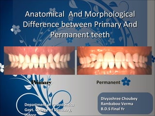
The presentation features the basic difference between primary and permanent dentition. The differences are tabulated under the headings of crown, roor and pulp.
A Complete presentation explaining the complete morphology of Maxillary first molar, for the benefit of people like me who tried and failed to find everything in one package
The document summarizes the development of teeth from the dental lamina. It discusses how the primary epithelial band forms and divides into the dental lamina and vestibular lamina. Tooth buds then develop from the dental lamina, forming the enamel organ, dental papilla, and dental follicle. Teeth progress through developmental stages including the bud stage, cap stage, bell stage, and root formation. The dental lamina gives rise to both primary and permanent teeth before degenerating.
This document provides an overview of cementum, including:
- Its physical characteristics, composition, classification, and formation process (cementogenesis).
- The cells involved in cementum formation and maintenance, including cementoblasts and cementocytes.
- Its locations and junctions with other tissues like enamel and dentin.
- The functions of cementum in anchoring teeth, adaptation, and repair.
- Some developmental anomalies and abnormalities that can affect cementum.
Radiographic Assessment of the Prevalence of Pulp Stones in Malaysians
Kannan et al.
JOE — Volume 41, Number 3, March 2015
Pulp stones are discrete calcified bodies found in the dental pulp.
They have calcium phosphorous ratios similar to dentin and can be seen in healthy, diseased, or even unerupted teeth
Radiographically, pulp stones appear as radiopaque structures in the pulp space that frequently act as an impediment during endodontic treatment
This document summarizes the process of tooth eruption. It discusses the pre-eruptive, eruptive, and post-eruptive phases of tooth movement. During the pre-eruptive phase, tooth germs move within the jaw before eruption. The eruptive phase involves tooth movement from within the bone to the oral cavity. Post-eruptive movements maintain tooth position as the jaws grow. Theories on the mechanisms controlling eruption and resorption are also presented, along with cellular and molecular factors such as the dental follicle that regulate eruption.
It is a presentation in detail about the strongest structure of the oral cavity "ENAMEL". It is a simple topic but people find it difficult to learn about it. I hope my presentation is a simple method to learn about it. I would like to thank my professors for assign me this project and i learn't a lot from it and still learning my basics daily.
A detailed look at the differences between the human primary and permanent dentition. Hope you find this informative. for further queries, please contact at dr.mathewthomasm@gmail.com.
This document provides an overview of anatomical landmarks in the maxilla that are important for complete denture construction. It discusses intraoral landmarks like the labial and buccal frenums, as well as maxillary arch structures like the residual alveolar ridge, hard palate, palatal rugae, incisive papilla, hamular notch, maxillary tuberosity, and fovea palatinae that serve as stress bearing or relief areas. The document emphasizes understanding the histology and functions of these structures to ensure dentures are designed and placed to avoid placing undue pressure on supporting tissues.
This document defines key terms related to cavity preparation and outlines the objectives and basic principles and steps of cavity preparation. It discusses definitions of cavities and tooth preparation. The objectives of cavity preparation are to remove caries and create a foundation for the restoration. The basic principles outlined by GV Black include biologic, mechanical, and esthetic principles. The main steps are 1) outline form, 2) resistance and retention form, 3) convenience form, 4) removal of remaining caries, 5) finishing cavity walls, and 6) toilet of the cavity. Resistance and retention forms are designed to resist forces and retain the restoration.
The document discusses the structure and development of dentin. It describes dentin as the layer beneath enamel that provides shape and structure to teeth. Dentin forms in stages that mirror tooth development from the lamina bud stage through late bell stage. Key features of dentin include dentinal tubules that contain odontoblastic processes and layers like peritubular dentin, intertubular dentin, and predentin near the pulp. Dentin is laid down in primary, secondary, and tertiary forms throughout life.
Impression compounds are thermoplastic materials used for dental impressions. They are composed mainly of rosin, copal resin, carnauba wax, stearic acid, and talc. There are two main types - lower fusing impression compound and higher fusing tray compound. Impression compound is a viscous material that is softened in hot water before making impressions, allowing it to flow and capture detail but also maintain shape. While able to displace soft tissue, its high viscosity limits fine detail capture. Impression compound requires careful heating and cooling to avoid distortion, and constructs must be poured promptly due to its marginal dimensional stability.
This document discusses various theories of tooth eruption and the phases of tooth eruption. It summarizes six main theories of tooth eruption: root elongation theory, bone remodeling theory, periodontal ligament contraction theory, hydrostatic pressure theory, pulp constriction theory, and dental follicle theory. It states that the periodontal ligament contraction theory, whereby fibroblasts in the periodontal ligament contract to apply an axial force, is the most widely accepted. It also outlines the three phases of tooth eruption: pre-eruptive, eruptive, and post-eruptive phases.
This document provides information on the steps of cavity preparation, including defining cavity preparation, the objectives and principles. It describes Black's classification system for cavities in 6 classes. The steps of cavity preparation outlined include obtaining the outline form and initial depth, primary resistance and retention forms, and convenience form. It also discusses final cavity preparation steps like removing remaining decay, providing pulp protection, and finishing enamel walls and margins.
The periodontal ligament is a connective tissue that connects the tooth to the alveolar bone. It contains collagen fibers, fibroblasts, cementoblasts, osteoblasts and other cells. The principal collagen fibers of the periodontal ligament originate on the cementum and insert into the alveolar bone in different orientations to provide structural support to the tooth and resist various forces. The periodontal ligament is essential for functions such as tooth eruption and maintains the space between the tooth and bone.
A number of theories have been put forward for impressions. each having its own advantage and disadvantage.
Different spacers guide and aid in in making the desired impression with adequate pressure in the desired region of the arch in maxilla and mandible. different materials are used for spacers depending on the need.
The pulp is a soft connective tissue located within the tooth. It has several unique features, including being surrounded by rigid dentin walls and susceptible to changes in pressure. The pulp contains odontoblasts, fibroblasts, undifferentiated cells, and defense cells. It is highly vascularized and innervated. During development, dental papilla forms the pulp through proliferation and differentiation of cells. The pulp cavity is divided into coronal and radicular regions. Nerves and blood vessels enter through the apical foramen, supplying the pulp.
The periodontal ligament is the soft connective tissue between the cementum and alveolar bone. It has an hourglass shape that is thinnest in the middle and widens coronally and apically. During tooth eruption, fibroblasts produce collagen fibers that develop into principal fiber groups including the transseptal, alveolar crest, horizontal, oblique, apical, and interradicular fibers. The periodontal ligament contains collagen fibers, cellular elements like fibroblasts, and ground substances such as glycosaminoglycans. It functions to support the tooth, sense pressure, and maintain attachment through Sharpey's fibers embedded in the cementum and bone.
Hypercementosis is characterized by the excessive deposition of cementum on tooth roots. It can be localized, affecting a single tooth due to conditions like periapical osteitis, or generalized, affecting many teeth as an age-related factor or due to diseases like Paget's disease of bone. Radiographically, it appears as thickening and blunting of roots with a bulbous or irregular apex. Diagnosis is clinical based on the bulbous root appearance. Treatment focuses on managing any underlying primary causes.
This document summarizes tooth eruption, including the physiological phases and mechanisms involved. It discusses preeruptive, eruptive, and posteruptive tooth movement and the histological changes that occur during each phase, such as root formation, remodeling of the bony crypt, and traction of the periodontal ligament. Key cellular and molecular events like the roles of PTHrP, EGF, and TGF-α are outlined. The chronology of eruption of the primary and permanent dentition is presented, as well as clinical considerations like natal teeth, teething, and impacted or submerged teeth.
- The document describes the anatomy and morphology of the mandibular first and second premolars.
- It discusses the chronology, number of lobes/cusps, relationships to surrounding teeth, number of surfaces/roots, and anatomical features of the buccal, lingual, mesial, distal, and occlusal aspects of each tooth.
- There are differences highlighted between the first and second premolars, such as the number of cusps and anatomical details of specific surfaces.
Aging causes irreversible changes to the dental hard tissues over time. The three main tissues - enamel, dentin, and cementum - all undergo changes as part of the aging process. Enamel becomes less permeable and more discolored with age. Dentin develops more dead tracts and sclerotic dentin. Cementum may experience hypercementosis and the formation of cementicles. The alveolar bone also undergoes resorption, decreasing in height and width over time. These morphological and functional changes to the dental tissues are a natural part of the biological aging process.
Fundamentals in tooth preparation (conservative dentistry)Adwiti Vidushi
Tooth preparation involves altering a tooth to receive a restorative material and reestablish health. It has initial and final stages. The initial stage establishes an outline form and primary resistance and retention forms. The outline form removes weakened enamel and extends to sound margins. Primary resistance form uses a box shape to resist forces, while primary retention form uses converging walls for amalgam and bonding for composites. Convenience form provides access and ease of operation.
Coronal and radicular pulp
Apical foramen
Accessory canal
Functions of dental pulp
Components of dental pulp
Functions of pulpal extracellular matrix
Organization of cells in the pulp
The principle cells of the pulp
The pathways of collagen synthesis
Matrix and ground substances
Vasculature and lymphatic supply
Innervation of Dentin- pulp complex
Disorders of the dental pulp
Advances in pulp vitality testing
The mandibular central incisor is the narrowest tooth in the dentition. It has sharp mesioincisal and distoincisal angles and a shallow lingual fossa. The root is typically single and straight, tapering to a relatively sharp apex. The crown is wider labiolingually than mesiodistally. It erupts between ages 7-8 and completes root development by age 10.
Differences between Primary and Perm teeth.pptxDentalYoutube
There are key differences between primary and permanent teeth. [1] Primary teeth, also called baby teeth or milk teeth, are the first set of teeth that start erupting around 6 months of age and are replaced by permanent teeth. [2] Primary teeth have thinner enamel and are lighter in color, while permanent teeth have thicker enamel and are darker in color. [3] Primary teeth are eventually replaced by 32 permanent teeth that develop larger and stronger over time from ages 6 to 21 years.
Differences between primary and permanent teeth and importanceKarishma Sirimulla
The document compares and contrasts primary and permanent teeth. Some key differences include:
- Primary teeth are smaller with shorter crowns and thinner enamel and dentin layers.
- Permanent teeth have larger crowns and thicker enamel and dentin.
- The first permanent molar is an important tooth that erupts around 6 years of age and bears significant occlusal forces.
- It plays a key role in arch development and tooth movement, so preserving it is important to prevent problems with spacing, function, and occlusion.
Differences between primary and permanent dentitionAkshMinhas
A longitudinal radiological study of children (N = 549) who participated in a comprehensive preventive maintenance program showed that caries related events in the approximal surfaces of permanent teeth differed from those in deciduous teeth. Changes in the approximal surfaces of the younger permanent teeth were more pronounced than of the older primary teeth and differed significantly from 1 year to 2.5 years. These findings can be explained by posteruptive maturation of tooth enamel.
Class I malocclusion is the most common type of malocclusion, accounting for 60% of cases. It is characterized by a Class I incisor relationship with the canine and molar relationships usually being Class I as well. Crowding is the most common problem associated with Class I malocclusion. Crowding can be due to the tooth size being larger than the jaw size (hereditary) or due to loss of arch length from premature loss of primary teeth or caries. Treatment of crowding depends on its severity and can include space maintenance, expansion, serial extraction, or orthodontic treatment with extraction of premolars.
- Anterior teeth are primarily selected based on esthetic requirements while posterior teeth are selected based on masticatory function and occlusion.
- Factors to consider in anterior tooth selection include morphology, size, color, placement based on anatomical landmarks and manufacturers' guides.
- Careful evaluation of the patient's existing dentition, facial characteristics, and preferences should guide tooth selection.
- Anterior teeth are primarily selected based on esthetic requirements while posterior teeth are selected based on masticatory function and occlusion.
- Factors to consider in anterior tooth selection include morphology, size, color, placement based on anatomical landmarks and guides.
- Careful evaluation of the patient's existing dentition, facial characteristics, and preferences are important in selecting the right anterior teeth.
Human dentition refers to the arrangement and structure of teeth in the human mouth. Key features include:
- Thecodont - Teeth are socketed into the jawbone
- Diphodont - Humans have two sets of teeth: 20 deciduous (baby) teeth that are replaced by 32 permanent teeth
- Heterodont - Teeth have different shapes suited to their functions, such as incisors for cutting, canines for tearing, and molars for grinding
- Teeth are made up of three parts: crown, root, and neck
- Human dental formulas describe the number and type of teeth in each jaw: deciduous is 2/2, 1/1, 2/2 and
This document provides an overview of dental anatomy and physiology for new dentistry students. It begins with an introduction and list of learning objectives. It then describes the different parts of the oral cavity including lips, gingiva, cheeks, tongue, floor of mouth, and roof of mouth. It discusses the two dental arches and the classification of teeth into incisors, canines, premolars, and molars. It also covers the primary and permanent dentitions, their eruption periods, and the dental formulas. The objectives are to provide knowledge of dental structures, tooth identification, and serve as a foundation for clinical courses.
Difference between primary and permanent dentitiongyana ranjan
This document compares the anatomical, morphological, histological, and applied aspects of primary and permanent teeth. It outlines key differences between primary and permanent teeth, including their duration, number, enamel thickness, occlusal plane, cusps, roots, pulp chamber, dentin, and periodontal ligament structures. The morphological and histological differences between primary and permanent teeth have important applications in procedures like cavity preparation, extraction, endodontic treatment, and pulp therapy.
This document discusses young permanent teeth and their characteristics compared to mature teeth. It notes that young permanent teeth are those that have recently erupted and have not completed root development and closure of the apical foramen. The root development process can take 2-3 years after eruption. These young teeth are still developing and possess stem cells that can aid in continued root development. Factors like deep caries or trauma can lead to pulp necrosis in an immature tooth and result in an open apex. The document also discusses various classifications and stages of root development in young permanent teeth.
The document discusses tooth development from bud stage through eruption. It describes the sequential eruption times of deciduous teeth from 6.5 months to 20-30 months. The 20 deciduous teeth allow proper chewing and guide permanent tooth eruption. Principal differences between deciduous and permanent teeth include the number (20 vs 32), size, enamel thickness, and root characteristics such as lack of trunk in deciduous molars and shorter, weaker roots.
The Indian Dental Academy is the Leader in continuing dental education , training dentists in all aspects of dentistry and
offering a wide range of dental certified courses in different formats.for more details please visit
www.indiandentalacademy.com
This document discusses various types of tooth eruption disorders, including:
1) Premature eruption which includes natal and neonatal teeth. Natal teeth erupt at birth while neonatal teeth erupt within 30 days of birth.
2) Eruption sequestrum which is a small irregular bone fragment overlying an erupting permanent molar.
3) Delayed eruption which occurs when a tooth erupts more than 6 months after the normal eruption time. Local and systemic factors can cause delays.
4) Multiple unerupted teeth which is uncommon and sometimes associated with genetic syndromes.
5) Embedded or impacted teeth which fail to erupt into normal positions and includes lack of erup
7 selection of teeth and esthetics in complete dentureTalal Al-Dham
This document provides guidelines for selecting artificial teeth for edentulous patients. It discusses selecting teeth based on esthetics, function, jaw relations, facial measurements, and other anatomical landmarks. Anterior tooth selection considers width, length, contour, form, and shade. Posterior tooth selection involves shade, size, form (anatomic, semi-anatomic, non-anatomic), and material (porcelain, acrylic, combination). Factors like age, gender, personality, jaw relations are also considered to achieve natural appearance and function.
Tooth development occurs in stages within the jaw bone, beginning with the bud stage and progressing to the cap and bell stages as the crown and root form. Teeth then erupt through the gums into the mouth. Deciduous teeth, or baby teeth, begin erupting around 6.5 months and are all replaced by age 3. There are 20 deciduous teeth that allow chewing, guide permanent tooth eruption, and maintain spacing. Deciduous teeth have shorter, weaker roots than permanent teeth and are eventually resorbed to make way for the 32 permanent teeth.
Development of dentition. /certified fixed orthodontic courses by Indian dent...Indian dental academy
The Indian Dental Academy is the Leader in continuing dental education , training dentists in all aspects of dentistry and offering a wide range of dental certified courses in different formats.
This document provides information about a dental anatomy course including:
1. The course title, code, contact hours, and director.
2. Dental anatomy studies the gross structure of the oral cavity and teeth without a microscope.
3. It discusses the structures of the oral cavity, dental arches, quadrants, and functions of teeth.
This document discusses dental pit and fissure sealants. It begins by defining pits and fissures, then provides a brief history of sealants. It describes the ideal requirements, materials used, indications and contraindications. It discusses which teeth should be sealed and the appropriate age ranges. The document concludes by outlining the technique for applying sealants.
The presentation features the pulp reparative and regenerative procedures which can be carried out in immature teeth. It involves development of mature tooth from an immature one by root formation and root fixation as a preparatory phase for root canal treatment.
The presentation features the understanding of a special child i.e. a physically or mentally challenged child for better assessment of his/her medical and dental problems to provide a proper approach for the specific treatment.
The presentation features the types, advantages, disadvantages, objectives, indications, contraindications, factors involved, clinical procedure, modifications and complications of Stainless Steel Crown.
Stages of deglutition and tongue thrustingprincesoni3954
The presentation features the types and stages of deglutition; types, etiology, classification, diagnosis, clinical findings and management of tongue thrusting.
This document discusses gingival diseases that can affect children. It begins by describing normal pediatric periodontium and then classifies and describes various gingival diseases including eruption gingivitis, dental plaque-induced gingivitis, acute conditions like herpes gingivostomatitis and recurrent aphthous ulcers, and gingival diseases modified by systemic factors. Treatment options are provided for each condition with an emphasis on prevention, improved oral hygiene and dental care, and management of predisposing factors.
The document discusses the development of occlusion from birth through adulthood. It begins by defining occlusion and describing an ideal occlusion. It then outlines the major periods of occlusal development: the neonatal period involving gum pads in infants; the primary dentition period when baby teeth erupt; the mixed dentition period involving both primary and permanent teeth; and the permanent dentition period when all adult teeth erupt. Key processes discussed include tooth eruption sequences, transitions between dentition periods, and changes to the dental arches that allow proper alignment of teeth.
This document provides information on conscious sedation techniques for pediatric dental patients. It defines conscious sedation and describes the different levels of sedation from minimum to general anesthesia. Common agents used for sedation like nitrous oxide, sevoflurane and midazolam are discussed along with their indications, benefits and limitations. Requirements for providing safe sedation like pre-sedation assessment, monitoring equipment and recovery are outlined. Inhalation sedation using nitrous oxide and oxygen is described in detail including administration techniques and planes of sedation. The document concludes by listing some references.
Kosmoderma Academy, a leading institution in the field of dermatology and aesthetics, offers comprehensive courses in cosmetology and trichology. Our specialized courses on PRP (Hair), DR+Growth Factor, GFC, and Qr678 are designed to equip practitioners with advanced skills and knowledge to excel in hair restoration and growth treatments.
- Video recording of this lecture in English language: https://youtu.be/Pt1nA32sdHQ
- Video recording of this lecture in Arabic language: https://youtu.be/uFdc9F0rlP0
- Link to download the book free: https://nephrotube.blogspot.com/p/nephrotube-nephrology-books.html
- Link to NephroTube website: www.NephroTube.com
- Link to NephroTube social media accounts: https://nephrotube.blogspot.com/p/join-nephrotube-on-social-media.html
Travel vaccination in Manchester offers comprehensive immunization services for individuals planning international trips. Expert healthcare providers administer vaccines tailored to your destination, ensuring you stay protected against various diseases. Conveniently located clinics and flexible appointment options make it easy to get the necessary shots before your journey. Stay healthy and travel with confidence by getting vaccinated in Manchester. Visit us: www.nxhealthcare.co.uk
Adhd Medication Shortage Uk - trinexpharmacy.comreignlana06
The UK is currently facing a Adhd Medication Shortage Uk, which has left many patients and their families grappling with uncertainty and frustration. ADHD, or Attention Deficit Hyperactivity Disorder, is a chronic condition that requires consistent medication to manage effectively. This shortage has highlighted the critical role these medications play in the daily lives of those affected by ADHD. Contact : +1 (747) 209 – 3649 E-mail : sales@trinexpharmacy.com
Clinic ^%[+27633867063*Abortion Pills For Sale In Tembisa Central19various
Clinic ^%[+27633867063*Abortion Pills For Sale In Tembisa Central Clinic ^%[+27633867063*Abortion Pills For Sale In Tembisa CentralClinic ^%[+27633867063*Abortion Pills For Sale In Tembisa CentralClinic ^%[+27633867063*Abortion Pills For Sale In Tembisa CentralClinic ^%[+27633867063*Abortion Pills For Sale In Tembisa Central
10 Benefits an EPCR Software should Bring to EMS Organizations Traumasoft LLC
The benefits of an ePCR solution should extend to the whole EMS organization, not just certain groups of people or certain departments. It should provide more than just a form for entering and a database for storing information. It should also include a workflow of how information is communicated, used and stored across the entire organization.
The skin is the largest organ and its health plays a vital role among the other sense organs. The skin concerns like acne breakout, psoriasis, or anything similar along the lines, finding a qualified and experienced dermatologist becomes paramount.
Osteoporosis - Definition , Evaluation and Management .pdfJim Jacob Roy
Osteoporosis is an increasing cause of morbidity among the elderly.
In this document , a brief outline of osteoporosis is given , including the risk factors of osteoporosis fractures , the indications for testing bone mineral density and the management of osteoporosis
8 Surprising Reasons To Meditate 40 Minutes A Day That Can Change Your Life.pptxHolistified Wellness
We’re talking about Vedic Meditation, a form of meditation that has been around for at least 5,000 years. Back then, the people who lived in the Indus Valley, now known as India and Pakistan, practised meditation as a fundamental part of daily life. This knowledge that has given us yoga and Ayurveda, was known as Veda, hence the name Vedic. And though there are some written records, the practice has been passed down verbally from generation to generation.
1. Anatomical And MorphologicalAnatomical And Morphological
Difference between Primary AndDifference between Primary And
Permanent teethPermanent teeth
Divyashree ChoubeyDivyashree Choubey
Rambaboo VermaRambaboo Verma
B.D.S Final YrB.D.S Final Yr
Primary Permanent
Department of PedodonticsDepartment of Pedodontics
Govt. College of Dentistry,Govt. College of Dentistry,
2. INTRODUCTION:INTRODUCTION:
• A. The human dentition is termed heterodont
• In comparison, a homodont dentition is one in which all of the teeth
are the same in form and type. This sort of dentition is found in some
of the lower vertebrates.
• B. Sets of teeth:
Diphyodont (Human): 2 sets of teeth – 1. Decidous and
2. Permanent.
Monophyodont
Polyphyodont
3. • 1. Deciduous dentition –
Eruption : about six months to two years of age
No. of teeth presents : 20
Other non-scientific name : "milk" teeth/" baby" teeth/ "temporary"
teeth.
• 2. Permanent dentition –
Eruption: from 6-21 years of age.
No. of teeth presents: 32
4. Dentition Periods and SuccedaneousDentition Periods and Succedaneous
Teeth:Teeth:
Three periods of dentition, since the deciduous and permanent dentitions
overlap in time. These periods are summarized in the following
manner:
1. Primary dentition period –
2. Mixed dentition period –
3. Permanent dentition Period-
6. Primary Teeth
- Thinner enamel and
dentin layers.
- Enamel rods in the
cervical area directed
Occlusally.
- Broad and flat contacts.
- Color is usually lighter.
Permanent Teeth
- Thick enamel and dentin
layer.
- Enamel rod in the cervical
area directed Gingivally.
- Point contacts.
- Color is much darker.
7. Primary Teeth
- Prominent mesio-buccal
cervical bulge seen in
primary molars.
- Incisors have no
developmental grooves
or mammelons.
Permanent Teeth
- Less prominent cervical
bulge seen in permanent
molars.
- Incisors have
developmental grooves or
mamelons on newly
erupted teeth
8. Primary Teeth
- Mandibular Incisors-
central is symmetrically
flat when viewed from
buccal, lateral has a more
rounded DI angle
Maxillary Incisors-
central is only tooth that
has a greater width than
height
Permanent Teeth
- Mandibular Incisors-
Narrowest teeth
mesio-
distally
- Maxillary Incisors-
Widest teeth
mesiodistally
having
two developmental
9. Primary Teeth
-Maxillary 1st Molar-
unique look, 3 cusps
-Mandibular 1st Molar 4
cusps, transverse ridge
dividing occlusal surface
-Canines-
maxillary is long and
sharp,
mandibular has similar
shape but smaller.
Permanent Teeth
-Maxillary 1st Molar-
Roughly Trapezoidal, MB
& DB are two Buccal cusps.
ML & DL line angle obtuse.
Buccal developmental groov
divides the two Buccal cusp.
-Mandibular 1st Molar-
5 cusps, the tip of lingual
cusp are higher then other.
- -Canines-
maxillary is also sharp and
long , the mesial slope is
shorter then the distal slope
10. Primary Teeth
Maxillary 2nd Molar –
resembles permanent
maxillary first molar but
smaller.
Mandibular 2nd Molar-
resembles permanent
mandibular first molar
but smaller.
Permanent Teeth
Maxillary 2nd Molar
5th
cusp is less evident
Both distal cusp are
less developed and
crown is smaller in
dimension
Mandibular 2nd Molar
Crown is shorter and
narrower then the 1st
molar.
Buccal developmental
groove b/w MB &DB.
Root is distally tilted.
12. PULPPULP Primary Teeth Permanent Teeth
1. Pulp Chamber Larger Smaller
2. Root Canals More Ribbon like
(hour glass appearance)
Well defined with less
branching
3. Accessory Canals Present May be Absent
4. Cellularity and
Vascularity
High degree Less degree
5. Potential High potential Low potential
13. Other Key PointsOther Key Points
Primary Teeth
- Develops directly from
dental lamina.
- Premolars –Absent
- Relation b/w upper and
lower teeth is tooth-to
tooth relation (Edge to
edge).
- Mesiodistal diameter of
crown is more then
cervico incisal length.
Permanent Teeth
- Develops as lingual or
distal extension of dental
lamina.
- Premolars-Present
- Intercuspation relation.
- Cervico incisal length is
more then the mesiodistal
dimension.
14. Other Key PointsOther Key Points
Primary Teeth
- More prone to acid
attack, thus rapidly
demineralised to dental
caries.
- Neonatal lines are seen.
- Dentin is less
mineralised .
- Lamina dura is relatively
Permanent Teeth
- Less prone to caries
attack.
- Neonatal lines is not seen
in Permanent teeth
except in Permanent 1st
molars.
- Dentin is more
mineralised.
- Lamina dura is relatively
thin.
15. CONCLUSIONCONCLUSION
• As per pedodontics point of view the things
that need to be remembered are-
• 1. enamel and dentin in child patient are
thinner as compaired to adult.
• 2.pulp chamber are wider in children.
• 3.pulp horn are more prominent.
• 4.smaller root trunk.
• 5.ribbon like root canal.