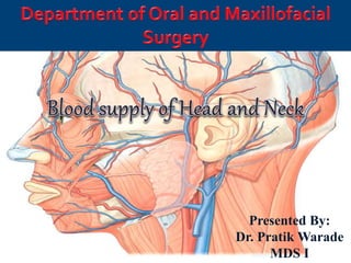
Blood supply of head and neck
•Download as PPTX, PDF•
18 likes•809 views

complete blood supply of head and neck along with the ligation of important arteries useful for oral and maxillofacial surgeon

Recommended
More Related Content
What's hot
What's hot (20)
Similar to Blood supply of head and neck
Similar to Blood supply of head and neck (20)
Recently uploaded
Recently uploaded (20)
Blood supply of head and neck
- 1. Presented By: Dr. Pratik Warade MDS I7/03/2018
- 2. Contents Functional morphology Arterial supply of head and neck Veinous supply of head and neck Veinous sinuses Applied anatomy
- 3. Functional morphology Arteries: 1. Blood flows away from the heart. 2. Possess thick elastic walls. 3. Carry oxygenated blood except the pulmonary artery. 4. Do not possess valve except in aorta. 5. Arteries are deeper in the flesh as veins. 6. Pulse is detectable. 7. Have narrower lumen.
- 4. Veins 1. Blood flow towards the heart. 2. Possess thin walls. 3. Carry deoxygenated blood except the pulmonary vein. 4. Have valves to prevent backflow of the blood. 5. Veins are nearer the surface of the skin. 6. Pulse is not detectable. 7. Have wider lumen.
- 5. Features Artery Vein Tunica Intima-Endothelium Usually rippled due vessel constriction Often smooth Internal elastic membrane Present Absent Tunica media Thick, dominated by smooth muscle cells and elastic fibers Thin, Dominated by smooth muscle cells and collagen fibers External elastic membrane Present Absent Tunica externa Collagen and elastic fibers Collagen, elastic fibers and smooth muscle cells
- 7. Arteries of head and neck Common carotid artery External carotid artery Internal carotid artery Subclavian artery
- 9. Common carotid artery CCA runs lateral to trachea and larynx to bifurcate at upper border of thyroid cartilage i.e., at the level of the disc b/w 3rd and 4th cervical vertebrae CCA gives rise to ICA posteromedially and ECA anterolaterally ICA is slightly widened at division to formCAROTID SINUS Common carotid artery
- 10. Relations to common carotid artery Anterolaterally: • Sternocleidomastoid • Sternohyoid •Sternothyroid •Superior belly of omohyoid Posteriorly: • Prevertebral muscles Medially: • Larynx • Pharynx Laterally: • Internal jugular vein
- 11. Applied anatomy of Common carotid artery The CCA can be compressed against the carotid tubercle i.e the anterior tubercle of the transverse process of the vertebra C6 which lies at the level of cricoid cartilage Patency of the carotid system can be investigated by angiography
- 12. Ligation of carotid arteries ECA can be ligated at 2 points: Origin from CCA Retromandibular fossa
- 13. 1. Origin from CCA Ligated usually above omohyoid muscle in carotid triangle i.e., between superior thyroid and lingual artery. Ligation can be used in bleeding from any one or more arteries or in injuries of ECA itself. Surgical procedure Incision over the skin starts at the level of angle of mandible just behind anterior border of Sternocleidomastoid muscle, downward and forward till level of cricoid cartilage Skin, platysma, superficial sheath of SCM is incised Blunt dissection over the anterior border of muscle is done to expose the deep layer of SCM and IJV Fascia in front of IJV is cut to expose arteries ECA is identified by it’s first ant. branch, sup. thyroid artery Isolated and ligated few mm above the origin of Superior thyroid artery.
- 14. 2) Retromandibular fossa Retromandibular fossa lies behind the angle of mandible ECA crosses stylomandibular ligament on it’s lateral side, therefore method known as “ligation of ECA at stylomandibular ligament.” Recommended if one deals with hemorrhage from one of the branches of maxillary artery As artery supplies both U/L jaws this should be method of choice in injuries of jaws and preparatory operations.
- 15. Surgical procedure 1. Less dangerous procedure than exposure of artery in neck 2. Incision placed over the skin at the tip of mastoid process, circling the mandibular angle, about 1 inch below mandible 3. Blunt dissection to locate retromandibular vein or EJV, tied and cut 4. Branches of greater auricular cut to permit mobilization of cervical lobe of parotid 5. Parotid capsule detached from anterior border of SCM 6. Helps skin retraction with parotid, anteriorly and upward 7. Exposes posterior belly of digastric and stylohyoid muscle above which lies stylomandibular ligament 8. Ligament can be palpated on mandibular protrusion 9. Pulse of ECA can be felt, isolated and cut even accompanied by larger vein.
- 16. Variations in branching of External carotid artery A. Most common arrangement B. Superior thyroid arising from CC and lingual and facial having common stem C. Posterior auricular artery branch of occipital and asc. pharyngeal arises higher than normal
- 17. Branches of common carotid artery External carotid artery Internal carotid artery
- 18. External carotid atery •External carotid artery is the terminal branch of common carotid artery. •Chief artery of supply to structures in front of the neck and face. •Generally arises medial and anterior to the ICA. Begins at the level of upper border of thyroid cartilage. •In 15% ECA originates lateral to the ICA, this variation occurs more frequently on the right (3:1)
- 19. Branches of external carotid artery Anterior 1. Superior thyroid 2. Facial 3. Lingual Posterior 1. Posterior auricular 2. Occipital Medial 1. Ascending pharyngeal Terminal 1. Superficial temporal 2. Maxillary
- 20. Superior thyroid artery First branch from ECA arises immediately above the bifurcation of CC Arises just below the greater cornu of hyoid bone. A diagnostic landmark in surgical exposure of EC Ends in thyroid gland.
- 21. Lingual artery Lingual Artery arises from the ECA opposite the tip of greater cornu of the hyoid bone. Second part of artery lies deep to the hyoglossus muscle which separates it from the hypoglossal nerve. Third Part or deep part : runs upwards along the anterior margin of the hyoglossus.
- 22. Lingual artery gives 2 dorsal-lingual branches as it passes under cover of hypoglossal muscle Sub-lingual artery is smaller branch, passes b/w genioglossus muscle and sublingual gland and supplies them At ant. floor of mouth, this artery anastomizes with opposite side and gives off artery to frenulum Cutting of frenulum in tongue-tie needs ligation of sub-lingual artery. Deep lingual artery lies in the inferior surface of the tongue Excision Involving only portion of tongue, artery can be identified through incision of mucous membrane in lateral part of floor of mouth and ligated
- 23. Ligation of lingual artery At origin stem from ECA gives good collateral circulation (1/5th of the cases have common stem for lingual and facial art.) Usually above hyoid bone, if interruption of entire vessel desired Hypoglossal n. identified after pushing sub-mandibular gland upwards as it runs across digastric LINGUAL OR LESSER’S triangle is formed by pushing hypoglossal n. upwards Lingual art. lies deeply in the fibers of hyoglossus muscle, posterior within the triangle
- 24. Facial artery Arises opp. the great cornu of hyoid bone, enters digastric triangle by passing deep to tendon Lies deeply embedded in the substance of submandibular salivary gland and then enters it’s facial course by curling around the inferior border of mandible immediately anterior to masseter muscle, i.e. about 1½ inch in front of angle of mandible At the anteroinferior angle of the masseter muscle, it can be palpated here and is called as an “anaesthetist’s artery”
- 25. Branches of cervical part: 1. Ascending palatine – supplies tonsil, root of tongue,soft palate and the pharyngotympanic tube 2. Tonsillar - mainly supplies the tonsils 3. Sub mental – large artery, accompanies mylohyoid nerve, supplies sub mental triangle and sublingual salivary gland. 4. Glandular branches – 3 or 4 large vessels ,supply the submandibular salivary gland and the lymph node.
- 26. Branches of facial part 1. Inferior labial 2. Superior labial 3. Lateral Nasal 4. Angular
- 27. Ligation of facial artery Easily exposed where it crosses the lower border of mandible to pass from submandibular region into face i.e. anterior to attachment of masseter muscle on mandible Care should be taken to prevent marginal mandibular branch of facial nerve.Therefore incision should be taken 1-2 cm below the border of mandible Skin, platysma muscle and deep fascia are cut, soft tissue bluntly retracted upwards until palpating finger can feel pulse of facial art. Artery isolated, tied and cut
- 28. Occipital artery Arises from post. surface of ECA about same level as facial Runs obliquely backwards, upwards, crosses ICA and IJV At it’s origin, hypoglossal n. lies b/w IC & IJ Branches – SCM (muscular) – Auricular – Mastoid – Meningeal
- 29. Posterior auricular artery Arises from posterior surface of EC in retro- mandibular fossa above stylohyoid muscle. Largely covered by parotid gland Ascends along the styloid process b/w external ear and mastoid process May arise as a branch of occipital rather than independent Supplies to outer ear and partly adjacent area of scalp Anastomosis with branches of occipital art. and auricular branch of superficial temporal
- 30. Ascending pharyngeal artery • Smallest branch, arises from post. surface often close to origin • Ascends vertically, anteromedially to ICA and pharynx • Supplies – to pharynx (several branches) – palate and tonsils – inf. tympanic branch to tympanum and – several meningeal branches • Larger terminal br. usually post. meningeal enters cranium through jugular foramen Approx. 14% of individuals asc. pharyngeal arises not from ECA but from occipital art.
- 31. Applied anatomy The ascending pharyngeal artery plays an important role in healing process of Le Fort I osteotomies, because it supplies the attached posterior palatal soft tissue pedicle.
- 32. Maxillary artery ECA artery gives 2 terminal branches (maxillary & sup. temporal) at approx. ½ to 2/3rd distance between lower border of angle andTMJ Leaves ECA at right angles passing almost horizontally b/w ramus and sphenomandibular ligament Acc. to Lasker et al maxillary artery may have varying relations to lateral pterygoid muscle in different individuals in slightly more than 50% individuals the artery is found on outer side of muscle,in remaining artery lies at medial side.
- 33. Branches 1st part (mandibular) Lies medial to mandible, it runs along the lower border of lateral pterygoid muscle Deep auricular artery Ant.tympanic artery Middle meningeal artery Accessory meningeal artery Inferior alveolar artery
- 34. Middle maningeal artery Largest artery that supplies the dura It ascends to the foramen spinosum through which it enters the cranium Divides into two branches,anterior and posterior. It supplies the dura mater (the outermost meninges) and the calvaria.
- 35. Inferior alveolar artery Runs downword & forward medial to ramus of mandible to reach mandibular foramina Before entering mandibular foramina gives off lingual and mylohyoid arteries In canal gives branches to mandibular teeth After coming out of canal supply chin via mental artery.Inferior alveolar artery
- 36. 2nd part (pterygoid part) Artery runs forward & upward superficial to the lower head of the lateral pterygoid muscle
- 37. 3rd part (pterygopalatine) Terminal portion of the artery passes between the two heads of the lateral pterygoid muscle
- 38. Superficial temporal artery Is 2nd terminal branch of ECA ascending vertically it crosses the posterior root of zygomatic arch immediately in front of outer ear Pulse of this artery can be felt as is covered by superficial fascia & skin Releases transverse facial artery before it leaves parotid gland Transverse facial lies beween zygomatic arch & parotid duct Sends branches to masseter muscle, parotid and terminates below outer corner of eye where anastomosis with palpebral artery.
- 39. ST divides above zygomatic arch into 2 main branches – parietal and frontal Parietal continues vertically upwards and supplies wide lateral area of scalp Frontal runs obliquely and forward, is often tortuous Superficial branch – zygomatico- orbital arises either from main stem or anterior branch which runs horizontally towards outer corner of eye supplies orbicularis oculi & anastomosis with lacrimal art.
- 40. Internal carotid artery Principal artery of brain and eye. It is one of the terminal branch of common carotid artery originates along with external carotid artery at the upper border of thyroid cartilage at the disk of third and fourth cervical vertebra. Divided into 4 parts: 1. Cervical part – in neck (Branchless) 2. Petrous part – within the petrous temporal bone a. Caroticotympanic b. Pterygoid 3. Cavernous part – within cavernous sinus a. Branch to trigeminal ganglion b. Sup. & inf. hypophyseal 4. Cerebral part – in relation to base of brain a. Opthalmic c. Middle cerebral e. Ant. chorodial b. Ant. cerebral d. Post. communicating
- 41. Cervical part It ascends vertically in the neck from its origin to the base of skull to reach the lower end of the carotid canal. This part is enclosed in carotid sheath along with internal jugular and vagus nerve. No branches arises from the internal carotid artery in the neck. Its initial part shows slight dilation, carotid sinus.Which acts as a baroreceptor
- 42. Petrous part Within the petrous part of the temporal bone,in the carotid canal runs upward forward & medially at right angle. Branches 1) Caroticotympanic- enter middle ear & anastomose with ant. & post.Tympanic branches 2) Artery of the Pterygoid Canal anastomose with greater palatine artery
- 43. Cavernous part Within the Cavernous Sinus Branches 1) Artery to trigeminal ganglion 2) Superior & inferior Hypophyseal artery
- 44. Cerebral part Lies at the base of the brain after emerging from the cavernous sinus Branches 1.Ophthalmic. 2.Anterior Cerebral. 3.Middle Cerebral. 4.Posterior Communicating. 5. Ant. choroidal
- 45. Subclavian artery Right Subclavian Artery: Arises from brachiocephalic artery (Behind right sternoclavicular joint) At outer border of 1st rib it becomes Axillary Artery Left Subclavian Artery: Arises from Arch of Aorta in the thorax Runs upwards to the root of the neck & arches laterally At outer border of 1st rib it becomes Axillary Artery
- 46. Subclavian artery Scalenus Anterior muscle passes anterior to the artery on each side and divides it into 3 parts. 1. 1st part of subclavian artery 2. 2nd part of subclavian artery 3. 3rd part of subclavian artery
- 47. 1st part of Subclavian artery Extends from the origin of the subclavian artery to the medial border of the Scalenus anterior muscle. Branches: 1.Vertebral artery 2.ThyrocervicalTrunk 3. Internal thoracic artery
- 48. 2nd part of subclavian artery Lies behind the Scalenus anterior muscle. Branches: 1. Costocervical trunk 2. Superior intercostal artery 3.Deep cervical artery
- 49. 3rd part of subclavian artery Extends from the lateral border of the Scalenus anterior muscle to the lateral border of 1st rib. Branches: (Occasional) 1. Superficial cervical artery 2. Suprascapular artery
- 50. Veins of the head and neck
- 51. Venous drainage from the face is entirely superficial All the venous drainage from the head and neck terminate in the internal jugular vein which join the subclavian vein to form the brachiocephalic vein behind the medial end of the clavicle
- 52. Internal Jugular vein It receive blood from the brain, face and the neck. It emerges through the jugular foramen, as a continuation of the sigmoid sinus descend down in the neck, first behind then lateral to the internal carotid artery inside the carotid sheath
- 53. Tributaries
- 54. Facial Vein Is formed by the union of the supraorbital and supratrochlear veins to form the angular vein Communicate with the cavernous sinus through the ophthalmic vein via the supraorbital
- 55. Runs downwards and backwards behind the facial artery to the lower border of the mandible To be joined by the anterior division of the retromandibular vein Joins the: 1. Pterygoid plexus through deep facial vein 2. Cavernous sinus through superior ophthalmic vein
- 56. Retromandibular vein Formed by the union of superficial temporal and maxillary vein from the pterygoid plexus Passes downwards in the substance of the parotid gland emerging from its lower border & divide into two divisions
- 57. Retromandibular vein Anterior division joins the facial vein Posterior division pierces the deep fascia and join the posterior auricular to form the external jugular. It empty into the subclavian vein
- 58. The maxillary vein A short trunk accompany the first part of the artery. Formed by confluence of the veins of the pterygoid plexus. It passes backward between the sphenomandibular ligament and the neck of the mandible Unite with the superficial temporal vein to form the retromandibular vein
- 59. Pterygoid plexus A network of very small veins, lie around and within the lateral pterygoid muscle in the infratemporal region Receive some of the veins that correspond to the maxillary vein, inferior ophthalmic vein (internal carotid blood) and the deep facial vein.
- 60. Drain into a pair of large, short maxillary veins which join the superficial temporal vein to form the retromandibular. Deep facial vein drain the plexus into the facial vein if the maxillary is occluded
- 61. External jugular vein Begins behind the angle of the mandible by the union of the posterior auricular and posterior division of the retromandibular veins. It descend obliquely, deep to the platysma, receive the posterior external jugular vein Pierce the deep fascia just above the clavicle and drain into the subclavian vein
- 62. Tributaries Posterior auricular vein Posterior division of retro mandibular vein Posterior external jugular vein Transverse cervical vein Suprascapular vein Anterior jugular vein
- 63. Venous sinuses The blood of the brain and the eye is collected by a system of specialized veins in the dura matter called sinuses. These sinuses are not collapsible, and their lumen is unchangeable because their walls are formed by the dense, rigid, and inelastic tissues of the dura matter. They drain eventually into the internal jugular vein, but there are numerous communications between the sinuses and extracranial veins.
- 65. Superior saggital sinus It lies within the convex attached margin of the falx cerebri. The sinus begins at the crista galli and is continuous with the right transeverse sinus.
- 66. Communications With the veins of the scalp through the parietal emissary vein. A vein from the nose through the foramen caecum. Cavernous sinus through superior anastomotic vein. Thrombosis of the superior sagittal sinus may take place due to spread of infection from the nose and scalp. This will lead to increased intracranial tension resulting in defective absorption of C. S. F.
- 67. Inferior saggital sinus It occupies the posterior two thirds of the lower free margin of the falx cerebri. It collects blood from the falx ceribri, medial surfase of the cerebrum and terminates into the straight sinus
- 68. Cavernous sinus These paired sinuses are situated on each side of the body of sphenoid bone Extend from superior orbital fissure in front to the apex of petrous temporal behind.
- 69. Structure passing through the sinus Internal carotid artery Abducent nerve Occulomotor nerve Trochlear nerve Ophthalmic nerve Maxillary nerve
- 70. Septic thrombosis of cavernous sinus may be caused by the numerous communications from the dangerous area of face, orbit and pharynx. If the internal carotid artery is ruptured as a result of fracture of the base of skull. Manifested by pulsating exophthalmos, oedema of the eye lids and loud systolic murmur
- 71. Dangerous area of face The facial vein communicates with the cavernous sinus through superior opthalmic vein. Infections from upper lip and lower part of nose can spread in a retrogade direction and cause thrombosis of cavernous sinus. So this area is known as Dangerous area of Face.
- 72. References: Gray’s Anatomy 41st Edition Sicher and Dubrul’s Oral Anatomy 8th edition BD Chaurasia’s Human Anatomy 5th edition.
- 73. Thank you.