
Recomendados
Recomendados
Mais conteúdo relacionado
Mais procurados
Mais procurados (20)
Semelhante a Breathing and Exchange of Gases
Semelhante a Breathing and Exchange of Gases (20)
Mais de pooja singh
Mais de pooja singh (20)
Último
Último (20)
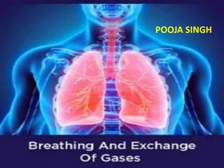
Breathing and Exchange of Gases
- 1. POOJA SINGH
- 2. RESPIRATORY ORGANS • Lower invertebrates like sponges, coelenterates, flatworms, etc., exchange O2 with CO2 by simple diffusion over their entire body surface. • Earthworms use their moist cuticle • insects have a network of tubes (tracheal tubes) to transport atmospheric air within the body. • Special vascularised structures called gills (branchial respiration) are used by most of the aquatic arthropods and molluscs • vascularised bags called lungs (pulmonary respiration) are used by the terrestrial forms for the exchange of gases.
- 4. Human Respiratory System Nasal Passage Pharynx Larynx Trachea Bronchi
- 5. • During swallowing glottis can be covered by a thin elastic cartilaginous flap called epiglottis to prevent the entry of food into the larynx. • Each bronchi undergoes repeated divisions to form the secondary and tertiary bronchi and bronchioles ending up in very thin terminal bronchioles. • The tracheae, primary, secondary and tertiary bronchi, and initial bronchioles are supported by incomplete cartilaginous rings. • Each terminal bronchiole gives rise to a number of very thin, irregular-walled and vascularised bag-like structures called alveoli. • The branching network of bronchi, bronchioles and alveoli comprise the lungs
- 7. • lungs are covered by a double layered pleura, with pleural fluid between them. • It reduces friction on the lung-surface • The part starting with the external nostrils up to the terminal bronchioles constitute the conducting part • alveoli and their ducts form the respiratory or exchange part of the respiratory system. • The conducting part transports the atmospheric air to the alveoli • Exchange part is the site of actual diffusion of O2 and CO2 between blood and atmospheric air.
- 8. • The lungs are situated in the thoracic chamber which is anatomically an air-tight chamber. • The thoracic chamber is formed dorsally by the vertebral column, ventrally by the sternum, laterally by the ribs and on the lower side by the dome-shaped diaphragm.
- 9. • Respiration involves the following steps: • Breathing or pulmonary ventilation by which atmospheric air is drawn in and CO2 rich alveolar air is released out. • Diffusion of gases (O2 and CO2 ) across alveolar membrane. • Transport of gases by the blood. • Diffusion of O2 and CO2 between blood and tissues. • Utilisation of O2 by the cells for catabolic reactions and resultant release of CO2
- 10. MECHANISM OF BREATHING • Breathing involves two stages : inspiration during which atmospheric air is drawn in and expiration by which the alveolar air is released out • Inspiration can occur if the pressure within the lungs (intra-pulmonary pressure) is less than the atmospheric pressure, i.e., there is a negative pressure in the lungs with respect to atmospheric pressure • Expiration takes place when the intra-pulmonary pressure is higher than the atmospheric pressure
- 11. • Inspiration is initiated by the contraction of diaphragm which increases the volume of thoracic chamber • contraction of inter-costal muscles lifts up the ribs and the sternum causing an increase in the volume of the thoracic chamber • An increase in pulmonary volume decreases the intra- pulmonary pressure to less than the atmospheric pressure which forces the air from outside to move into the lungs, i.e., inspiration
- 12. • Relaxation of the diaphragm and the inter- costal muscles returns the diaphragm and sternum to their normal positions and reduce the thoracic volume and thereby the pulmonary volume • This leads to an increase in intra-pulmonary pressure to slightly above the atmospheric pressure causing the expulsion of air from the lungs, i.e., expiration
- 13. Respiratory Volumes and Capacities • Tidal Volume (TV)- Volume of air inspired or expired during a normal respiration. It is approx. 500 mL., i.e., a healthy man can inspire or expire approximately 6000 to 8000 mL of air per minute • Inspiratory Reserve Volume (IRV): Additional volume of air, a person can inspire by a forcible inspiration. This averages 2500 mL to 3000 mL • Expiratory Reserve Volume (ERV): Additional volume of air, a person can expire by a forcible expiration. This averages 1000 mL to 1100 mL.
- 14. • Residual Volume (RV): Volume of air remaining in the lungs even after a forcible expiration. This averages 1100 mL to 1200 mL. • Inspiratory Capacity (IC): Total volume of air a person can inspire after a normal expiration. This includes tidal volume and inspiratory reserve volume ( TV+IRV). • Expiratory Capacity (EC): Total volume of air a person can expire after a normal inspiration. This includes tidal volume and expiratory reserve volume (TV+ERV).
- 15. • Functional Residual Capacity (FRC): Volume of air that will remain in the lungs after a normal expiration. This includes ERV+RV • Vital Capacity (VC): The maximum volume of air a person can breathe in after a forced expiration. This includes ERV, TV and IRV or the maximum volume of air a person can breathe out after a forced inspiration • Total Lung Capacity (TLC): Total volume of air accommodated in the lungs at the end of a forced inspiration. This includes RV, ERV, TV and IRV or vital capacity + residual volume.
- 16. EXCHANGE OF GASES • Alveoli are the primary sites of exchange of gases. • Exchange of gases also occur between blood and tissues. • O2 and CO2 are exchanged in these sites by simple diffusion mainly based on pressure/concentration gradient • Pressure contributed by an individual gas in a mixture of gases is called partial pressure and is represented as pO2 for oxygen and pCO2 for carbon dioxide
- 20. TRANSPORT OF GASES • About 97 per cent of O2 is transported by RBCs in the blood. • The remaining 3 per cent of O2 is carried in a dissolved state through the plasma. • Nearly 20-25 per cent of CO2 is transported by RBCs whereas 70 per cent of it is carried as bicarbonate ions • About 7 per cent of CO2 is carried in a dissolved state through plasma
- 21. Transport of Oxygen • Haemoglobin is a red coloured iron containing pigment present in the RBCs. • O2 can bind with haemoglobin in to form oxyhaemoglobin. • Each haemoglobin molecule can carry a maximum of four molecules of O2 . • Binding of oxygen with haemoglobin is primarily related to partial pressure of O2 .
- 22. • A sigmoid curve is obtained when percentage saturation of haemoglobin with O2 is plotted against the pO2 • This curve is called the Oxygen dissociation curve and is highly useful in studying the effect of factors like pCO2 , H+ concentration, etc., on binding of O2 with haemoglobin
- 23. • In the alveoli, where there is high pO2 low pCO2 lesser H+ concentration lower temperature • the factors are all favourable for the formation of oxyhaemoglobin,
- 24. • whereas in the tissues, where low pO2 high pCO2 high H+ concentration higher temperature exist • the conditions are favourable for dissociation of oxygen from the oxyhaemoglobin. • This clearly indicates that O2 gets bound to haemoglobin in the lung surface and gets dissociated at the tissues. • Every 100 ml of oxygenated blood can deliver around 5 ml of O2 to the tissues under normal physiological conditions.
- 25. Transport of Carbon dioxide • CO2 is carried by haemoglobin as carbamino- haemoglobin (about 20-25 per cent) • When pCO2 is high and pO2 is low as in the tissues, more binding of carbon dioxide occurs • when the pCO2 is low and pO2 is high as in the alveoli, dissociation of CO2 from carbamino-haemoglobin takes place
- 26. • RBCs contain a very high concentration of the enzyme, carbonic anhydrase and minute quantities of the same is present in the plasma too. • This enzyme facilitates the following reaction in both directions
- 27. • At the tissue site CO2 diffuses into blood and forms HCO3 – and H+,. • At the alveolar site the reaction proceeds in the opposite direction leading to the formation of CO2 and H2O. • Thus, CO2 trapped as bicarbonate at the tissue level and transported to the alveoli is released out as CO2 • Every 100 ml of deoxygenated blood delivers approximately 4 ml of CO2 to the alveoli.
- 28. REGULATION OF RESPIRATION • A specialised centre present in the medulla region of the brain called respiratory rhythm centre is primarily responsible for respiratory regulation • Another centre present in the pons region of the brain called pneumotaxic centre can moderate the functions of the respiratory rhythm centre • Receptors associated with aortic arch and carotid artery also can recognise changes in CO2 and H+ concentration and send necessary signals to the rhythm centre for remedial actions.
- 29. DISORDERS OF RESPIRATORY SYSTEM • Asthma is a difficulty in breathing causing wheezing due to inflammation of bronchi and bronchioles. • Emphysema is a chronic disorder in which alveolar walls are damaged due to which respiratory surface is decreased • One of the major causes of this is cigarette smoking • Occupational Respiratory Disorders- In certain industries, especially those involving grinding or stone-breaking, so much dust is produced • Long exposure can give rise to inflammation leading to fibrosis (proliferation of fibrous tissues) and thus causing serious lung damage • Workers in such industries should wear protective masks.