SlideShare uma empresa Scribd logo
1 de 66
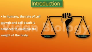
• In humans, the rate of cell
growth and cell death is
balanced to maintain the
weight of the body. Mitosis Apoptosis
Introduction Of Apoptosis
 The word "apoptosis" comes from the
ancient Greek, meaning the:
 "falling of petals from a flower" or "of
leaves from a tree in autumn"
 In 1964 Lockshin, study on programmed cell
death. The term apoptosis (a-po-toe-sis)
was first used in a now-classic paper by
Kerr et al 1972 to describe a
morphologically distinct form of cell death.
• Apoptosis is the process of programmed cell death.
• Biochemical events lead to characteristic cell changes (morphology)
and death. These changes include blebbing, cell shrinkage, nuclear
fragmentation, chromatin condensation, and chromosomal DNA
fragmentation.
• Between 50 and 70 billion cells die each day due to apoptosis in the
average human adult. For an average child between the ages of 8 and
14, approximately 20 billion to 30 billion cells die a day.
• Apoptosis or programmed cell death (PCD) is a mode of cell death
that occurs under normal physiological conditions and the cell is an
active participant in its own demise ("cellular suicide").
• It is important for the development of multicellular organism
(embryonic development) and homeostasis of their tissues (adult).
German scientist Carl Vogt was first to describe the
principle of apoptosis in 1842.
In 1972 Kerr first introduced the term apoptosis in a publication.
Kerr received the Paul Ehrlich and Ludwig Darmstaedter Prize
on March 14, 2000, for his description of apoptosis.
History
The 2002 Nobel Prize in Medicine was awarded to Sydney Brenner, Horvitz and
John E. Sulston for their work identifying genes
that control apoptosis.
Sydney Brenner
Horvitz John E. Sulston
A
P
O
P
T
O
S
I
S
IMPORTANCE OF APOPTOSIS
Important in normal physiology/development
Development: Immune systems maturation, Morphogenesis, Neural development
Adult: Immune privilege, DNA damage and wound repair.
Excess apoptosis
 Neurodegenerative diseases
Deficient apoptosis
 Cancer
 Autoimmunity
Importance of Apoptosis
Apoptosis is a beneficial and
important phenomenon:
In embryo
1. During embryonic development,
help to digit formation.
Lack of apoptosis in humans can lead
to webbed fingers called
"syndactyly".
Morphogenesis
(elimination excess cells)
Selection
(elimination non-
functional cells)
Apoptosis
Important in
embryogenesis
Organ size
Elimination
Excess Cells
Immunity
Elimination
dangerous
Cells
Self antigen
Recognizing cell
 Normal cell turn over
 Tissue homeostasis
 Induction and maintenance of immune tolerance
 Development of the nervous system
 Endocrine-dependent tissue atrophy
 Elimination of activated, damaged and abnormal cells
In adult
Mechanism of cell death
Apoptosis = “normal” or “programmed” cell death
Apoptosis is the physiological cell death which unwanted or useless cells are
eliminated during development and other normal biological processes.
Necrosis “accidental” or “ordinary” cell death
Necrosis is the pathological cell death which occurs when cells are exposed to a
serious physical or chemical insult (hypoxia, hyperthermia, ischemia).
Necrosis Apoptosis
Accidental /Premature cell death and
it is not programed.
Programmed cell death
Energy independent cell death. Energy dependent cell death
It occur when Cell are exposed to
harmful chemical or physical
agents/conditions.
Unwanted cell get eliminated by biological process.
Necrotic events: cell swell, rupture,
affects surrounding cells too.
Apoptotic events: cells become more compact, blebbing
of membrane, chromatin condensation, DNA
Fragmentation, cell shrink, release of apoptotic bodies.
They are inflammatory. Phagocytes involved.
Two form of cell death
Apoptosis VS Necrosis
Necrosis
1) Cellular swelling
2) Membranes are broken
3) ATP is depleted
4) Cell lyses, eliciting an
inflammatory reaction
5) DNA fragmentation is random, or
smeared
6) In vivo, whole areas of the tissue
are affected Apoptosis
Apoptosis
1) Cellular condensation
2) Membranes remain intact
3) Requires ATP
4) Cell is phagocytosed, no tissue
reaction
5) Ladder-like DNA fragmentation
6) In vivo, individual cells appear
affected
Apoptosis
VS
Necrosis
1) Cell shrinkage
2) Organelle reduction
3) Mitochondrial leakage
4) Chromatin condensation
5) Nuclear fragmentation
6) Membrane blebbing & changes
Apoptosis- Morphological
Changes
Apoptosis-Biochemical changes
1)Chromosomal DNA cleaved into fragments
2)Change in the plasma membrane – phosphatidylserine In the outer leaflet
3)Loss of electrical potential across the inner membrane of the mitochondria
4)Relocation of cytochrome c from the intermembrane space of the
mitochondria to the cytosol
Characteristic biochemical changes in cells undergoing apoptosis
Regulation Of Apoptosis
1.Anti-apoptotic factors
2.Pro-apoptotic factors
Example- BCI2 family proteins.
BCl2 Family
Protein
Anti-Apoptotic
Factor
BCl2, BCl-XL
Pro-Apoptotic
Factor
Bax, Bad, Bik, Bim,
PUMA, NOXA
What are caspases?
Types of caspases
How caspases activates?
Role in apoptosis/importance
What happen in the absence of
caspases?
Evolution Of Caspase over time
What are
caspases?
Caspases (Cysteine dependent
aspartate specific protease)
family of protease enzymes.
Contain cysteine residue in the catalytic
site and selectively cleave proteins at a
C-terminal of aspartate.
More than 12 caspases are
identified
1
2
3
4
Caspases
Caspases Cysteinyl aspartate specific proteases
A family of intracellular cysteine proteases
that play a pivotal role in the initiation and
execution of apoptosis.
At least 14 different members of caspases in
mammalian cells have been identified
All are synthesized as inactive proenzymes
(zymogen) with 32-56 kDa
Small subunit
(10-13 kDa)
Large subunit
(17-21 kDa)
Prodomain
(2-25 kDa)
Asp-X Asp-X
QACXG
32-56 kDa
Effector caspases
Initiator caspases
Caspase-2
Caspase-8
Caspase-9
Caspase-10
Caspase-12
Caspase-3
Caspase-6
Caspase-7
Active Caspase
C
N
Caspase structure
Caspase subgroups
To date, ten major caspases have been
identified and broadly categorized into:
Signaling or Initiator
caspases (2, 8, 9, 10)
Effector or Executioner
caspases (3, 6, 7)
Inflammatory
caspases (1, 4, 5)
 The other caspases that have been identified
include: Caspases 11, 12, 13, 14
 Central role in cascade of apoptotic events is played by caspase 3 (CPP32)
TYPES OF CASPASES
Initiator caspases
• Caspase-2, 8, 9, 10
• Self activating by
autocatalysis
EFFECTOR/ EXECUTIONERS
Caspase-3, 6, 7
Activated by initiator
caspases.
How caspases activates?
• Caspases are synthesized as inactive
zymogens /pro-caspases that are only
activated following an appropriate stimulus
• This post-translational level of control allows
rapid and tight regulation of the enzyme.
Pro-caspase split into small and large subunits
that form a heterodimer and two such dimers
assembles to form the active tetramer.
Role in apoptosis/importance
Extrinsic pathway of
apoptosis
Granzyme B pathway.
Intrinsic pathway of
apoptosis
What happen in the absence of caspases?
 Caspase deficiency has been identified as a cause of tumour development.
 Tumour growth can occur by a combination of factors, including a mutation in
a cell cycle gene and in apoptotic proteins such as Caspases.
Evolution Of Caspase
 In animals apoptosis is induced by caspases and in fungi and plants, apoptosis
is induced by arginine and lysine-specific caspase like proteases called
metacaspases
Recent studies on caspases
1. Over activation of caspase-3 can lead to excessive
programmed cell death.
2. Over activation of caspase-3 playing role in causing
Alzheimer's disease.
3. Inflammatory caspases- caspase-1, 4, 5, 11, 12
APOPTOSIS IN
C. elegans
( Nematodes)
 Four genes whose encoded proteins play an essential role in controlling programmed cell
death during C. elegans development: ced-3, ced-4, ced-9, and egl-1.
 The human Bcl-2 protein and worm CED-9 protein are homologous; even though the two
proteins are only 23 percent identical in sequence
 The first mammalian apoptotic gene to be cloned, bcl-2.
 Bcl-2 gene can block the extensive cell death found in ced-9 mutant worms. Thus both
proteins act as regulators that suppress the apoptotic pathway.
INHIBITORS OF APOPTOSIS(IAPs)
• Inhibitors of Apoptosis Proteins (IAPs) are a class of highly conserved proteins
known for the regulation of caspases and immune signaling.
• Inhibitor of apoptosis proteins (IAPs), also known as BIRCS (BIR domain
containing proteins) are a class of proteins characterized by the presence of
Baculovirus IAP Repeat (BIR) domain, a Zn²+ ion coordinating protein-protein
interaction motif.
• They are highly conserved from viruses to mammals.
• Yeasts and plants undergo a form of programmed cell death
(PCD) by caspase homologs known as metacaspases.
• Yet no IAP homologs are found in prokaryotes and plants.
• In Yeast the known IAP is BIR1p.
• There are eight known mammalian IAPS/BIRCS
INHIBITORS OF APOPTOSIS(IAPs)
• There are eight known mammalian IAPS/BIRCS
1. BIRC1 (neuronal IAP/NAIP),
2. BIRC2 (cellular IAP1/clAP1),
3. BIRC3 (cellular IAP2/clAP2),
4. BIRC4 (X-linked IAP/XIAP),
5. BIRC5
6. BIRC6
7. BIRC7 (Melanoma IAP/ML-IAP)and
8. BIRC8 (IAP-like protein 2).
They function by blocking
caspase-3, -7, and -9
• Induction of apoptosis in Drosophila requires the activity of three
closely linked genes, reaper, hid and grim.
• proteins encoded by reaper, hid and grim activate cell death by
inhibiting the anti-apoptotic activity of the Drosophila IAP1 (diap1)
protein.
• Gain-of-function mutations in diap1 strongly suppressed reaper hid-
and grim-so no apoptosis.
Anti- IAPs in Drosophila
Apoptosis pathways
Extrinsic pathway (death receptor-mediated events)
Death trigger signal from extracellular stimulus and they require the
receptor for receiving the signal.
Intrinsic pathway (mitochondria-mediated events)
Internal damage, So they don’t require Receptor.
Reason behind extrinsic response
• The extrinsic pathway triggers apoptosis in response to external
stimuli, namely by ligand binding at ‘death’ receptors on the cell
Surface.
• These receptors are typically members of the Tumour Necrosis Factor
Receptor (TNFR) gene family, such as TNFR1 or FAS. Binding at these
receptors leads to receptor molecules grouping up on the cell surface
to initiate downstream caspase activation
Components:
Death
Receptors
Death
Ligands
Adaptor
Proteins
Caspaseses
Death Receptors
"Death receptors" that are members of the tumor necrosis factor (TNF)
receptor superfamily.
• Death receptors have a cytoplasmic domain of about 80 amino acids
called the "death domain".
• This death domain plays a critical role in transmitting the death signal
from the cell surface to the intracellular signaling pathways.
Receptor Ligand
FasR (CD95/APOI)
DR3
DR4 (TRAIL-RI)
DRS (TRAIL-R2)
TNFRI
TNFR2
Fasl.
Apo3L
Apo21
Apo2L
TNF-α
TNF-β
The best characterized
receptors & ligands
corresponding Death
receptors include:
Adaptor proteins
Apoptotic adaptor proteins play a critical role in regulating pro-
and anti-apoptotic signalling pathways
1)FADD (Fas-associated death domain)
2) TRADD (TNF receptor-associated death domain), are
recruited to ligand-activated, oligomerized death receptors to
mediate apoptotic signalling pathways.
Two theories of the direct initiation of apoptotic mechanisms in
mammals have been suggested: the TNF-induced (tumor
necrosis factor) model and the Fas-Fas ligand-mediated model,
both involving receptors of the TNF receptor (TNFR) family
coupled to extrinsic signals.
EXTRINSIC PATHWAY
Killer lymphocytes
Caspase 8
Caspase 3
Death
domain
FAS receptor
Disc
Extracellular signal
Ligand and receptor binding
FADD recruitment
Activation of procaspase 8
Disc formation
Activation of executioner caspase
Apoptosis of target cell
Ligand- Cytokines, TNF, FAS Ligand
Receptors-Cytokines receptors, TNF receptors etc
Extrinsic
Pathway of
Apoptosis
INTRINSIC PATHWAY/•CASPASE-DEPENDENT
INTRINSIC PATHWAY/•MITOCHONDIAL APOPTOSIS
REASON OF INTRENSIC PATHWAY
Intracellular death signal like DNA damage, Biochemical stress, ROS
generation, oncogenic stress, Virus attack or Lack of growth factors.
Component
• Bcl-2 family proteins
• Cytochrome c
• Apoptosome
• Caspaseses
 The control & regulation of apoptotic mitochondrial events occurs
through members of the Bcl-2 family of proteins
 Anti-apoptotic proteins include Bcl-2, Bcl-x, Bcl-XL, Bel-w Pro-
apoptotic proteins include Bax, Bak, Bid, Bad, Bim, Bik
 The main mechanism of action of the Bcl-2 family of proteins is the
regulation of cytochrome c release from the mitochondria via
alteration of mitochondrial membrane permeability.
BH4 BH3 BH1 BH2 TM
Bcl-2
Bax
Bid
Bik
GROUP I
GROUP II
GROUP III
Group I- Bcl-2 and bcl-xl possess anti apoptotic activity
Group II and III- Bax and Bid , Bak possess pro apoptotic activity
Bcl2 Homology (BH) domains of Bcl-2 Protein
Intrinsic Pathway
 The stimuli that initiate the intrinsic pathway produce intracellular signals such as radiation (DNA
damage), absence of certain growth factors, hormones and cytokines.
 All of these stimuli cause changes in the mitochondrial outer membrane permeabilization (MOMP)
 Release of pro-apoptotic proteins such as cytochrome c, Smac/DIABLO, AIF, endonuclease
G and CAD from the intermembrane space into the cytosol. Cytochrome c binds and activates Apaf-1 as
well as procaspase-9, forming an "apoptosome Caspase-9 activation, subsequent caspase-3 activation
and cell death.
• Internal damage signal
• Inhibition of anti apoptotic
protein
• Oligomerization of Bax/Bak
• Mitochondrial changes/channel
formation of bak or bax
• Cytochrome C release
• Activation of Apaf-1
• Apoptosome formation
• Caspase 9 activation
• Caspase 3 activation
Apoptosis assay methods
1. Cytomorphological alterations
2. DNA fragmentation
3. Detection of caspases, cleaved Substrates, regulators and inhibitors
4. Membrane alterations
5. Detection of apoptosis in whole mounts
6. Mitochondrial assays
Apoptosis assays, based on methodology, can be classified into six
Major groups:
1) Cytomorphological Alterations
• The evaluation of hematoxylin and eosin-stained tissue sections with light
microscopy does allow the visualization of apoptotic cells.
• This method detects the later events of apoptosis
• TEM is considered the
gold standard to
confirm apoptosis:
Electron-dense nucleus
Nuclear fragmentation
Disorganized
cytoplasmic organelles
Large clear Vacuoles
Intact cell membrane
Blebs at the cell
surface
DNA Fragmentation
 DNA fragmentation occurs in the later phase of apoptosis.
 TUNEL (Terminal dUTP Nick End-Labeling) assay quantified The incorporation
of deoxyuridine triphosphate (dUTP) at single and double stranded DNA
breaks in a reaction catalyzed by the template independent enzyme,
terminal deoxynucleotidyl transferase (TdT).
 Incorporated dUTP is labeled such that breaks can be quantified either by
flowcytometry, fluorescent microscopy, or light
Membrane Alterations
• During the process of apoptosis, one of the earliest events is Externalization
of Phosphatidylserine (PS) from the inner to the outer plasma membrane of
apoptotic cells.
• These cells can be demonstrated by bound with Fluorescein isothiocyanate
(FITC)-labeled Annexin V and detected with Fluorescent microscopy.
• The vital dye propidium iodide (PI) should be used in combination of annexin
V that help in distinguish viable, apoptotic & necrotic cell populations at the
same time.
 Mitochondrial assays and cytochrome c release allow the detection of
changes in the early phase of the intrinsic pathway.
 The mitochondrial outer membrane (MOM) collapses during apoptosis,
allowing detection with a fluorescent cationic dye.
 Cytochrome e release from the mitochondria can also be assayed using
fluorescence and electron microscopy in living or fixed cells.
 Apoptotic or anti-apoptotic regulator proteins such as Bax, Bid, and Bcl-2 can
also be detected using fluorescence and confocal microscopy
BCL2 inhibitors;
1)G3139 is an antisense oligodeoxynucleotide targeting BCL2 mRNA
resulting in RNAse H activation.
2)ABT263 is a small molecule mimetic of the BH3 domain of the pro-
apoptotic BAD protein that is currently in clinical trial in chronic
lymphatic leukaemia.
1) VX-765 is an orally active, reversible caspase-1 inhibitor that was being developed for
the treatment of inflammatory disorders.
2) Emricasan is a novel, irreversible, orally active pan-caspase inhibitor that has been
investigated for the treatment of chronic HCV infection and liver transplantation rejection.
3) NCX-1000, a small-molecule inhibitor that selectively inhibits caspase-3, -8 and -9 in the
micromolar range, was in phase II clinical trials for the treatment of chronic liver disease.
4) PAC-1, a small molecule that induces both procaspase-3 activation in vitro and apoptosis
in several cancer cell lines

Mais conteúdo relacionado

Mais procurados (20)

Apoptosis
ApoptosisApoptosis
Apoptosis
 
Programmed Cell death or Apoptosis
Programmed Cell death or ApoptosisProgrammed Cell death or Apoptosis
Programmed Cell death or Apoptosis
 
Apoptosis
ApoptosisApoptosis
Apoptosis
 
Apoptosis (Programm of cell death)
Apoptosis (Programm of cell death)Apoptosis (Programm of cell death)
Apoptosis (Programm of cell death)
 
Cell death
Cell death Cell death
Cell death
 
Apoptosis
Apoptosis Apoptosis
Apoptosis
 
Apoptosis
ApoptosisApoptosis
Apoptosis
 
Apoptosis and neurological disorders
Apoptosis and neurological disordersApoptosis and neurological disorders
Apoptosis and neurological disorders
 
Apoptosis
ApoptosisApoptosis
Apoptosis
 
PROGRAMMED CELL DEATH
PROGRAMMED CELL DEATHPROGRAMMED CELL DEATH
PROGRAMMED CELL DEATH
 
Apoptosis (presentation)
Apoptosis (presentation)Apoptosis (presentation)
Apoptosis (presentation)
 
Apoptosis
ApoptosisApoptosis
Apoptosis
 
Apoptosis my ppt
Apoptosis my pptApoptosis my ppt
Apoptosis my ppt
 
Apoptosis
ApoptosisApoptosis
Apoptosis
 
Apoptosis
ApoptosisApoptosis
Apoptosis
 
Apoptosis
Apoptosis Apoptosis
Apoptosis
 
Oncogenes
Oncogenes Oncogenes
Oncogenes
 
Role of apoptosis in cancer progression
Role of apoptosis in cancer progressionRole of apoptosis in cancer progression
Role of apoptosis in cancer progression
 
Apoptosis and the response to chemotherapy
Apoptosis and the response to chemotherapyApoptosis and the response to chemotherapy
Apoptosis and the response to chemotherapy
 
Apoptosis
Apoptosis Apoptosis
Apoptosis
 

Semelhante a Apoptosis slide

Semelhante a Apoptosis slide (20)

Apoptosis Pathway
 Apoptosis Pathway Apoptosis Pathway
Apoptosis Pathway
 
Apoptosis in plant cell
Apoptosis in plant cellApoptosis in plant cell
Apoptosis in plant cell
 
Apoptosis
ApoptosisApoptosis
Apoptosis
 
Apoptosis, program cell death
Apoptosis, program cell deathApoptosis, program cell death
Apoptosis, program cell death
 
Apoptosis
ApoptosisApoptosis
Apoptosis
 
Apoptosis
ApoptosisApoptosis
Apoptosis
 
Apoptosis
ApoptosisApoptosis
Apoptosis
 
Programmed cell death
Programmed cell death  Programmed cell death
Programmed cell death
 
physiology of apoptosis first year mbbs medical.pdf
physiology of apoptosis first year mbbs medical.pdfphysiology of apoptosis first year mbbs medical.pdf
physiology of apoptosis first year mbbs medical.pdf
 
Apoptosis / dental crown & bridge courses
Apoptosis / dental crown & bridge coursesApoptosis / dental crown & bridge courses
Apoptosis / dental crown & bridge courses
 
apoptosisII.pdf
apoptosisII.pdfapoptosisII.pdf
apoptosisII.pdf
 
Apoptosis.ppt
Apoptosis.pptApoptosis.ppt
Apoptosis.ppt
 
APOPTOSIS by dr Alfarah.pptx
APOPTOSIS by dr Alfarah.pptxAPOPTOSIS by dr Alfarah.pptx
APOPTOSIS by dr Alfarah.pptx
 
Cell Death and Cancer
Cell Death and CancerCell Death and Cancer
Cell Death and Cancer
 
Apoptosis
ApoptosisApoptosis
Apoptosis
 
Intrinsic and Extrinsic Pathway of Apoptosis
Intrinsic and Extrinsic  Pathway of ApoptosisIntrinsic and Extrinsic  Pathway of Apoptosis
Intrinsic and Extrinsic Pathway of Apoptosis
 
APOPTOSIS MBBS.pdf
APOPTOSIS MBBS.pdfAPOPTOSIS MBBS.pdf
APOPTOSIS MBBS.pdf
 
Apoptosis
ApoptosisApoptosis
Apoptosis
 
Apoptosis
ApoptosisApoptosis
Apoptosis
 
apopwqgdcbhuredgbcfyhhvhcctosis (1).pptx
apopwqgdcbhuredgbcfyhhvhcctosis (1).pptxapopwqgdcbhuredgbcfyhhvhcctosis (1).pptx
apopwqgdcbhuredgbcfyhhvhcctosis (1).pptx
 

Mais de vidan biology

ABC Model of Flower development
ABC Model of Flower developmentABC Model of Flower development
ABC Model of Flower developmentvidan biology
 
Cytokinin Plant Hormones
Cytokinin Plant HormonesCytokinin Plant Hormones
Cytokinin Plant Hormonesvidan biology
 
POLYMORPHISM IN COELENTERATA
POLYMORPHISM IN COELENTERATAPOLYMORPHISM IN COELENTERATA
POLYMORPHISM IN COELENTERATAvidan biology
 
Larval Stage in Different Animal
Larval Stage  in Different AnimalLarval Stage  in Different Animal
Larval Stage in Different Animalvidan biology
 
Biodiversity Conservation
Biodiversity ConservationBiodiversity Conservation
Biodiversity Conservationvidan biology
 
Lichens and its types
Lichens and its typesLichens and its types
Lichens and its typesvidan biology
 
Terminology of plant Taxonomy.pdf
Terminology of plant Taxonomy.pdfTerminology of plant Taxonomy.pdf
Terminology of plant Taxonomy.pdfvidan biology
 
Vernalization (Revernalization, Devernalization) gene
Vernalization (Revernalization, Devernalization) geneVernalization (Revernalization, Devernalization) gene
Vernalization (Revernalization, Devernalization) genevidan biology
 
Invertebrates (Plant Kingdom) complete
Invertebrates (Plant Kingdom) completeInvertebrates (Plant Kingdom) complete
Invertebrates (Plant Kingdom) completevidan biology
 
Cytokinin plant hormones complete
Cytokinin plant hormones completeCytokinin plant hormones complete
Cytokinin plant hormones completevidan biology
 
Gibberellic Acid or Gibberellin Hormones
Gibberellic Acid or Gibberellin HormonesGibberellic Acid or Gibberellin Hormones
Gibberellic Acid or Gibberellin Hormonesvidan biology
 
Animal Kingdom phylum Detail
Animal Kingdom phylum DetailAnimal Kingdom phylum Detail
Animal Kingdom phylum Detailvidan biology
 
Innate Behavior (Animal Behavior) Taxis, Fixed Action Pattern, reflex.
Innate Behavior (Animal Behavior) Taxis, Fixed Action Pattern, reflex.Innate Behavior (Animal Behavior) Taxis, Fixed Action Pattern, reflex.
Innate Behavior (Animal Behavior) Taxis, Fixed Action Pattern, reflex.vidan biology
 
Fish migration (Animal Behavior) Catadromous, Anadromous
Fish migration (Animal Behavior) Catadromous, AnadromousFish migration (Animal Behavior) Catadromous, Anadromous
Fish migration (Animal Behavior) Catadromous, Anadromousvidan biology
 
BIRD MIGRATION (ANIMAL BEHAVIOR)
BIRD MIGRATION (ANIMAL BEHAVIOR) BIRD MIGRATION (ANIMAL BEHAVIOR)
BIRD MIGRATION (ANIMAL BEHAVIOR) vidan biology
 
Photoperiodism Detail slide
Photoperiodism Detail slidePhotoperiodism Detail slide
Photoperiodism Detail slidevidan biology
 
Phloem loading ( Food Transported)
Phloem loading ( Food Transported)Phloem loading ( Food Transported)
Phloem loading ( Food Transported)vidan biology
 

Mais de vidan biology (20)

ABC Model of Flower development
ABC Model of Flower developmentABC Model of Flower development
ABC Model of Flower development
 
Cytokinin Plant Hormones
Cytokinin Plant HormonesCytokinin Plant Hormones
Cytokinin Plant Hormones
 
RNA Interference
RNA InterferenceRNA Interference
RNA Interference
 
Abscisic Acid.pptx
Abscisic Acid.pptxAbscisic Acid.pptx
Abscisic Acid.pptx
 
POLYMORPHISM IN COELENTERATA
POLYMORPHISM IN COELENTERATAPOLYMORPHISM IN COELENTERATA
POLYMORPHISM IN COELENTERATA
 
Larval Stage in Different Animal
Larval Stage  in Different AnimalLarval Stage  in Different Animal
Larval Stage in Different Animal
 
Biodiversity Conservation
Biodiversity ConservationBiodiversity Conservation
Biodiversity Conservation
 
Lichens and its types
Lichens and its typesLichens and its types
Lichens and its types
 
Terminology of plant Taxonomy.pdf
Terminology of plant Taxonomy.pdfTerminology of plant Taxonomy.pdf
Terminology of plant Taxonomy.pdf
 
Root
RootRoot
Root
 
Vernalization (Revernalization, Devernalization) gene
Vernalization (Revernalization, Devernalization) geneVernalization (Revernalization, Devernalization) gene
Vernalization (Revernalization, Devernalization) gene
 
Invertebrates (Plant Kingdom) complete
Invertebrates (Plant Kingdom) completeInvertebrates (Plant Kingdom) complete
Invertebrates (Plant Kingdom) complete
 
Cytokinin plant hormones complete
Cytokinin plant hormones completeCytokinin plant hormones complete
Cytokinin plant hormones complete
 
Gibberellic Acid or Gibberellin Hormones
Gibberellic Acid or Gibberellin HormonesGibberellic Acid or Gibberellin Hormones
Gibberellic Acid or Gibberellin Hormones
 
Animal Kingdom phylum Detail
Animal Kingdom phylum DetailAnimal Kingdom phylum Detail
Animal Kingdom phylum Detail
 
Innate Behavior (Animal Behavior) Taxis, Fixed Action Pattern, reflex.
Innate Behavior (Animal Behavior) Taxis, Fixed Action Pattern, reflex.Innate Behavior (Animal Behavior) Taxis, Fixed Action Pattern, reflex.
Innate Behavior (Animal Behavior) Taxis, Fixed Action Pattern, reflex.
 
Fish migration (Animal Behavior) Catadromous, Anadromous
Fish migration (Animal Behavior) Catadromous, AnadromousFish migration (Animal Behavior) Catadromous, Anadromous
Fish migration (Animal Behavior) Catadromous, Anadromous
 
BIRD MIGRATION (ANIMAL BEHAVIOR)
BIRD MIGRATION (ANIMAL BEHAVIOR) BIRD MIGRATION (ANIMAL BEHAVIOR)
BIRD MIGRATION (ANIMAL BEHAVIOR)
 
Photoperiodism Detail slide
Photoperiodism Detail slidePhotoperiodism Detail slide
Photoperiodism Detail slide
 
Phloem loading ( Food Transported)
Phloem loading ( Food Transported)Phloem loading ( Food Transported)
Phloem loading ( Food Transported)
 

Último

MS4 level being good citizen -imperative- (1) (1).pdf
MS4 level   being good citizen -imperative- (1) (1).pdfMS4 level   being good citizen -imperative- (1) (1).pdf
MS4 level being good citizen -imperative- (1) (1).pdfMr Bounab Samir
 
4.9.24 School Desegregation in Boston.pptx
4.9.24 School Desegregation in Boston.pptx4.9.24 School Desegregation in Boston.pptx
4.9.24 School Desegregation in Boston.pptxmary850239
 
BIOCHEMISTRY-CARBOHYDRATE METABOLISM CHAPTER 2.pptx
BIOCHEMISTRY-CARBOHYDRATE METABOLISM CHAPTER 2.pptxBIOCHEMISTRY-CARBOHYDRATE METABOLISM CHAPTER 2.pptx
BIOCHEMISTRY-CARBOHYDRATE METABOLISM CHAPTER 2.pptxSayali Powar
 
ICS 2208 Lecture Slide Notes for Topic 6
ICS 2208 Lecture Slide Notes for Topic 6ICS 2208 Lecture Slide Notes for Topic 6
ICS 2208 Lecture Slide Notes for Topic 6Vanessa Camilleri
 
CHEST Proprioceptive neuromuscular facilitation.pptx
CHEST Proprioceptive neuromuscular facilitation.pptxCHEST Proprioceptive neuromuscular facilitation.pptx
CHEST Proprioceptive neuromuscular facilitation.pptxAneriPatwari
 
4.11.24 Poverty and Inequality in America.pptx
4.11.24 Poverty and Inequality in America.pptx4.11.24 Poverty and Inequality in America.pptx
4.11.24 Poverty and Inequality in America.pptxmary850239
 
Indexing Structures in Database Management system.pdf
Indexing Structures in Database Management system.pdfIndexing Structures in Database Management system.pdf
Indexing Structures in Database Management system.pdfChristalin Nelson
 
DIFFERENT BASKETRY IN THE PHILIPPINES PPT.pptx
DIFFERENT BASKETRY IN THE PHILIPPINES PPT.pptxDIFFERENT BASKETRY IN THE PHILIPPINES PPT.pptx
DIFFERENT BASKETRY IN THE PHILIPPINES PPT.pptxMichelleTuguinay1
 
Beauty Amidst the Bytes_ Unearthing Unexpected Advantages of the Digital Wast...
Beauty Amidst the Bytes_ Unearthing Unexpected Advantages of the Digital Wast...Beauty Amidst the Bytes_ Unearthing Unexpected Advantages of the Digital Wast...
Beauty Amidst the Bytes_ Unearthing Unexpected Advantages of the Digital Wast...DhatriParmar
 
Concurrency Control in Database Management system
Concurrency Control in Database Management systemConcurrency Control in Database Management system
Concurrency Control in Database Management systemChristalin Nelson
 
Grade Three -ELLNA-REVIEWER-ENGLISH.pptx
Grade Three -ELLNA-REVIEWER-ENGLISH.pptxGrade Three -ELLNA-REVIEWER-ENGLISH.pptx
Grade Three -ELLNA-REVIEWER-ENGLISH.pptxkarenfajardo43
 
Daily Lesson Plan in Mathematics Quarter 4
Daily Lesson Plan in Mathematics Quarter 4Daily Lesson Plan in Mathematics Quarter 4
Daily Lesson Plan in Mathematics Quarter 4JOYLYNSAMANIEGO
 
Scientific Writing :Research Discourse
Scientific  Writing :Research  DiscourseScientific  Writing :Research  Discourse
Scientific Writing :Research DiscourseAnita GoswamiGiri
 
4.16.24 Poverty and Precarity--Desmond.pptx
4.16.24 Poverty and Precarity--Desmond.pptx4.16.24 Poverty and Precarity--Desmond.pptx
4.16.24 Poverty and Precarity--Desmond.pptxmary850239
 
Active Learning Strategies (in short ALS).pdf
Active Learning Strategies (in short ALS).pdfActive Learning Strategies (in short ALS).pdf
Active Learning Strategies (in short ALS).pdfPatidar M
 
How to Manage Buy 3 Get 1 Free in Odoo 17
How to Manage Buy 3 Get 1 Free in Odoo 17How to Manage Buy 3 Get 1 Free in Odoo 17
How to Manage Buy 3 Get 1 Free in Odoo 17Celine George
 
Q-Factor General Quiz-7th April 2024, Quiz Club NITW
Q-Factor General Quiz-7th April 2024, Quiz Club NITWQ-Factor General Quiz-7th April 2024, Quiz Club NITW
Q-Factor General Quiz-7th April 2024, Quiz Club NITWQuiz Club NITW
 
Sulphonamides, mechanisms and their uses
Sulphonamides, mechanisms and their usesSulphonamides, mechanisms and their uses
Sulphonamides, mechanisms and their usesVijayaLaxmi84
 
Mythology Quiz-4th April 2024, Quiz Club NITW
Mythology Quiz-4th April 2024, Quiz Club NITWMythology Quiz-4th April 2024, Quiz Club NITW
Mythology Quiz-4th April 2024, Quiz Club NITWQuiz Club NITW
 
Textual Evidence in Reading and Writing of SHS
Textual Evidence in Reading and Writing of SHSTextual Evidence in Reading and Writing of SHS
Textual Evidence in Reading and Writing of SHSMae Pangan
 

Último (20)

MS4 level being good citizen -imperative- (1) (1).pdf
MS4 level   being good citizen -imperative- (1) (1).pdfMS4 level   being good citizen -imperative- (1) (1).pdf
MS4 level being good citizen -imperative- (1) (1).pdf
 
4.9.24 School Desegregation in Boston.pptx
4.9.24 School Desegregation in Boston.pptx4.9.24 School Desegregation in Boston.pptx
4.9.24 School Desegregation in Boston.pptx
 
BIOCHEMISTRY-CARBOHYDRATE METABOLISM CHAPTER 2.pptx
BIOCHEMISTRY-CARBOHYDRATE METABOLISM CHAPTER 2.pptxBIOCHEMISTRY-CARBOHYDRATE METABOLISM CHAPTER 2.pptx
BIOCHEMISTRY-CARBOHYDRATE METABOLISM CHAPTER 2.pptx
 
ICS 2208 Lecture Slide Notes for Topic 6
ICS 2208 Lecture Slide Notes for Topic 6ICS 2208 Lecture Slide Notes for Topic 6
ICS 2208 Lecture Slide Notes for Topic 6
 
CHEST Proprioceptive neuromuscular facilitation.pptx
CHEST Proprioceptive neuromuscular facilitation.pptxCHEST Proprioceptive neuromuscular facilitation.pptx
CHEST Proprioceptive neuromuscular facilitation.pptx
 
4.11.24 Poverty and Inequality in America.pptx
4.11.24 Poverty and Inequality in America.pptx4.11.24 Poverty and Inequality in America.pptx
4.11.24 Poverty and Inequality in America.pptx
 
Indexing Structures in Database Management system.pdf
Indexing Structures in Database Management system.pdfIndexing Structures in Database Management system.pdf
Indexing Structures in Database Management system.pdf
 
DIFFERENT BASKETRY IN THE PHILIPPINES PPT.pptx
DIFFERENT BASKETRY IN THE PHILIPPINES PPT.pptxDIFFERENT BASKETRY IN THE PHILIPPINES PPT.pptx
DIFFERENT BASKETRY IN THE PHILIPPINES PPT.pptx
 
Beauty Amidst the Bytes_ Unearthing Unexpected Advantages of the Digital Wast...
Beauty Amidst the Bytes_ Unearthing Unexpected Advantages of the Digital Wast...Beauty Amidst the Bytes_ Unearthing Unexpected Advantages of the Digital Wast...
Beauty Amidst the Bytes_ Unearthing Unexpected Advantages of the Digital Wast...
 
Concurrency Control in Database Management system
Concurrency Control in Database Management systemConcurrency Control in Database Management system
Concurrency Control in Database Management system
 
Grade Three -ELLNA-REVIEWER-ENGLISH.pptx
Grade Three -ELLNA-REVIEWER-ENGLISH.pptxGrade Three -ELLNA-REVIEWER-ENGLISH.pptx
Grade Three -ELLNA-REVIEWER-ENGLISH.pptx
 
Daily Lesson Plan in Mathematics Quarter 4
Daily Lesson Plan in Mathematics Quarter 4Daily Lesson Plan in Mathematics Quarter 4
Daily Lesson Plan in Mathematics Quarter 4
 
Scientific Writing :Research Discourse
Scientific  Writing :Research  DiscourseScientific  Writing :Research  Discourse
Scientific Writing :Research Discourse
 
4.16.24 Poverty and Precarity--Desmond.pptx
4.16.24 Poverty and Precarity--Desmond.pptx4.16.24 Poverty and Precarity--Desmond.pptx
4.16.24 Poverty and Precarity--Desmond.pptx
 
Active Learning Strategies (in short ALS).pdf
Active Learning Strategies (in short ALS).pdfActive Learning Strategies (in short ALS).pdf
Active Learning Strategies (in short ALS).pdf
 
How to Manage Buy 3 Get 1 Free in Odoo 17
How to Manage Buy 3 Get 1 Free in Odoo 17How to Manage Buy 3 Get 1 Free in Odoo 17
How to Manage Buy 3 Get 1 Free in Odoo 17
 
Q-Factor General Quiz-7th April 2024, Quiz Club NITW
Q-Factor General Quiz-7th April 2024, Quiz Club NITWQ-Factor General Quiz-7th April 2024, Quiz Club NITW
Q-Factor General Quiz-7th April 2024, Quiz Club NITW
 
Sulphonamides, mechanisms and their uses
Sulphonamides, mechanisms and their usesSulphonamides, mechanisms and their uses
Sulphonamides, mechanisms and their uses
 
Mythology Quiz-4th April 2024, Quiz Club NITW
Mythology Quiz-4th April 2024, Quiz Club NITWMythology Quiz-4th April 2024, Quiz Club NITW
Mythology Quiz-4th April 2024, Quiz Club NITW
 
Textual Evidence in Reading and Writing of SHS
Textual Evidence in Reading and Writing of SHSTextual Evidence in Reading and Writing of SHS
Textual Evidence in Reading and Writing of SHS
 

Apoptosis slide

  • 1. • In humans, the rate of cell growth and cell death is balanced to maintain the weight of the body. Mitosis Apoptosis
  • 2. Introduction Of Apoptosis  The word "apoptosis" comes from the ancient Greek, meaning the:  "falling of petals from a flower" or "of leaves from a tree in autumn"  In 1964 Lockshin, study on programmed cell death. The term apoptosis (a-po-toe-sis) was first used in a now-classic paper by Kerr et al 1972 to describe a morphologically distinct form of cell death.
  • 3. • Apoptosis is the process of programmed cell death. • Biochemical events lead to characteristic cell changes (morphology) and death. These changes include blebbing, cell shrinkage, nuclear fragmentation, chromatin condensation, and chromosomal DNA fragmentation. • Between 50 and 70 billion cells die each day due to apoptosis in the average human adult. For an average child between the ages of 8 and 14, approximately 20 billion to 30 billion cells die a day.
  • 4. • Apoptosis or programmed cell death (PCD) is a mode of cell death that occurs under normal physiological conditions and the cell is an active participant in its own demise ("cellular suicide"). • It is important for the development of multicellular organism (embryonic development) and homeostasis of their tissues (adult).
  • 5. German scientist Carl Vogt was first to describe the principle of apoptosis in 1842. In 1972 Kerr first introduced the term apoptosis in a publication. Kerr received the Paul Ehrlich and Ludwig Darmstaedter Prize on March 14, 2000, for his description of apoptosis. History
  • 6. The 2002 Nobel Prize in Medicine was awarded to Sydney Brenner, Horvitz and John E. Sulston for their work identifying genes that control apoptosis. Sydney Brenner Horvitz John E. Sulston
  • 8.
  • 9. IMPORTANCE OF APOPTOSIS Important in normal physiology/development Development: Immune systems maturation, Morphogenesis, Neural development Adult: Immune privilege, DNA damage and wound repair. Excess apoptosis  Neurodegenerative diseases Deficient apoptosis  Cancer  Autoimmunity
  • 10. Importance of Apoptosis Apoptosis is a beneficial and important phenomenon: In embryo 1. During embryonic development, help to digit formation. Lack of apoptosis in humans can lead to webbed fingers called "syndactyly".
  • 11. Morphogenesis (elimination excess cells) Selection (elimination non- functional cells) Apoptosis Important in embryogenesis
  • 13.
  • 14.  Normal cell turn over  Tissue homeostasis  Induction and maintenance of immune tolerance  Development of the nervous system  Endocrine-dependent tissue atrophy  Elimination of activated, damaged and abnormal cells In adult
  • 15. Mechanism of cell death Apoptosis = “normal” or “programmed” cell death Apoptosis is the physiological cell death which unwanted or useless cells are eliminated during development and other normal biological processes. Necrosis “accidental” or “ordinary” cell death Necrosis is the pathological cell death which occurs when cells are exposed to a serious physical or chemical insult (hypoxia, hyperthermia, ischemia).
  • 16. Necrosis Apoptosis Accidental /Premature cell death and it is not programed. Programmed cell death Energy independent cell death. Energy dependent cell death It occur when Cell are exposed to harmful chemical or physical agents/conditions. Unwanted cell get eliminated by biological process. Necrotic events: cell swell, rupture, affects surrounding cells too. Apoptotic events: cells become more compact, blebbing of membrane, chromatin condensation, DNA Fragmentation, cell shrink, release of apoptotic bodies. They are inflammatory. Phagocytes involved. Two form of cell death
  • 17. Apoptosis VS Necrosis Necrosis 1) Cellular swelling 2) Membranes are broken 3) ATP is depleted 4) Cell lyses, eliciting an inflammatory reaction 5) DNA fragmentation is random, or smeared 6) In vivo, whole areas of the tissue are affected Apoptosis Apoptosis 1) Cellular condensation 2) Membranes remain intact 3) Requires ATP 4) Cell is phagocytosed, no tissue reaction 5) Ladder-like DNA fragmentation 6) In vivo, individual cells appear affected
  • 19. 1) Cell shrinkage 2) Organelle reduction 3) Mitochondrial leakage 4) Chromatin condensation 5) Nuclear fragmentation 6) Membrane blebbing & changes Apoptosis- Morphological Changes
  • 20. Apoptosis-Biochemical changes 1)Chromosomal DNA cleaved into fragments 2)Change in the plasma membrane – phosphatidylserine In the outer leaflet 3)Loss of electrical potential across the inner membrane of the mitochondria 4)Relocation of cytochrome c from the intermembrane space of the mitochondria to the cytosol Characteristic biochemical changes in cells undergoing apoptosis
  • 21. Regulation Of Apoptosis 1.Anti-apoptotic factors 2.Pro-apoptotic factors Example- BCI2 family proteins.
  • 23. What are caspases? Types of caspases How caspases activates? Role in apoptosis/importance What happen in the absence of caspases? Evolution Of Caspase over time
  • 24. What are caspases? Caspases (Cysteine dependent aspartate specific protease) family of protease enzymes. Contain cysteine residue in the catalytic site and selectively cleave proteins at a C-terminal of aspartate. More than 12 caspases are identified 1 2 3 4
  • 25. Caspases Caspases Cysteinyl aspartate specific proteases A family of intracellular cysteine proteases that play a pivotal role in the initiation and execution of apoptosis. At least 14 different members of caspases in mammalian cells have been identified All are synthesized as inactive proenzymes (zymogen) with 32-56 kDa
  • 26. Small subunit (10-13 kDa) Large subunit (17-21 kDa) Prodomain (2-25 kDa) Asp-X Asp-X QACXG 32-56 kDa Effector caspases Initiator caspases Caspase-2 Caspase-8 Caspase-9 Caspase-10 Caspase-12 Caspase-3 Caspase-6 Caspase-7 Active Caspase C N Caspase structure
  • 27. Caspase subgroups To date, ten major caspases have been identified and broadly categorized into: Signaling or Initiator caspases (2, 8, 9, 10) Effector or Executioner caspases (3, 6, 7) Inflammatory caspases (1, 4, 5)  The other caspases that have been identified include: Caspases 11, 12, 13, 14  Central role in cascade of apoptotic events is played by caspase 3 (CPP32)
  • 28. TYPES OF CASPASES Initiator caspases • Caspase-2, 8, 9, 10 • Self activating by autocatalysis EFFECTOR/ EXECUTIONERS Caspase-3, 6, 7 Activated by initiator caspases.
  • 29. How caspases activates? • Caspases are synthesized as inactive zymogens /pro-caspases that are only activated following an appropriate stimulus • This post-translational level of control allows rapid and tight regulation of the enzyme. Pro-caspase split into small and large subunits that form a heterodimer and two such dimers assembles to form the active tetramer.
  • 30. Role in apoptosis/importance Extrinsic pathway of apoptosis Granzyme B pathway. Intrinsic pathway of apoptosis
  • 31. What happen in the absence of caspases?  Caspase deficiency has been identified as a cause of tumour development.  Tumour growth can occur by a combination of factors, including a mutation in a cell cycle gene and in apoptotic proteins such as Caspases. Evolution Of Caspase  In animals apoptosis is induced by caspases and in fungi and plants, apoptosis is induced by arginine and lysine-specific caspase like proteases called metacaspases
  • 32. Recent studies on caspases 1. Over activation of caspase-3 can lead to excessive programmed cell death. 2. Over activation of caspase-3 playing role in causing Alzheimer's disease. 3. Inflammatory caspases- caspase-1, 4, 5, 11, 12
  • 33. APOPTOSIS IN C. elegans ( Nematodes)  Four genes whose encoded proteins play an essential role in controlling programmed cell death during C. elegans development: ced-3, ced-4, ced-9, and egl-1.  The human Bcl-2 protein and worm CED-9 protein are homologous; even though the two proteins are only 23 percent identical in sequence  The first mammalian apoptotic gene to be cloned, bcl-2.  Bcl-2 gene can block the extensive cell death found in ced-9 mutant worms. Thus both proteins act as regulators that suppress the apoptotic pathway.
  • 34.
  • 35.
  • 36.
  • 37.
  • 38. INHIBITORS OF APOPTOSIS(IAPs) • Inhibitors of Apoptosis Proteins (IAPs) are a class of highly conserved proteins known for the regulation of caspases and immune signaling. • Inhibitor of apoptosis proteins (IAPs), also known as BIRCS (BIR domain containing proteins) are a class of proteins characterized by the presence of Baculovirus IAP Repeat (BIR) domain, a Zn²+ ion coordinating protein-protein interaction motif. • They are highly conserved from viruses to mammals.
  • 39. • Yeasts and plants undergo a form of programmed cell death (PCD) by caspase homologs known as metacaspases. • Yet no IAP homologs are found in prokaryotes and plants. • In Yeast the known IAP is BIR1p. • There are eight known mammalian IAPS/BIRCS INHIBITORS OF APOPTOSIS(IAPs)
  • 40. • There are eight known mammalian IAPS/BIRCS 1. BIRC1 (neuronal IAP/NAIP), 2. BIRC2 (cellular IAP1/clAP1), 3. BIRC3 (cellular IAP2/clAP2), 4. BIRC4 (X-linked IAP/XIAP), 5. BIRC5 6. BIRC6 7. BIRC7 (Melanoma IAP/ML-IAP)and 8. BIRC8 (IAP-like protein 2). They function by blocking caspase-3, -7, and -9
  • 41. • Induction of apoptosis in Drosophila requires the activity of three closely linked genes, reaper, hid and grim. • proteins encoded by reaper, hid and grim activate cell death by inhibiting the anti-apoptotic activity of the Drosophila IAP1 (diap1) protein. • Gain-of-function mutations in diap1 strongly suppressed reaper hid- and grim-so no apoptosis. Anti- IAPs in Drosophila
  • 42. Apoptosis pathways Extrinsic pathway (death receptor-mediated events) Death trigger signal from extracellular stimulus and they require the receptor for receiving the signal. Intrinsic pathway (mitochondria-mediated events) Internal damage, So they don’t require Receptor.
  • 43.
  • 44. Reason behind extrinsic response • The extrinsic pathway triggers apoptosis in response to external stimuli, namely by ligand binding at ‘death’ receptors on the cell Surface. • These receptors are typically members of the Tumour Necrosis Factor Receptor (TNFR) gene family, such as TNFR1 or FAS. Binding at these receptors leads to receptor molecules grouping up on the cell surface to initiate downstream caspase activation
  • 46. Death Receptors "Death receptors" that are members of the tumor necrosis factor (TNF) receptor superfamily. • Death receptors have a cytoplasmic domain of about 80 amino acids called the "death domain". • This death domain plays a critical role in transmitting the death signal from the cell surface to the intracellular signaling pathways.
  • 47. Receptor Ligand FasR (CD95/APOI) DR3 DR4 (TRAIL-RI) DRS (TRAIL-R2) TNFRI TNFR2 Fasl. Apo3L Apo21 Apo2L TNF-α TNF-β The best characterized receptors & ligands corresponding Death receptors include:
  • 48. Adaptor proteins Apoptotic adaptor proteins play a critical role in regulating pro- and anti-apoptotic signalling pathways 1)FADD (Fas-associated death domain) 2) TRADD (TNF receptor-associated death domain), are recruited to ligand-activated, oligomerized death receptors to mediate apoptotic signalling pathways.
  • 49. Two theories of the direct initiation of apoptotic mechanisms in mammals have been suggested: the TNF-induced (tumor necrosis factor) model and the Fas-Fas ligand-mediated model, both involving receptors of the TNF receptor (TNFR) family coupled to extrinsic signals. EXTRINSIC PATHWAY
  • 50. Killer lymphocytes Caspase 8 Caspase 3 Death domain FAS receptor Disc Extracellular signal Ligand and receptor binding FADD recruitment Activation of procaspase 8 Disc formation Activation of executioner caspase Apoptosis of target cell Ligand- Cytokines, TNF, FAS Ligand Receptors-Cytokines receptors, TNF receptors etc
  • 52.
  • 53. INTRINSIC PATHWAY/•CASPASE-DEPENDENT INTRINSIC PATHWAY/•MITOCHONDIAL APOPTOSIS REASON OF INTRENSIC PATHWAY Intracellular death signal like DNA damage, Biochemical stress, ROS generation, oncogenic stress, Virus attack or Lack of growth factors. Component • Bcl-2 family proteins • Cytochrome c • Apoptosome • Caspaseses
  • 54.  The control & regulation of apoptotic mitochondrial events occurs through members of the Bcl-2 family of proteins  Anti-apoptotic proteins include Bcl-2, Bcl-x, Bcl-XL, Bel-w Pro- apoptotic proteins include Bax, Bak, Bid, Bad, Bim, Bik  The main mechanism of action of the Bcl-2 family of proteins is the regulation of cytochrome c release from the mitochondria via alteration of mitochondrial membrane permeability.
  • 55. BH4 BH3 BH1 BH2 TM Bcl-2 Bax Bid Bik GROUP I GROUP II GROUP III Group I- Bcl-2 and bcl-xl possess anti apoptotic activity Group II and III- Bax and Bid , Bak possess pro apoptotic activity Bcl2 Homology (BH) domains of Bcl-2 Protein
  • 56.
  • 57. Intrinsic Pathway  The stimuli that initiate the intrinsic pathway produce intracellular signals such as radiation (DNA damage), absence of certain growth factors, hormones and cytokines.  All of these stimuli cause changes in the mitochondrial outer membrane permeabilization (MOMP)  Release of pro-apoptotic proteins such as cytochrome c, Smac/DIABLO, AIF, endonuclease G and CAD from the intermembrane space into the cytosol. Cytochrome c binds and activates Apaf-1 as well as procaspase-9, forming an "apoptosome Caspase-9 activation, subsequent caspase-3 activation and cell death.
  • 58. • Internal damage signal • Inhibition of anti apoptotic protein • Oligomerization of Bax/Bak • Mitochondrial changes/channel formation of bak or bax • Cytochrome C release • Activation of Apaf-1 • Apoptosome formation • Caspase 9 activation • Caspase 3 activation
  • 59. Apoptosis assay methods 1. Cytomorphological alterations 2. DNA fragmentation 3. Detection of caspases, cleaved Substrates, regulators and inhibitors 4. Membrane alterations 5. Detection of apoptosis in whole mounts 6. Mitochondrial assays Apoptosis assays, based on methodology, can be classified into six Major groups:
  • 60. 1) Cytomorphological Alterations • The evaluation of hematoxylin and eosin-stained tissue sections with light microscopy does allow the visualization of apoptotic cells. • This method detects the later events of apoptosis • TEM is considered the gold standard to confirm apoptosis: Electron-dense nucleus Nuclear fragmentation Disorganized cytoplasmic organelles Large clear Vacuoles Intact cell membrane Blebs at the cell surface
  • 61. DNA Fragmentation  DNA fragmentation occurs in the later phase of apoptosis.  TUNEL (Terminal dUTP Nick End-Labeling) assay quantified The incorporation of deoxyuridine triphosphate (dUTP) at single and double stranded DNA breaks in a reaction catalyzed by the template independent enzyme, terminal deoxynucleotidyl transferase (TdT).  Incorporated dUTP is labeled such that breaks can be quantified either by flowcytometry, fluorescent microscopy, or light
  • 62.
  • 63. Membrane Alterations • During the process of apoptosis, one of the earliest events is Externalization of Phosphatidylserine (PS) from the inner to the outer plasma membrane of apoptotic cells. • These cells can be demonstrated by bound with Fluorescein isothiocyanate (FITC)-labeled Annexin V and detected with Fluorescent microscopy. • The vital dye propidium iodide (PI) should be used in combination of annexin V that help in distinguish viable, apoptotic & necrotic cell populations at the same time.
  • 64.  Mitochondrial assays and cytochrome c release allow the detection of changes in the early phase of the intrinsic pathway.  The mitochondrial outer membrane (MOM) collapses during apoptosis, allowing detection with a fluorescent cationic dye.  Cytochrome e release from the mitochondria can also be assayed using fluorescence and electron microscopy in living or fixed cells.  Apoptotic or anti-apoptotic regulator proteins such as Bax, Bid, and Bcl-2 can also be detected using fluorescence and confocal microscopy
  • 65. BCL2 inhibitors; 1)G3139 is an antisense oligodeoxynucleotide targeting BCL2 mRNA resulting in RNAse H activation. 2)ABT263 is a small molecule mimetic of the BH3 domain of the pro- apoptotic BAD protein that is currently in clinical trial in chronic lymphatic leukaemia.
  • 66. 1) VX-765 is an orally active, reversible caspase-1 inhibitor that was being developed for the treatment of inflammatory disorders. 2) Emricasan is a novel, irreversible, orally active pan-caspase inhibitor that has been investigated for the treatment of chronic HCV infection and liver transplantation rejection. 3) NCX-1000, a small-molecule inhibitor that selectively inhibits caspase-3, -8 and -9 in the micromolar range, was in phase II clinical trials for the treatment of chronic liver disease. 4) PAC-1, a small molecule that induces both procaspase-3 activation in vitro and apoptosis in several cancer cell lines

Notas do Editor

  1. Electron-dense nucleus